Most Common Form of Scoliosis: Idiopathic Adolescent Scoliosis (IAS)
Scoliosis affects about 3–5% of the population. A distinction is made between idiopathic and congenital scoliosis, with the idiopathic form being the most common type of scoliosis.
Congenital scoliosis results from malformation and/or abnormal development of individual vertebrae.
Idiopathic adolescent scoliosis (IAS) is a deviation from the normal shape of the spine that occurs in all three planes of the spine—the coronal, sagittal, and transverse planes.
Scoliosis is most readily recognizable by its lateral curvature (coronal plane). This curvature in the coronal plane is always associated with rotation of the vertebral bodies. Since the ribs are attached to the individual vertebral bodies, this rotation leads to the formation of a more or less pronounced rib bulge—depending on the degree of rotation—or to the formation of a lumbar bulge.
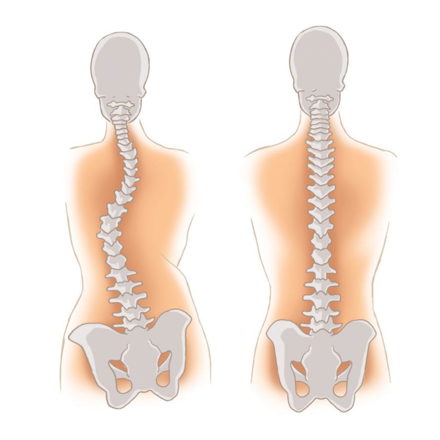
Left: Schematic illustration of scoliosis. Right: Healthy spine © Koterka Studio | AdobeStock
When describing scoliosis, however, the sagittal profile (side view of the spine) must not be overlooked, as IAS is often accompanied by a significant change in the sagittal profile as well, particularly in the form of a flat back.
At what age does scoliosis occur?
Women are affected by scoliosis significantly more often than men, at a ratio of 80:20. Scoliosis usually first becomes apparent between the ages of 10 and 12, often following a significant growth spurt.
The most noticeable features that play a role in the diagnosis of IAS are the lateral curvature and rotation of the vertebral bodies, leading to the development of the aforementioned deformities in the thoracic and/or lumbar spine.
Cause of Scoliosis
We currently assume that IAS arises from a genetic disorder, although the “scoliosis gene” has not yet been identified. This hypothesis is also supported by the fact that in families with a history of scoliosis, IAS frequently develops in subsequent generations, often skipping a generation.
In addition, recent studies show that, in addition to genetic factors, hormonal influences and spinal growth may also play a role. Idiopathic scoliosis can worsen particularly during childhood and adolescence, when the spine is developing rapidly.
In this condition, the spine curves sideways and rotates, which can lead to a curved spine over the long term. The exact cause of scoliosis remains the subject of ongoing research, as multiple mechanisms interact and the severity of scoliosis varies greatly from person to person.
Diagnosis of IAS
If a spinal curvature is suspected, the following examinations are necessary.
As part of the clinical examination, the doctor observes the child from behind and palpates the spinous processes of the spine. A curvature of the spine is often visible relatively early on, either in the thoracic spine (T-spine) or the lumbar spine (L-spine).
If a rotational misalignment of the vertebral bodies has already occurred at this stage, a more or less pronounced rib or lumbar bulge is present.
This formation of a rib bulge is of considerable importance in the early diagnosis of scoliosis, since the scoliosis often straightens out during an actively performed inclination movement (bending the trunk forward), causing the rib bulge to become more prominent. This occurs because, while the lateral curvature is partially corrected during the forward bend, the axial rotation is not, which then makes the rib bulge clearly visible.
During the clinical examination, it is important to note whether the waist triangle is flattened or indented. In cases of scoliosis in the lumbar spine, the curvature of the spine—usually to the left—causes the waist triangle to bulge on the left side and to be indented on the opposite side.
If these clinical changes are evident, radiological imaging should be performed as soon as possible. Care should be taken to ensure that a full-spine X-ray is always performed during the initial X-ray examination, in both the coronal and sagittal planes (side view).
It is important that the initial radiological evaluation be performed in both planes, as changes in the lateral profile can provide very good indications of the further progression of idiopathic scoliosis (IAS).
Progression and Treatment of Scoliosis Symptoms for a Better Quality of Life—Conservative Therapy and Surgical Treatment
Progression: How does scoliosis develop?
Once IAS has been definitively confirmed clinically and radiologically, regular follow-up examinations are necessary to detect progression.
Not every mild case of scoliosis worsens during further growth. At this stage—when the scoliosis curve measures between 15 and 20° on radiographic imaging—there are three possibilities:
- The scoliosis remains stable at this stage.
- The scoliosis worsens, with an increase in the curvature in all three planes.
- In a small percentage of cases, scoliosis of the magnitude described above may even show a reduction in the curvature.
Treatment: Further Diagnosis and Therapy for Scoliosis
If scoliosis of 15–20° is diagnosed, physical therapy is initiated as soon as possible, and many patients are recommended to wear a brace.
However, in cases of scoliosis with a curvature angle of 30° or more, the muscle strengthening achieved through physical therapy can no longer bring about any improvement. Nevertheless, continued, targeted exercise is still beneficial, and physical therapy should be combined with appropriate breathing therapy.
Physical therapy—not even the Lehnert-Schroth method—is definitely not capable of halting the progression of scoliosis. Furthermore, the patient and the treating physician should be aware that bracing therapy is never capable of halting the progression of scoliosis.
However, we must caution against bracing treatment, which is often initiated without sufficient consideration; there are several reasons for this:
- To date, there is no evidence that bracing can permanently halt the progression of scoliosis. It is already considered a very favorable outcome if the scoliosis does not curve any further, although I have already pointed out the possible progression of scoliosis above.
- A key reason against bracing is its negative impact on the sagittal (lateral) profile of the spine, which in most cases is already altered in the sense of lordosis (in most cases of thoracic scoliosis) and in the sense of deckyphosis of the spine (apical, thoracic lordosis).
- The third important point is that corset treatment is based on the fact that it does not involve a direct corrective intervention on the spine itself, but rather supports the spine via the ribs. As a result, long-term brace treatment—which, in cases of early detection of spinal abnormalities, can often last 4–5 years—leads to significant changes in the area where the ribs attach, specifically at the costotransverse and costovertebral joints (the ribs are connected to the corresponding vertebral body via two joints).
This pressure-induced change in the area of the costovertebral joints leads to a significant increase in rigidity. If, after many years of bracing, surgical intervention is eventually recommended or required, these changes pose a significant problem for surgical correction. Progressive ankylosis (stiffening of the vertebral joints) poses a very significant problem in the surgical treatment of scoliosis.
Surgical Treatment for Scoliosis
When is surgery necessary?
Depending on the progression (worsening) of the scoliosis, surgical treatment may be necessary; the peak age for surgery is between 12 and 16 years of age, depending on the rate of growth. Surgery should be delayed as long as possible to minimize its negative impact on further longitudinal growth of the spine.
A general guideline for surgery is a curvature angle of more than 40° in the coronal plane.
However, the progression of scoliosis is not limited to the coronal plane but also extends to the sagittal and rotational planes; therefore, these two planes must always be taken into account when determining the indication for surgery. An indication for surgery may be, for example, scoliosis with an angle of 30° accompanied by very severe rotation, since in such cases it can be predicted with a high degree of certainty that the scoliosis will significantly worsen over time.
When determining the indication for surgery, it should also be noted that the progression of scoliosis does not cease once spinal growth is complete (at 16 or 17 years of age). This is quite easy to explain: particularly in the lumbar spine, the scoliotic misalignment leads to massive abnormal stress on the intervertebral discs (increased pressure in the intervertebral disc on the concave side of the scoliosis), which then leads to premature, pathological degeneration of the intervertebral discs—a process that, in turn, is accompanied by a worsening of the curvature.
This is particularly evident in so-called thoracolumbar or lumbar scoliosis, where an increase in scoliosis is predictably accompanied by an increase in pain due to the mechanism described above.
The purpose of surgery to correct the misalignment in all three planes is not only to improve the cosmetic appearance—which, in severe cases, can certainly lead to psychological distress—but also to prevent secondary complications (premature degeneration of the scoliosis with resulting significant pain in adulthood), which often necessitates a more extensive and complex surgical procedure in adulthood.
Surgical Techniques
Various surgical approaches are possible to achieve surgical correction of scoliosis:
- The most commonly used posterior approach to the spine, which, however, is associated with various disadvantages (more extensive instrumentation, insufficient correction of rotational and sagittal deformities).
- The anterior approach to the spine, which in this case is an anterior-lateral approach. When indicated appropriately, this approach allows for very good correction in all three planes of the spine, typically combined with shorter instrumentation extending into the lumbar spine. Minimizing the length of instrumentation and fusion in the lumbar spine is of great functional importance, as approximately 70–80% of total spinal motion originates from the lumbar spine.
- In cases of very rigid scoliosis or double curves, a dual approach (from the back and the front-lateral sides) may be necessary to achieve optimal cosmetic and functional outcomes.
In the context of the anterior (from the front) approach, the tethering technique—which has been in use for about 5 years—must also be mentioned. This technique aims to correct scoliosis without fusion (stabilization) via an anterior lateral approach. The goal is to achieve correction by restricting growth on the convex side of the scoliosis and promoting further growth on the concave side.
The initial mid-term results indicate good corrections in some cases. However, it is important to note that this technique has no significant effect on the lateral profile or on axial rotational deformity; correction is primarily achieved in the frontal plane.
At this point in time, it is not yet possible to definitively evaluate this “tethering technique.” However, it offers an interesting approach that, to a limited extent, warrants cautious optimism.
FAQ on Scoliosis
What is scoliosis and how does it develop?
Scoliosis is a three-dimensional curvature of the spine that is often idiopathic. The exact cause is usually unknown, but spinal growth plays a central role.
What are the symptoms of scoliosis?
Typical symptoms of scoliosis include uneven shoulders or a tilted pelvis, a rib hump, back pain, and, in severe cases, impaired lung function.
How is scoliosis diagnosed?
Scoliosis is diagnosed through a physical examination and imaging techniques such as X-rays. The so-called Cobb angle is used to determine the curvature and the severity of the condition.
When is treatment necessary?
Treatment depends on the severity of the scoliosis, the patient’s age, and their growth. Mild cases of scoliosis are often treated conservatively, while severe cases may require surgery.
Can scoliosis worsen in adulthood?
Yes, untreated or severe scoliosis in particular can worsen in adulthood and lead to chronic symptoms or limited mobility.
Share this article