Cavernomas are benign vascular malformations of the brain and are classified as cerebral vascular malformations. A cavernoma is also known as a cerebral cavernous malformation (CCM). Many cavernomas remain asymptomatic and are diagnosed incidentally during an MRI scan. Depending on their location, however, cavernomas can cause neurologically significant symptoms, including epileptic seizures, hemorrhages, or headaches.
Cavernomas in the brainstem, in particular, are considered critical due to their increased risk of hemorrhage. Diagnosis is usually made using MRI (magnetic resonance imaging), as it clearly visualizes minor hemorrhages and blood breakdown products. In neurosurgery, a decision is made as to whether a cavernoma should be surgically removed or whether follow-up monitoring is sufficient. The management and treatment of cerebral cavernomas depend on the symptoms, location, and individual risk associated with the condition.
What is a cavernoma?
Cavernomas are also commonly referred to as “blood sponges.” They are a vascular malformation, also known as an arteriovenous malformation (AVM). In this condition, very small veins and arteries cluster together to form a tangle of blood vessels. This leads to frequent minor or even major bleeding episodes.
Cavernomas can occur in any tissue. However, only those that develop in the central nervous system (brain and spinal cord) are clinically significant. For this reason, the English-language scientific literature generally uses the term “cavernoma” exclusively to refer to cerebral cavernomas.
Blood pressure around cavernomas is low, so blood loss and tissue damage are limited. For this reason, they are usually asymptomatic in most tissue structures (e.g., legs, arms).
The situation is different with cerebral cavernomas. These are surrounded by brain or spinal cord tissue. Here, even minor bleeding can lead to pronounced symptoms such as severe headaches and seizures.
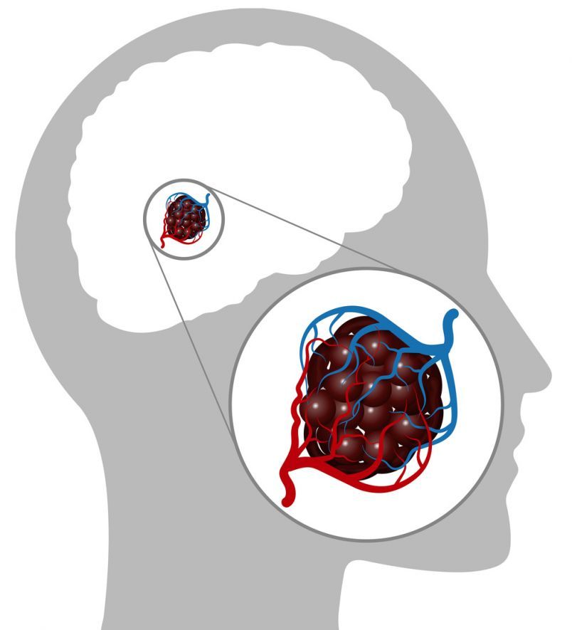
Cerebral cavernomas are benign malformations consisting of dilated blood vessels in the brain @ Naeblys /AdobeStock
Do cavernomas generally cause problems?
From asymptomatic to life-threatening: The symptoms of cavernomas vary greatly from case to case. About half of all cavernomas in the brain are asymptomatic. In the other half of cases, however, cerebral cavernomas cause a whole range of symptoms due to bleeding and compression.
The most common consequences are:
Major hemorrhages are particularly problematic.
Their symptoms closely resemble those of a stroke:
- Signs of paralysis
- Dizziness
- Visual and sensory disturbances, or
- Headaches
The incidence of such an event is low: Experts estimate the annual risk of bleeding at 1–5 percent. The exact risk depends on the location of the hemorrhage within the brain.
However, 30 percent of those who have experienced such a hemorrhage once will suffer another one within two years. These severe hemorrhages not only significantly impair the patient but can also be life-threatening.
In cases of minor bleeding, it is not the bleeding itself but the blood deposits in adjacent areas of the cerebral cortex that cause the symptoms. Cavernomas in the temporal lobe, in particular, can cause epilepsy in this way.
How common are cavernomas, and what causes them?
Cerebral cavernous malformations occur in about 0.5 percent of the German population, often without causing any symptoms. Exactly how they develop remains unclear.
What is certain is that there is a hereditary form. According to estimates, one-third of cavernomas are caused by a genetic factor. Six percent of cases involve a special form. In this familial form, also known as “multiple cerebral cavernomas,” cavernomas occur in clusters.
Consequently, it is likely that those affected will develop symptoms, especially since the cavernomas often grow more rapidly. This form primarily affects children.
Inheritance follows an autosomal dominant pattern. Consequently, there is a 50% probability that children will be affected by the same condition. Multiple cerebral cavernomas are less common in Europe than in other regions. However, the mechanisms by which cavernomas develop and the reasons behind their formation remain unclear. Apart from a genetic predisposition, there are no other known risk factors that prevent the development of cavernomas.
Diagnosis of a Cavernoma
In many cases, cavernomas are incidental findings because fewer than half of them cause symptoms. If a patient presents with symptoms at a doctor’s office or hospital, imaging is usually performed for further evaluation.
The following imaging techniques are used:
- Computed tomography (CT) or
- Magnetic resonance imaging (MRI)
CT is used in emergency situations such as stroke or acute bleeding. The scan and interpretation take only a few seconds.
MRI is suitable for situations where time is not of the essence, such as during planned procedures or after the emergency situation has been stabilized.
Performing an MRI is more time-consuming than a CT scan and usually takes 30 to 40 minutes. One advantage of MRI is that it provides significantly better resolution of morphology and localization.
A so-called “popcorn pattern” and the typical appearance of a “brom” or “mulberry” are often observed. Also typical are calcifications and signals indicating microbleeds that occurred at varying times in the past. Doctors generally look for other vascular malformations, which frequently occur alongside cavernomas. Examples include congenital vascular malformations or neoplastic conditions.
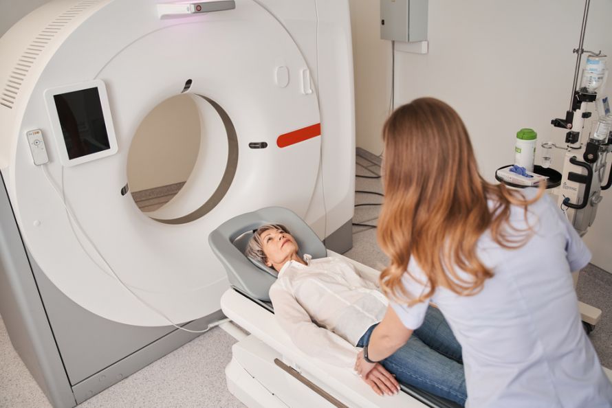 A cavernoma is diagnosed using imaging tests such as magnetic resonance imaging (MRI) or computed tomography (CT) @ Yakobchuk Olena /AdobeStock
When is treatment necessary?
Exactly how and when treatment for a cavernoma is necessary depends on many factors.
Key factors include:
- Location in the brain
- Accessibility during surgery
- Risk of bleeding
- Severity of symptoms
If the cavernoma is an incidental finding and asymptomatic, it is usually monitored annually via MRI.
Exceptions include cavernomas whose location necessitates intervention, for example, because they are very close to the brainstem. Very large cavernomas also frequently require treatment. A genetic cause of the cavernoma in cases of familial disease also often leads to a broad indication for treatment.
There is some debate regarding the avoidance of medications that affect blood clotting. This seems logical, as reduced blood clotting could increase the risk of bleeding. However, there are studies on other cerebrovascular diseases that actually view the use of anticoagulants positively. For this reason, there is still no clear recommendation on this matter. In cases of epilepsy or a single seizure without symptoms, the International League Against Epilepsy recommends treatment with anticonvulsant medications.
However, there are factors that favor direct removal.
These include:
- Young patient age
- Increased risk of bleeding, e.g., due to
- taking blood-thinning medications due to other medical conditions
- Epilepsy that is difficult to treat with medication
- Prone to falls
- High-risk sports (boxing, ski jumping)
- Rapid growth of the cavernoma
- Severe symptoms
What happens during surgery?
For cavernoma surgery, microsurgical removal using MRI-guided neuronavigation is the treatment of choice.
In this procedure, surgeons remove the diseased tissue using special instruments and light sources through small incisions (microsurgery).
It may also be necessary to sparingly remove the surrounding gliosis zone—that is, scar tissue in the brain and nerve sheaths.
Neurosurgeons, who are specialists in disorders of the central nervous system, typically perform the surgery.
Minimally invasive catheter procedures fall within the scope of neuroradiologists. German experts generally do not recommend radiation therapy (radiation) or chemotherapy.
What is the prognosis for cavernomas?
Cavernomas often remain asymptomatic for a lifetime. When symptoms do appear, this typically occurs between the ages of 10 and 40 in the majority of affected individuals.
The course of the disease depends primarily on the location and type of the cavernoma. For many patients, living with the risk of cerebral hemorrhage is often a significant burden. Symptoms such as headaches, seizures, and other neurological deficits can significantly impair quality of life. For young patients, their professional careers are often at risk. It can even lead to an inability to work.
One problem is the dynamic nature of cavernomas. This means that their growth patterns are difficult to predict. In some cases, they remain stable for years and change very little. At other times, they experience sudden growth spurts that can cause severe symptoms and are associated with a high risk of hemorrhage. Once the cavernoma has been surgically removed, the prognosis is often very good. Especially when surgeons can easily access the cavernoma, surgery carries comparatively few risks.
Bleeding or the consequences of epilepsy would often be more severe and more complicated to treat without surgery. Therefore, the majority of patients who undergo surgery experience fewer symptoms. Consequently, they are more satisfied. In the case of a single small cavernoma, surgery can often cure the condition.
In the form that runs in families, additional spontaneously occurring cavernomas are likely. This form is not curable. For those affected, ongoing monitoring of the cavernomas and, possibly, medication are necessary. The psychological distress experienced by those affected can be quite high due to a variety of symptoms and growing cavernomas. For this reason, additional psychological support is helpful.
FAQ
What are cavernomas?
Cavernomas are benign vascular malformations, or cavernous malformations, in the brain. The condition is also known as cerebral cavernous malformations (CCM). Cavernomas are characterized by dilated blood vessels that carry an increased risk of minor hemorrhages or bleeding.
What symptoms can cavernomas cause?
Depending on their location, cavernomas can cause symptoms such as epileptic seizures, neurological deficits, or headaches. A cavernoma in functionally important areas or cavernomas in the brainstem can be particularly critical. However, many asymptomatic cerebral cavernomas remain undetected for a long time and are discovered incidentally.
How are cavernomas diagnosed?
The primary diagnostic method is MRI (magnetic resonance imaging). This technique is particularly effective at visualizing cerebral cavernous malformations, blood breakdown products, and minor hemorrhages. In acute and emergency diagnostics, additional imaging techniques are also used.
When is surgery necessary?
Surgical treatment is considered when cavernomas cause recurrent bleeding or are symptomatic. The closer a cavernoma is to functionally important regions of the brain, the more carefully the neurosurgical treatment is planned. The goal is to prevent neurologically related damage.
How high is the risk of bleeding with a cavernoma?
The risk of bleeding depends on the location, size, and history of previous bleeds. Multiple cerebral cavernomas or cavernous hemangiomas in the brainstem often carry a higher risk. Regular follow-up MRI scans help detect changes at an early stage.
Share this article

Sources
- https://www.angiodysplasie.de/de/syndrome/Intracerebrale-Kavernome.php
- Mouchtouris N et al.: Management of Cerebral Cavernous Malformations: From Diagnosis to Treatment. The Scientific World Journal 2015; Art. 808314: doi.org/10.1155/2015/808314