Antireflux surgery is a procedure used to treat reflux disease. Antireflux surgery is performed via laparoscopy and is divided into three surgical steps: hiatal repair, gastropexy, and fundoplication. Antireflux surgery typically requires a one-week hospital stay. This is followed by a recovery period of 6 to 8 weeks.
Here you will find further information as well as a selection of antireflux surgery specialists and centers.
Definition: Antireflux surgery
Antireflux surgery is a surgical treatment option for reflux disease. This condition is also known as “gastroesophageal reflux disease” and, colloquially, as heartburn. The following video shows how heartburn develops in reflux disease:
Causes of heartburn
Heartburn occurs when acidic stomach fluid flows back up into the esophagus—and in extreme cases, all the way to the throat.
The most common cause of heartburn is a weakened lower esophageal sphincter at the entrance to the stomach. As a result, the stomach no longer seals properly against the esophagus, allowing stomach contents to rise. This condition is known as reflux disease.
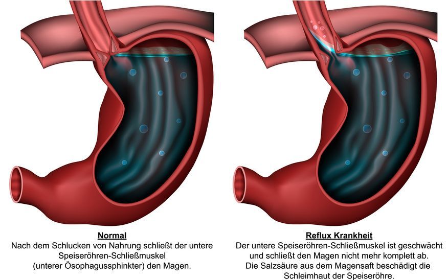
A hiatal hernia can also be a cause of heartburn. The diaphragm is a muscle shaped like an open umbrella that separates the chest cavity from the abdominal cavity.
The esophagus passes through an opening in the diaphragm from the throat into the abdominal cavity, where it connects to the stomach. This opening (known as the esophageal hiatus or diaphragmatic hiatus) is a natural weak point prone to herniation.
A hiatal hernia develops as the edges of this opening in the diaphragm slowly separate. Through this enlarged opening, the stomach may, under certain circumstances, move upward into the chest cavity.
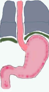
Fig. 1: In a normal situation, the stomach (pink) lies below the diaphragm (green), which separates the abdominal cavity from the chest cavity (thorax, gray).
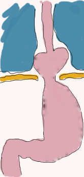
Fig. 2: In a diaphragmatic hernia, the opening in the diaphragm through which the esophagus passes is enlarged, allowing the esophagus to slide upward into the chest cavity, taking parts of the stomach (pink) with it.
Due to this change in position, the lower esophageal sphincter no longer functions properly. As a result, stomach acid can flow unimpeded into the esophagus, causing heartburn.
The aggressive gastric juice can lead to severe chemical burns of the esophagus (esophagitis). This alters the microscopic structure of the esophagus (metaplasia), which can lead to the development of cancer.
The main cause of a hiatal hernia is a congenital weakness of the connective tissue. Additionally, being overweight and a strong, persistent increase in abdominal pressure—for example, due to
- frequent straining,
- chronic coughing,
- strenuous physical labor, or
- pregnancy
contribute to the development of this hernia.
Eating habits can also contribute to the development of a hiatal hernia. Eating too quickly or hurriedly and frequently swallowing pieces of food that have not been chewed thoroughly increase the risk of developing the condition.
Diagnosis of Reflux Disease
The diagnosis of reflux disease is based on a physical examination and an endoscopy (gastroscopy). Additionally,
- an X-ray examination with contrast medium,
- an acid measurement (pH monitoring), and
- esophageal pressure measurement (manometry)
may also be used. If there is already significant inflammation of the esophageal lining caused by reflux (esophagitis), tissue samples are required. These allow the laboratory to rule out or confirm the presence of malignant changes.
Treatment of Reflux Disease with Antireflux Surgery
Heartburn is initially treated with medication. However, this only alleviates the symptom of reflux disease and does not address the underlying cause.
Antireflux surgery (also known as fundoplication or hemifundoplication) may be considered if
- medications are no longer effective (the so-called “tolerance effect”),
- side effects of the medication occur, or
- the patient does not wish to undergo long-term medication treatment.
Antireflux surgery puts an end to medication use that may have lasted for years. It can also help prevent the development of esophageal cancer (carcinogenesis prevention) to some extent.
Procedure for Antireflux Surgery
Antireflux surgery is performed via laparoscopy. During the procedure, the surgeons insert a camera and small instruments into the abdomen through 5 to 6 small incisions measuring 0.5 to 1 cm in length. The procedure is thus performed inside the body. The surgeon uses the camera to navigate directly within the surgical site.
In antireflux surgery, this mechanical change in the body is corrected in three surgical steps:
- Hiatoplasty
- Gastropexy
- Fundoplication
First surgical step of antireflux surgery: hiatoplasty
Hiatoplasty is used to reduce the size of the opening in the diaphragm—which has become too large—so that only the esophagus can pass through it.
The edges of the diaphragmatic opening are reattached using special sutures. Depending on the findings, a synthetic mesh is sometimes sutured over these sutures. This synthetic mesh remains permanently in the body and promotes more extensive scar tissue formation on the diaphragm. This significantly reduces the risk of a recurrence of the hernia.
Over time, the mesh becomes completely enclosed by scar tissue. It then lies “inside the body but outside the body” (extracorporeal mesh).
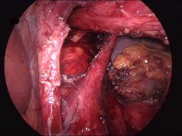
Fig. 3: View from the inside during a laparoscopy of the dilated diaphragmatic legs (V-shaped). The esophagus is at the top right of the image, and its gaping passageway between the diaphragmatic legs is visible.
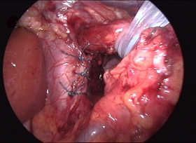
Fig. 4: In the so-called posterior pillar repair, the diaphragmatic legs are brought together using single-button sutures to narrow the passageway. The esophagus is held in place at the top right with a plastic sling.
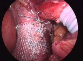
Fig. 5: To prevent a recurrent diaphragmatic hernia, the suture site (see Fig. 6) is additionally secured with a mesh. The esophagus is held out of the image toward the upper right with a suture.
Second surgical step of antireflux repair: gastropexy
During gastropexy, the surgeon pulls the portion of the stomach from the chest cavity back into the abdominal cavity. There, he secures it to the diaphragm on the right and left sides using staples or sutures.
This prevents the stomach from slipping upward again. It also reduces the strain on the sutures of the fundoplication cuff that is applied afterward.
Third step of antireflux surgery: fundoplication
Fundoplication involves forming a cuff from the gastric dome to reinforce the sphincter from the outside.
The so-called gastric dome—a bulge in the upper third of the stomach—is first freed from its adhesions to the spleen. The surgeon then pulls it beneath the esophagus and secures it to the esophagus on both the right and left sides. As a result, the esophagus lies as if in a “gastric cradle.”
The stomach now surrounds the esophagus at the level of the sphincter, thereby reinforcing it (Toupet procedure).
In another type of procedure, the esophagus is completely encircled by the gastric cuff (Nissen procedure).
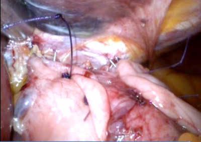
Fig. 6: Envelopment of the esophagus by the gastric dome (Toupet procedure). The two gastric folds (light-colored tissue)
converge again in front of the esophagus without touching.
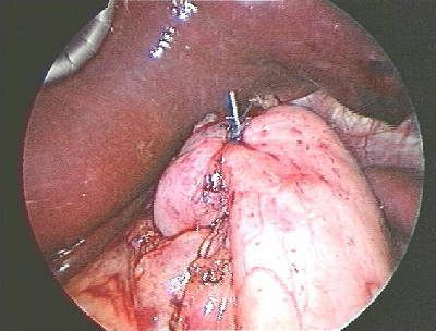
Fig. 7: In the Nissen procedure, the gastric fold is completely wrapped around the esophagus and the upper portion
of the gastric inlet. This ensures that the region is completely surrounded by gastric
tissue from the front. The liver is visible in the upper left of the image.
The doctor decides, based on the available findings, which type of procedure is most likely to yield the best results.
Complications and Risks of Antireflux Surgery
Complications occur less frequently in centers specializing in antireflux surgery.
However, as with any surgery, there is a risk of injuring neighboring organs. These include
- the liver,
- aorta,
- intestines, and
- spleen.
If the spleen is injured and bleeds, it may be necessary in very rare cases to remove it. This would mean that vaccinations would be required against diseases that the spleen would otherwise protect against.
If a large portion of the stomach has shifted into the chest cavity, its repositioning may result in injury to the pleura. In most cases, this heals without complications after the injury is properly sutured.
In rare cases, during antireflux surgery, a drainage tube is placed under the lung in addition to the sutures.
Consequences of Antireflux Surgery
Temporary difficulty swallowing may occur for a period of 6 to 8 weeks. This is due to the change in organ position and swelling in the surgical area. Patients should therefore eat more slowly and, of course, chew their food thoroughly. The difficulty swallowing usually resolves without any special treatment.
Some patients complain of a feeling of fullness in the upper abdomen for a period of 6 to 8 months. This is also a matter of getting used to it. Before antireflux surgery, patients often unconsciously swallow air repeatedly to “swallow away” heartburn. This unconscious swallowing does not stop immediately after surgery; the body adjusts only slowly.
Because the lower esophageal sphincter is now “functioning” again, the air swallowed can no longer easily escape upward. It must find its way through the intestines, resulting in a feeling of fullness.
Some patients are unable to vomit after antireflux surgery, or can do so only with difficulty. This is ultimately a sign that the surgery was successful, since the antireflux surgery is intended to prevent the reflux of stomach contents.
Postoperative Care Following Antireflux Surgery
Antireflux surgery typically requires a hospital stay of about one week. The patient can start drinking again on the day of the surgery. Many patients are already drinking coffee or orange juice again—drinks they had to go without for a long time due to heartburn.
On the first day after surgery, the patient is given a liquid diet, followed by soft foods the next day. After that, a normal, solid diet is provided.
After the procedure, the patient should avoid strenuous physical activity for 6 to 8 weeks. This allows the body to form a strong scar in the surgical area, which reduces the risk of a recurrent diaphragmatic hernia (“recurrent hernia”).
The patient should also change their eating habits to prevent another diaphragmatic hernia. This includes
- eating slowly and taking plenty of time
- chewing thoroughly, and
- not eating and drinking at the same time (i.e., not “washing down” food with a drink).
No further special follow-up care is necessary for this surgery.
After the required recovery period of 6 to 8 weeks, the patient can resume their normal life.
Conclusion on Antireflux Surgery
When performed at appropriately specialized surgical centers, antireflux surgery is a low-risk procedure for treating heartburn and hiatal hernias.
About the medical author
Dr. med. Susanne Röttgermann
Medical writer