Sclerotherapy (also known as sclerotic treatment) is a common treatment method for varicose veins and spider veins. Doctors also use it to treat hemorrhoids and dilated veins in the esophagus.
You can find more information on this topic, as well as a list of selected specialists in sclerotherapy, below.
What is sclerotherapy?
Sclerotherapy (also known as sclerotic treatment) is a common treatment for varicose veins and spider veins. These conditions occur when superficial veins dilate. Varicose veins and spider veins most often occur in the veins of the legs.
Varicose veins can cause blood to pool. This leads, on the one hand, to pain in the legs and, on the other hand, to a feeling of heaviness and congestion. In addition, varicose veins are a cosmetic concern for many people affected, as their bluish color makes them noticeable.
Veins may also bulge in cases of liver cirrhosis. These are also called esophageal varices. They occur in the area of the esophagus. Doctors treat these with sclerotherapy as well.
The same applies to hemorrhoids and varicose veins in the scrotum (varicocele). Doctors rarely use sclerotherapy to secure organs in place.
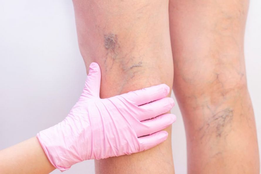 Varicose veins are knotty and winding, forming clusters or networks, and can protrude noticeably outward @ Elena /AdobeStock
How does sclerotherapy work?
During sclerotherapy, doctors destroy the tissue using special agents. The injected sclerosing agent damages the inner wall of the vein. This triggers an inflammatory reaction that leads to the vein becoming obstructed and eventually closed off (sclerotherapy). The veins then transform into a connective-tissue-like strand through which blood can no longer flow.
If there are multiple varicose veins, several sessions will be necessary. Careful preparation is important before beginning sclerotherapy.
The doctor performs various examinations:
- Ultrasound examination
- Venous occlusion plethysmography: This is a functional test in which doctors measure how quickly blood flows through the veins.
- Phlebography: In phlebography, the veins are visualized using a contrast agent. This helps to map the course of the veins and identify any narrowings, blockages, or dilations.
The sclerotherapy itself is performed using sclerosing agents in liquid or foamed form.
Sclerotherapy with Liquid Sclerosing Agents
Liquid sclerosing agents are typically used for smaller sections of veins and short-distance dilations.
Sclerotherapy is performed in several steps:
- First, the doctor draws the sclerosing agent into a sterile cannula using a syringe.
- The doctor performs an air block on the vein to ensure that the affected section no longer contains blood. This is important so that the sclerosing agent comes into direct contact with the vein wall. For the air block, a small, harmless amount of air or gas is injected into the relevant section of the vein. Carbon dioxide, for example, is suitable for this purpose. It displaces the blood in the vessel.
- The doctor then injects the medication into the vein wall. It is important that the needle is positioned precisely within the vessel and does not enter the surrounding tissue.
- After sclerotherapy, the doctor covers the injection site with a cotton pad and a bandage and applies a compression bandage. This compresses the affected area of the body. Compression stockings may also be used (compression therapy).
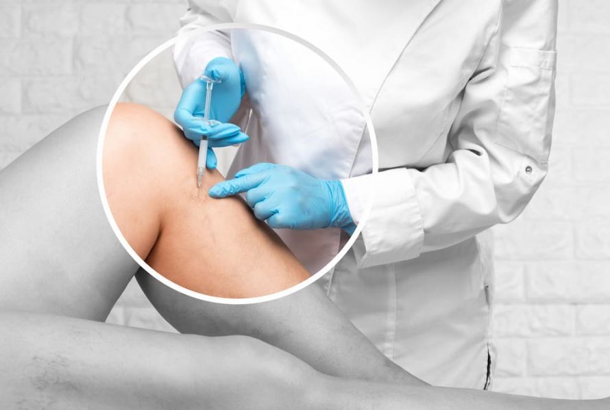 The sclerotherapy of varicose veins is performed by injecting various sclerosing agents, which can be liquid or foamed @ Dimid /AdobeStock
Sclerotherapy with Foamed Medications
In foam sclerotherapy, doctors mix the medication with a small amount of air or gas, such as carbon dioxide. This creates a fine-bubbled foam. Foamed medications are particularly suitable for long stretches of varicose veins.
The procedure for foam sclerotherapy is similar to that for sclerotherapy with liquid medications. Here, too, the doctor draws the medication into a syringe and then injects it into the vein. By drawing back a small amount of blood, doctors can verify that the syringe is positioned correctly. However, an air block is not necessary for foam sclerotherapy, as the blood is displaced by the foam. The medication can then reach the vessel walls and take effect there.
Risks of Sclerotherapy
Sclerotherapy is one of the standard procedures in modern medicine.
Nevertheless, problems can still arise:
- Damage to or perforation of the vessel wall, resulting in bleeding
- Infection at the injection site (which may require follow-up treatment with antibiotics or surgery)
- Permanent discoloration of the skin surrounding the blood vessel
- Tissue damage (abscesses or cell necrosis)
- Inflammatory reactions with skin redness
- Impaired wound healing
- Temporary visual disturbances (also known as flickering)
- Scab formation at the injection site
- Nerve damage (rarely permanent)
- Migraine attack (only in patients with a history of migraines)
- Allergic reaction to administered medications or intolerance to the materials used
- Formation of blood clots
- Lymphatic congestion
Follow-up Care After Sclerotherapy
Immediately after treatment, minor swelling, a feeling of tightness, bruising, and skin redness may occur at the injection site. These symptoms usually subside after a few days and are not a cause for concern. However, patients should definitely consult their doctor if the following symptoms occur:
- Increasing, throbbing pain
- Pain on pressure or a burning sensation on the skin caused by the bandages
- Severe redness, swelling, or warmth in the treated area
- Numbness or tingling in the foot
- Blue discoloration of the toes
- Fever above 38 degrees
To avoid such complications and incidents, patients must have their bandages checked and changed regularly. During these regular checkups, the doctor will determine how long the patient should wear compression stockings.
After sclerotherapy, patients may shower as usual. However, taking a long bath or visiting a sauna is not recommended until several weeks have passed. The exact timeframe depends on the extent of the varicose veins and the type of sclerotherapy performed.
Patients should avoid exposing the affected area to direct sunlight for four to six weeks. This increases the risk of discoloration.
To prevent lymphatic congestion, patients should elevate their legs as often as possible. They should also avoid sitting or standing for too long. Daily physical activity, such as cycling or walking, is recommended.