Umbilical hernias—commonly known as belly button hernias—are a common type of abdominal wall hernia. A hernial opening forms in the abdominal wall around the navel, through which the intestines or fatty tissue can protrude into the abdominal cavity.
While an umbilical hernia in children often heals on its own, surgery is almost always necessary for adults. Depending on the size of the hernia, the symptoms, and the risk of incarceration, doctors perform the surgery either on an outpatient basis or as an inpatient procedure.
In this article, patients will find all the important information about the umbilical hernia surgery procedure, surgical methods, risks, and postoperative care.
What Is an Umbilical Hernia? Causes & Typical Symptoms
An umbilical hernia—also known medically as an umbilical hernia—is a common type of abdominal wall hernia. It occurs in the area around the navel, which is a natural weak spot in the abdominal wall. A hernial opening can form there, through which abdominal contents protrude into the abdominal cavity. Parts of the intestine are often affected as well.
The navel is the spot where the umbilical cord ran during pregnancy. If the abdominal wall does not close completely as the body develops, a weak spot remains—and this is how an umbilical hernia develops.
Umbilical hernias occur in both children and adults:
- In newborns, the umbilical hernia usually resolves on its own by the age of two. Surgery is necessary at this age only if complications arise, as the risk of incarceration is very low.
- In adults, an umbilical hernia is usually caused by a weakening of the abdominal wall, overstretching, or increased pressure in the abdominal cavity (e.g., when lifting heavy loads or during pregnancy).
Many patients initially have no symptoms or only mild discomfort. Only when the umbilical hernia becomes incarcerated does a dangerous complication arise—in this case, immediate surgery is required.
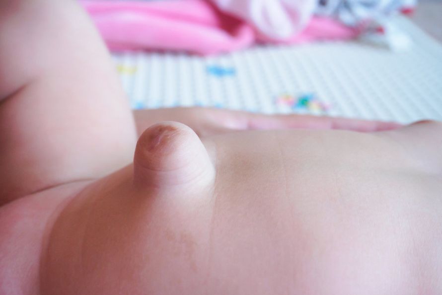
Risk Factors: When Does an Umbilical Hernia Develop?
An umbilical hernia occurs whenever abdominal pressure in the area around the navel becomes too high or there is a weak spot in the abdominal wall.
In newborns, a small umbilical hernia is initially normal, as the umbilical cord has not yet completely closed shortly after birth. Premature infants with very low birth weight are particularly affected—about two-thirds of these children develop an umbilical hernia.
In adults, there are various risk factors that contribute to an umbilical hernia. These primarily include situations associated with increased abdominal pressure, such as:
- severe obesity
- heavy lifting and handling heavy loads in daily life or at work
- liver cirrhosis with abdominal fluid (ascites)
- pregnancy
In addition, a family history of connective tissue weakness also increases the risk of developing an umbilical hernia.
Symptoms and Types of an Umbilical Hernia
An umbilical hernia can cause a wide range of symptoms—but it can also remain asymptomatic for a long time.
A typical symptom is a visible bulge in the navel area, which in many cases can be pushed back into the abdominal cavity. Especially in infants with an umbilical hernia, the bulge may persist briefly when the child cries or strains.
An umbilical hernia often worsens:
- when coughing,
- when feeling the urge to have a bowel movement,
- during physical exertion at work, or
- during sports.
The bulge may also remain visible even when lying down. Patients then report pain in the abdominal area or a feeling of pressure.
Warning signs:
If severe pain suddenly occurs along with fever, nausea, and vomiting, the intestine may be strangulated. If the bulge can no longer be pushed back into the abdominal cavity or if the umbilical hernia turns bluish, there is an acute danger.
In such cases, there is a risk of life-threatening intestinal obstruction. The intestine is no longer receiving adequate blood flow, tissue may die, and bacteria can enter the abdominal cavity.
If these symptoms are present, immediate surgery for an umbilical hernia is required.
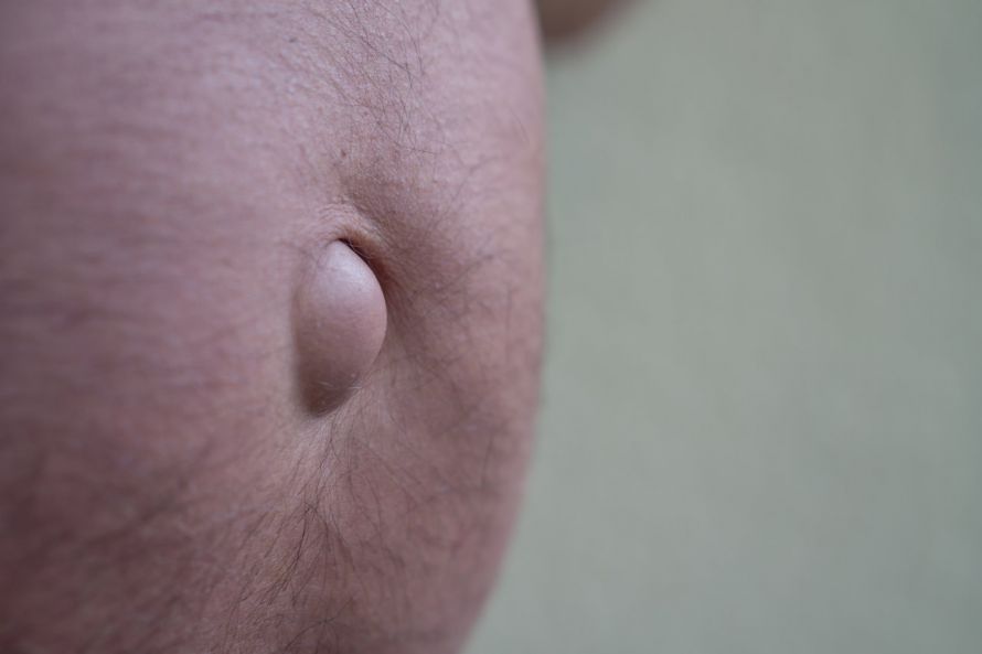
Diagnosis of an umbilical hernia: How an umbilical hernia is detected
An umbilical hernia can usually be diagnosed clinically. The soft bulge directly in the area of the navel is a typical sign for the doctor or surgeon.
During the physical examination, the surgeon palpates the hernial orifice. This allows them to check whether the contents of the hernial sac can be easily pushed back into the abdominal cavity.
Additional tests are sometimes necessary for an accurate assessment of an umbilical hernia. The most important factors are:
- the size of the hernial opening,
- the patient’s age and weight,
- any symptoms.
Ultrasound as the Most Important Test
Ultrasound is particularly informative, as it can visualize the hernial orifice and the contents of the hernia. Further diagnostic procedures are generally not required.
Treatment & Surgery: When Is Umbilical Hernia Surgery Necessary?
In the case of small hernias in children, the umbilical hernia often resolves spontaneously during the first few years of life. Therefore, doctors rarely need to perform surgery for an umbilical hernia.
An umbilical hernia that develops during pregnancy also often resolves on its own.
However, if the hernia opening is larger than 1.5 cm in diameter, it rarely closes on its own. In such cases, umbilical hernia surgery should be considered in a timely manner.
When is umbilical hernia surgery necessary?
Spontaneous healing does not occur in adults: an umbilical hernia does not resolve on its own. Additionally, there is a risk of intestinal obstruction. Therefore, surgery should be performed early for any umbilical hernia that causes symptoms.
However, even in the case of an umbilical hernia that is initially asymptomatic, specialists recommend scheduling surgery in a timely manner. This helps prevent the hernial opening from enlarging or the hernia from becoming incarcerated.
In children, doctors generally wait until the age of 6 to perform umbilical hernia surgery. If the hernia enlarges or symptoms develop, the procedure is performed earlier.
In acute cases—such as when the navel turns blue, severe pain is present, or the hernial sac can no longer be pushed back into the abdominal cavity—it is considered an emergency. In such cases, the surgical principle applies: “The sun must neither
rise nor set over the strangulated umbilical hernia.”
Surgical treatment depends on the size of the hernia:
- Doctors can repair small hernias under local anesthesia.
- For larger hernias, general anesthesia is usually used.
Depending on the findings, the surgery at the hospital may be performed on an outpatient basis. In that case, the patient may go home on the day of the surgery. In complicated cases (e.g., acute incarceration or pre-existing conditions), inpatient surgery is necessary.
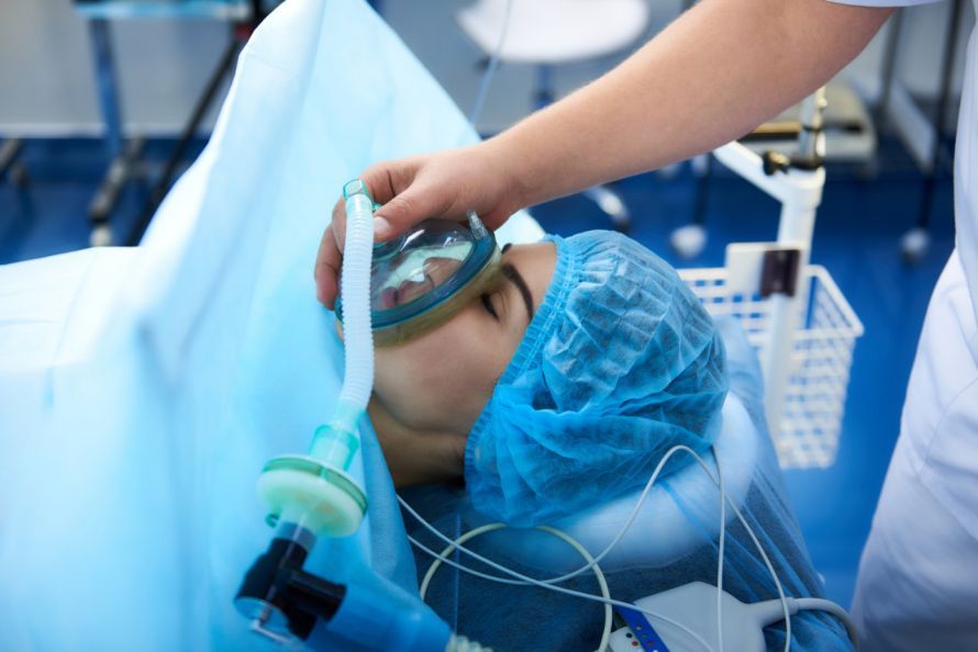
Before every surgery, the anesthesiologist explains the anesthesia procedure and potential risks to the patient. Hours before the procedure, the patient receives thrombosis prophylaxis and a mild laxative. Shortly before surgery, a sedative is administered.
Procedures and Methods for Umbilical Hernia Surgery: Open Surgery & Minimally Invasive Procedures
The procedure for an umbilical hernia operation depends on the size and severity of the hernia. First, the surgeon makes an arc-shaped incision around the navel to open the abdominal wall. Next, the skin over the hernial sac is lifted, and the hernial contents are carefully pushed back into the abdominal cavity. If necessary, the surgeon removes the hernial sac.
The edges of the hernial orifice are then sutured directly together to close the hernial opening.
- Small hernias are usually treated with sutures—doctors sew the edges of the hernia directly together.
- For larger umbilical hernias (2 cm or larger), doctors almost always insert a synthetic mesh. This stabilizes the abdominal wall and reduces the risk of the hernia recurring.
Depending on the location and size, different surgical methods are used:
- Sublay method
- Inlay method
- Onlay method
- IPOM method
Closed (laparoscopic) umbilical hernia surgery
In this minimally invasive procedure, access is gained through the abdominal wall using laparoscopy. To do this, the surgeon makes several small incisions in the abdominal wall, inserts a camera and surgical instruments into the abdominal cavity, and closes the hernia site. A mesh is often inserted as well to reinforce the abdominal wall.
Advantages:
- less pain after surgery,
- lower infection rate,
- Patients are able to resume physical activity within the first few days and can soon begin light exercise.
Open Umbilical Hernia Surgery
In open surgery, the surgeon exposes the hernial sac through a larger skin incision and pushes the contents of the hernial sac back into the abdominal cavity.
The incision is closed using traditional sutures—the so-called Spitzy procedure. Less commonly, a fascial flap is used.
For larger umbilical hernias, doctors permanently stabilize the abdominal wall using:
- non-absorbable synthetic mesh or
- a combination of mesh and the body’s own tissue.
This supports the abdominal wall and reduces the likelihood of a recurrence.
Spitzy procedure: Treatment of small umbilical hernias
In the case of a small umbilical hernia (hernia orifice < 2 cm), the Spitzy procedure involves directly suturing the edges of the hernia. Although this classic umbilical hernia surgery is often performed under general anesthesia, it can also be done on an outpatient basis. The procedure usually takes only 20 to 30 minutes.
Advantages:
- short procedure time,
- minimal trauma (hardly any tissue damage),
- very low complication rate.
However, this method does not provide stabilization through a synthetic mesh. As a result, the risk of a recurrent hernia is higher, and the patient must take it easy for several weeks after umbilical hernia surgery before resuming light exercise or full physical activity.
Fascial Double-Stitching: Umbilical Hernia Surgery According to the Mayo Method
In fascial doubling, surgeons suture the edges of the abdominal wall in two layers to create an overlap. This umbilical hernia surgery is also known as the Mayo method.
It was commonly used in the past but is rarely performed today. Reason: Recurrences occur significantly more frequently with this technique, as stability is lower without mesh reinforcement.
Techniques Using Synthetic Mesh: Modern Surgical Methods
For larger umbilical hernias, a synthetic mesh is almost always used. This polypropylene mesh is well-tolerated and is inserted either during open surgery or via minimally invasive umbilical hernia surgery.
The mesh stabilizes the abdominal wall and significantly reduces the risk of recurrence. This prevents another umbilical hernia from developing.
There are several different surgical methods:
- Sublay mesh technique (under the abdominal muscles),
- IPOM technique (intraperitoneal onlay mesh): mesh placed between the intestines and the peritoneum,
- Inlay technique: The mesh is placed directly into the hernial defect.
This allows the hernia defect to be optimally closed depending on its size and location. Once in place, the mesh ensures that the abdominal wall remains permanently stable.
Complications: Possible Risks Following Umbilical Hernia Surgery
The outcomes following umbilical hernia surgery are generally very good. Recurrences—that is, the reappearance of the umbilical hernia—usually occur only in cases of larger defects or in patients with additional risk factors.
However, as with any surgical procedure, there are potential complications associated with umbilical hernia surgery. While these are rare in elective surgeries, it is important to be aware of them:
- Wound infections or inflammatory reactions in the area of the wound,
- Seroma formation (accumulation of fluid in the surgical area),
- hematomas (bruising),
- temporary numbness due to nerve irritation.
Particularly when a synthetic mesh is used, the infection rate following umbilical hernia surgery is approximately 2 to 4 percent. Patients may also notice slight swelling or hardening in the surgical area during the first few days after surgery.
The fluid accumulations (seromas) usually resolve on their own within 2 to 6 weeks. A puncture is rarely necessary.
In isolated cases, patients may complain of limited mobility or more severe symptoms such as nausea and vomiting. However, these are rare and usually subside after a short time.
Postoperative Care & Healing Process: Weeks After Umbilical Hernia Surgery
Postoperative care following an umbilical hernia surgery is crucial for supporting a complication-free recovery. Patients can resume a normal diet immediately after surgery. The skin is either closed with adhesive strips or sutured with dissolvable sutures—allowing patients to shower again within the first few days after surgery.
In the first few weeks after surgery:
- Lifting up to 10 kg is permitted.
- Lifting heavy loads and intense abdominal exercises should be avoided.
- After 4 weeks to 3 months, activities such as jogging and light exercise are possible again.
In daily life, patients should pay particular attention to physical strain at work. A return to full activity depends on the individual’s healing process.
Most patients can resume their normal daily routines just a few weeks after umbilical hernia surgery. It is important to follow the recommendations of the treating surgeon to allow the hernia site to heal properly.
An experienced specialist in hernia surgery guides patients through the entire process, from the umbilical hernia surgery itself to postoperative care.

About the medical author
Prof. Dr. med. Ferdinand Köckerling
Medical Author · Director
Prof. Dr. med. Ferdinand Koeckerling – medical author: expert articles, professional insights and medical knowledge in the Leading Medicine Guide.
More about the medical author →