Cholangitis is an acute or chronic inflammation of the gallbladder, medically known as cholecystitis. In most cases, cholangitis is caused by gallstones that obstruct the flow of bile.
This results in acute inflammation, which can be accompanied by severe pain in the upper right abdomen, fever, and nausea. An inflamed gallbladder is a serious condition, as it can lead to complications if left untreated. Cholecystitis occurs particularly frequently when a gallstone blocks the bile duct and bile becomes backed up.
In addition to acute gallbladder inflammation, there are also chronic forms that cause recurring symptoms. Early diagnosis and targeted treatment are crucial for a favorable outcome.
Definition: Inflammation of the Gallbladder (Cholecystitis)
Gallbladder inflammation refers to inflammation of the gallbladder wall. In most cases (90 to 95 percent), gallstones are the cause of the inflammation.
Cholangitis can be acute or chronic.
In acute cholecystitis, the patient experiences colicky pain in the right upper abdomen. This pain develops into a persistent ache and may be accompanied by fever and nausea.
In chronic cholecystitis, the gallbladder wall is repeatedly irritated by gallstones or bile. Persistent dull or intermittent pain and digestive symptoms may occur.
Chronic cholecystitis increases the risk of gallbladder cancer.
Location and Function of the Gallbladder
The gallbladder is located in the gallbladder fossa at the lower edge of the liver in the right upper abdomen. The main function of the gallbladder is to store the bile produced by the liver. When food is ingested, it releases bile through the common bile duct into the duodenum. There, the bile emulsifies the chyme and plays a crucial role in the digestion of fats.
The gallbladder is equipped with several sphincter muscles that regulate the flow of bile through contraction.
Causes of Gallbladder Inflammation
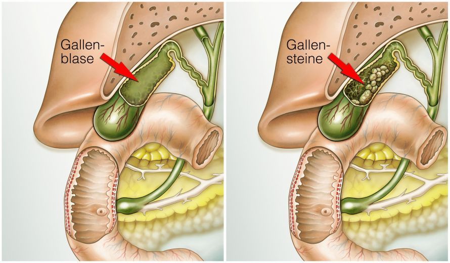
Cholangitis is caused in most cases by gallstones. They obstruct the flow of bile, causing the gallbladder to become overfilled.
This overfilling initially leads to cramp-like pain, known as colic. As the condition progresses, bacteria from the intestines can enter the gallbladder via the bile duct and infect it.
Gallstones form when
- the composition of bile—and thus the solubility of substances such as cholesterol—changes,
- the motility (movement) of the gallbladder is impaired, or
- bacteria cause the bile to crystallize.
Accordingly, different types of gallstones are distinguished. In most cases, these are cholesterol and mixed gallstones. Pigment gallstones, which consist mainly of bilirubin, are less common.
Risk factors for the development of gallstones include
- physical inactivity,
- an unhealthy diet (too high in calories and too low in fiber), and
- being overweight.
Estrogen-based hormone therapy also appears to increase the risk of gallstones.
Other risk factors include
- being around 40 years old,
- being female,
- pregnancy,
- metabolic disorders,
- rapid weight loss, and
- family history.
In rare cases, gallbladder inflammation is caused by
- surgery,
- inflammation of other internal organs,
- burns, or
- accidents
.
Symptoms of gallbladder inflammation
Gallbladder inflammation is primarily characterized by severe, cramping pain in the upper right abdomen. The pain may also radiate in a belt-like pattern toward the back.
- Nausea and the urge to vomit, occasionally accompanied by vomiting,
- a slight yellowing of the skin, and
- digestive problems
are additional symptoms of gallbladder inflammation.
If bacteria infect the accumulated bile, the symptoms listed above are accompanied by
- fever,
- chills,
- tenderness, and
- a tense abdominal wall in the upper right quadrant
.
Diagnosis of Cholecystitis
Various tests are used to diagnose gallbladder inflammation:
- physical examination,
- blood tests, and
- imaging tests.
Typical signs of gallbladder inflammation include
- local signs of inflammation: tenderness on ultrasound examination and tension in the abdominal muscles during palpation of the abdomen—known as defensive tension,
- general signs of inflammation: See below under “Laboratory Analysis,”
- thickening of the gallbladder wall, and
- in most cases, the detection of gallstones.
Laboratory Analysis
A blood test is an important part of the evaluation for gallbladder inflammation.
In cases of gallbladder inflammation,
- the white blood cell count is elevated,
- the erythrocyte sedimentation rate is accelerated, and
- the concentration of C-reactive protein (CRP) is elevated.
In addition, liver enzyme levels may be above the normal range.
Imaging Procedures
During an ultrasound examination (sonography), the gallbladder is examined using a transducer. With this procedure, the doctor can
- detect an enlarged gallbladder,
- swelling of the gallbladder wall, and
- subsequently a so-called three-layered structure of the gallbladder wall
. The gallstones causing the condition can also be visualized using ultrasound. A typical symptom of gallbladder inflammation is localized pain that occurs when the doctor applies pressure with the transducer. This phenomenon is also known in medicine as Murphy’s sign.
Sometimes the results of an ultrasound examination are inconclusive. In such cases, magnetic resonance imaging (MRI) or computed tomography (CT) may also be used.
For this, the patient is first given a contrast agent, which allows the doctor to better visualize the gallbladder wall and the surrounding area. This may provide evidence of inflammation.
Treatment of Gallbladder Inflammation
Gallbladder inflammation should always be treated early. If treatment is delayed, life-threatening complications can develop, such as a ruptured gallbladder. This causes infected bile to spread throughout the abdominal cavity. This can lead to peritonitis.
Once diagnosed, immediate treatment with antibiotics should be initiated to combat the bacterial infection. In addition, symptomatic therapy is provided with pain relievers and antispasmodic medications.
Surgical Treatment of Gallbladder Inflammation
Surgical removal of the gallbladder (cholecystectomy) and the stones it contains is then recommended and necessary. Otherwise, a recurrence of symptoms and inflammation would be expected.
The surgery can be performed immediately during the acute phase of inflammation. Alternatively, it is performed at least 6 weeks after the inflammation has subsided.
Surgical removal of the gallbladder is usually possible using a minimally invasive laparoscopic approach. Under general anesthesia, the surgeon makes 3–4 small incisions, each 0.5–2 cm long. Through these incisions, the surgeon inserts a tiny camera, a light source, and the necessary instruments. The surgery is then performed while the surgeon views a monitor that displays the camera’s image from inside the body.
Most patients are able to leave the hospital as early as the day after surgery.
Only in rare cases of severe inflammation is it necessary to open the abdomen with a large incision to remove the gallbladder.
If gallstones are lodged in the common bile duct, they can be removed endoscopically using an ERCP (endoscopic retrograde cholangiopancreatography).
Complications from Improper Treatment
Dissolving or breaking up (extracorporeal shock wave lithotripsy) gallstones is not recommended. Such attempts can lead to serious complications. For example, fragments of the gallstones can enter the common bile duct and block it.
Stones could also become lodged in the area of the papilla. The papilla is the common opening where the common bile duct and the pancreatic duct enter the duodenum. This can lead to life-threatening inflammation of the pancreas (pancreatitis).
How can gallbladder inflammation be prevented?
In the vast majority of cases, gallbladder inflammation is caused by gallstones. Therefore, prevention primarily involves measures to prevent gallstones.
A balanced diet tailored to one’s needs plays an important role. The following also appear to have a preventive effect:
- fruit,
- vegetables,
- legumes,
- olive oil,
- whole-grain products,
- Lettuce,
- nuts, and
- unsaturated fats.
The same applies to an adequate intake of vitamin C and magnesium. Regular physical activity also helps maintain a healthy body weight.
In contrast, saturated fats, sweets, and high-calorie beverages increase the risk. In certain situations, such as
- in cases of overweight,
- following weight loss, or
- after bariatric surgery,
medications can help lower the risk. However, there is no general recommendation for preventive medication.
FAQ
What exactly is gallbladder inflammation?
Gallbladder inflammation, also known as cholecystitis, is an inflammation of the gallbladder that is usually caused by gallstones. These block the flow of bile, leading to inflammation of the gallbladder.
What are the symptoms of gallbladder inflammation?
Typical symptoms of gallbladder inflammation include severe pain in the upper right abdomen, fever, nausea, and vomiting. The pain often radiates to the right shoulder and intensifies when pressure is applied to the upper right abdomen.
How is the diagnosis made?
If cholecystitis is suspected, a physical examination is performed first, followed by an ultrasound to visualize the inflamed gallbladder. A thickening of the gallbladder wall visible on ultrasound is considered a key indicator of acute cholecystitis.
How is gallbladder inflammation treated?
Treatment for gallbladder inflammation usually begins with antibiotics, pain relievers, and antispasmodic medications. In many cases, a cholecystectomy—the surgical removal of the gallbladder—is necessary. The surgical removal of the gallbladder is often performed using minimally invasive techniques.
Can gallbladder inflammation occur without gallstones?
Yes, acalculous cholecystitis is gallbladder inflammation without gallstones. This form is less common but can occur, particularly in cases of severe infection or in patients receiving intensive care.
Share this article
Sources
- Gutt C et al. (2017) Aktualisierte S3-Leitlinie der Deutschen Gesellschaft für Gastroenterologie, Verdauungs- und Stoffwechselkrankheiten (DGVS) und der Deutschen Gesellschaft für Allgemein- und Viszeralchirurgie (DGAV) zur Prävention, Diagnostik und Behandlung von Gallensteinen. AWMF-Register-Nr. 021/008. Z Gastroenterol 2018; 56: 912–966
- Lenzen H, Lankisch T (2014) Cholelithiasis. In: Lehnert H et al. (eds) SpringerReference Innere Medizin. Springer Reference Medizin. Springer, Berlin, Heidelberg
- Zylka-Menhorn V (2013) Akute Cholezystitis: Die frühzeitige Operation ist dem konservativen Vorgehen überlegen. Dtsch Arztebl 110(37): A-1683 / B-1489 / C-1464