In an umbilical hernia, tissue protrudes through a weak point by the navel and bulges out, taking a spherical form. Umbilical hernias are not rare in children and infants. But adults can also have umbilical hernias - though much less frequently. An umbilical hernia can be present at birth or acquired later. Whereas an umbilical hernia in a baby normally corrects itself, in adults umbilical hernia surgery is necessary.
Recommended specialists
Article overview
What is an umbilical hernia?
The abdominal wall consists of connective tissue, abdominal muscles and abdominal skin. It is not of the same consistency everywhere and has weak points. One of these weak points is the navel. The abdominal wall is always subject to enormous pressure - it contains our internal abdominal organs. When we cough or sneeze or lift something, the pressure increases significantly. Pregnancy is also a strain on the abdominal wall. An umbilical hernia forms if the connective tissue around the navel cannot withstand the pressure.
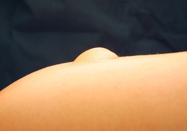
An umbilical hernia seen from the side © Prof. Dr. med. Thomas W. Kraus
A gap - known as a hernial ring - develops in the abdominal wall. The peritoneum (abdominal lining) then protrudes through this gap. The medical term for this protrusion is 'hernia sac'. Occasionally, some parts of the bowel also enter the hernia sac. In rare cases, the rupture causes the bowel loops to become strangulated, which means that blood can no longer circulate in them. A strangulated or incarcerated umbilical hernia is a medical emergency, especially if parts of the bowel have prolapsed. There is a risk of parts of the bowel dying so immediate surgery is essential.
Who is affected most often by an umbilical hernia?
Statistically, babies are foremost in the list - in this country around one in ten babies has this. Premature babies with a birth weight of less than 1,500 g are especially at risk - two thirds of them are born with a congenital umbilical hernia. In adults, women are more at risk then men - they suffer from umbilical hernias around four times as often as men. Women between the ages of 50 and 60 are especially at risk.
How does an umbilical hernia occur?
The navel is a natural weak point in the abdominal wall. During pregnancy, the umbilical cord, which supplies the embryo with all the essentials of life, starts developing. After birth, the exit point of the umbilical cord slowly closes up and develops into the navel. However, the navel remains as a permanent weak point because it consists of only thin muscle layers and connective tissue.
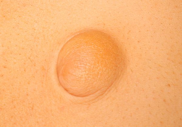
Umbilical hernia seen from above © Prof. Dr. med. Thomas W. Kraus
As well as that, the navel represents an interruption in the stabilizing line of connective tissue, which links the tendon layers of the abdominal muscles. This line, known as the white line or linea alba, does not pass through the navel area. Expectant mothers will be aware of this white line because during pregnancy it is clearly visible as a vertical 'seam'. If the tissue around the navel cannot withstand the increased pressure in the abdominal cavity an abdominal ring will develop. The peritoneum will then push through the opening, forming a protruding pouch of skin. If this hernia sac is big enough, bowel loops can force their way into it.
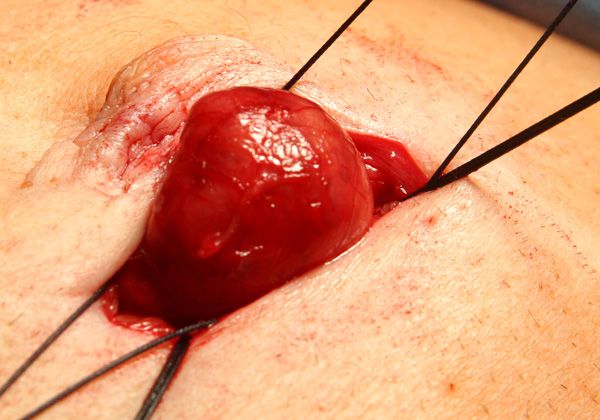
Hernia sac of an umbilical hernia with prolapsed tissue © Prof. Dr. med. Thomas W. Kraus
There are different reasons for umbilical hernias in babies. If the umbilical hernia is congenital, the causes lie in the way the embryo developed in the mother's uterus. After the 32nd day of pregnancy, the embryo develops a natural umbilical hernia. At this stage in the baby's development, the bowel loops develop faster than the abdominal cavity, so because there is insufficient room for them, parts of the bowel escape into the umbilical cord. Normally, this umbilical hernia retracts of its own accord by the ninth week of pregnancy. Occasionally, this does not happen and the baby is born with an umbilical hernia. Sometimes, an umbilical hernia does not develop until the first weeks after birth, before the tissue around the navel has closed up completely. The navel tissue, which is still weak, cannot always withstand the pressure of coughing, shouting or straining.
The following risk factors favor the formation of an umbilical hernia:
- lifting heavy weights
- significant overweight
- pregnancy
- genetically conditioned weakness of the connective tissue
- certain illnesses (for example, ascites, i.e. an accumulation of fluid in the abdomen, chronic lung diseases or diabetes)
What are the symptoms of an umbilical hernia?
In babies, an umbilical hernia can be seen as a soft swelling in the navel, which the doctor can push back into the abdomen. The size of the protrusion varies according to the size of the opening which has developed. Anything is possible, from the size of a cherry up to that of a tennis ball. Smaller umbilical hernias only occur through shouting or straining. In normal circumstances, an umbilical hernia does not cause any discomfort. It is sometimes painful only if the abdominal cavity is under increased pressure (through lifting or coughing). It is quite different if bowel loops are trapped in the umbilical hernia. Extreme pain is felt very quickly. Babies scream continuously and the navel turns a dark color. Immediate treatment is essential in the event of an incarcerated hernia. Even though an umbilical hernia in babies is generally harmless and causes no discomfort, it is still essential for a pediatrician or surgeon - generally a visceral surgeon or hernia surgeon - to identify the cause.
How is an umbilical hernia treated?
It is rare for a baby's umbilical hernia to need treatment. In 90% of all cases, the umbilical hernia retracts of its own accord by the second year of life. If this is not the case, surgery will be needed. However, there is no hurry as long as the umbilical hernia remains inconspicuous. In most cases, surgery is carried out before the child starts school.
Because umbilical hernias in adults do not generally retract naturally, the only treatment option is hernia surgery. In this, the surgeon removes the hernia sac and closes the hernial ring by suturing the separate tissue layers together.
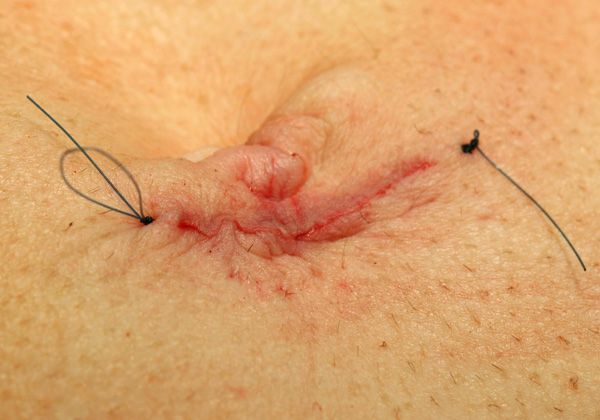
Suture after umbilical hernia surgery with good cosmetic result © Prof. Dr. med. Thomas W. Kraus
If the hernial ring is very big, the surgeon will also insert a reinforcing synthetic mesh to prevent the umbilical hernia from recurring. In most cases, hernia surgery can be a minimally invasive procedure, i.e. the keyhole technique. However, because the treatment only involves a single small incision right by the navel, MIS procedures have not been adopted convincingly across this country.


