A glenoid fracture is a fracture of the shoulder socket that often occurs in conjunction with a shoulder dislocation. It frequently leads to instability of the shoulder joint. However, glenoid fractures can be effectively treated using modern trauma surgery techniques. Here you will find further information as well as a selection of specialists and centers that treat glenoid fractures.
Anatomy of the Glenoid Cavity
The scapula bears the shoulder socket on its side, which is shaped like a small soup plate. The head of the humerus fits perfectly into the curve of this “plate.” The shoulder socket (medical term: glenoid) and the head of the humerus together form the main part of the shoulder joint.
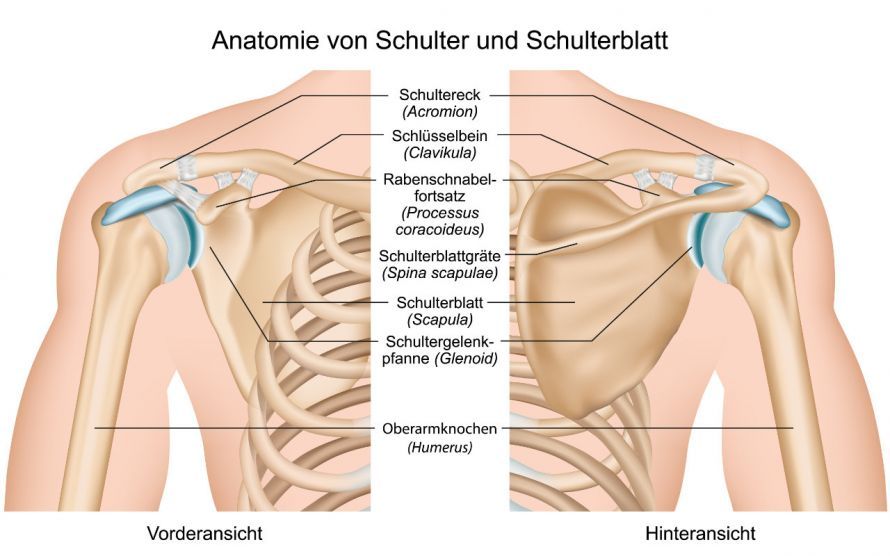
Glenoid Fracture: Fracture of the Shoulder Socket—Information on Symptoms, Diagnosis, Trauma Surgery, and Orthopedic Surgeons
Classification of Glenoid Fractures
Glenoid fractures are divided into two main categories.
There are smaller marginal fractures, usually located only along the front edge of the glenoid cavity, which occur in the context of shoulder dislocations, such as
- shoulder dislocations,
- dislocation fractures,
- osseous Bankart lesion
These fractures of the glenoid cavity are also called Bankart fractures. They are usually caused by an accident in which the impact energy is relatively low (low-energy mechanism). For example, the shoulder socket may partially fracture during a simple fall due to the dislocation mechanism.
In the case of glenoid fractures affecting the entire shoulder socket, the term “high-energy mechanism” is used because of the high forces involved. Traffic accidents are often the cause of such glenoid fractures.
Symptoms of a Glenoid Fracture
All glenoid fractures are relatively painful.
Although smaller Bankart fractures appear minor in terms of their extent, they have a significant functional impact.
The shoulder socket is much smaller than the corresponding humeral head. Much of the joint’s stability therefore comes from the surrounding soft tissues that are attached to the rim of the shoulder socket. Even if the surrounding soft tissues initially appear intact, mechanical stability is often significantly reduced. If this rim is fractured, the soft tissue complex can no longer maintain its stabilizing function. As a result, the shoulder joint often becomes unstable overall. In particular, the labrum—a cartilaginous ring at the rim of the glenoid cavity—plays an important stabilizing role.
This means that the joint frequently dislocates, and those affected experience an unpleasant feeling of instability. They feel as though they cannot rely on the shoulder joint.
This often leads to avoiding wide-ranging movements and a significant reduction in the range of motion. Furthermore, complex comminuted fractures are so painful that affected patients usually keep the shoulder joint completely still in a protective posture.
Diagnosis of a Glenoid Fracture
A shoulder fracture is diagnosed using X-rays. For more complex fractures, computed tomography (CT) or magnetic resonance imaging (MRI) may also be used.
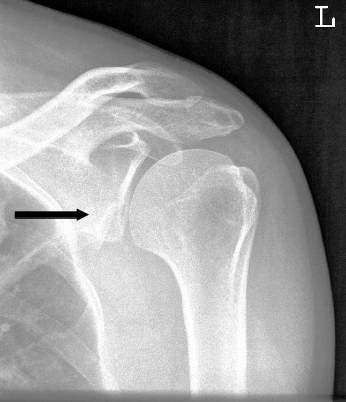
Treatment of a Glenoid Fracture
The common issue with all glenoid fractures is their direct impact on the stability of the shoulder joint.
Young, athletically active patients usually have high expectations regarding the mobility of their shoulder. For this reason, nearly all glenoid fractures require surgical treatment.
The goal of any surgical treatment is to reposition the displaced bone fragments into their anatomically correct position. They must be fixed in place there until they have healed back together. To achieve this fixation, the trauma surgeon often uses
- sutures,
- wires,
- screws, or
- plates.
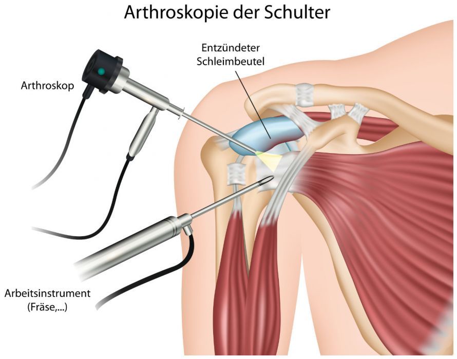
Smaller marginal fractures or Bankart fractures can often be treated through arthroscopic shoulder surgery. Shoulder arthroscopy is a minimally invasive procedure in which the surgeon makes only very small incisions in the skin. Minor marginal fractures can often be treated arthroscopically.
Through a small incision, the surgeon inserts a thin tube into the joint. A camera lens is attached to the tip of the tube. The surgeon views the camera’s image, magnified several times, on a monitor. Even tiny instruments can be guided to the surgical site through very small incisions.
Arthroscopy makes it possible to reposition the displaced bone back to its original location and secure it there.
In cases of more complex shoulder fractures, open surgery involving screws and plates may be necessary. This procedure is medically referred to as osteosynthesis or screw osteosynthesis.
Prognosis for Glenoid Fractures
A prerequisite for good outcomes is a reconstruction of the joint surface that is as anatomically accurate as possible. If the bone fragments can be repositioned in their original position and secured there, the prognosis for healing is very good. In most cases, these glenoid fractures result in only minimal restriction of movement.
The more complex the injury and the greater the force acting on the joint, the more extensive the damage to the cartilage and soft tissues. Therefore, all such joint fractures carry a higher risk of osteoarthritis for patients. In cases of severe malalignment, the humeral head may eventually become displaced relative to the glenoid cavity.
Without surgery, the prognosis for glenoid fractures is very poor. Bankart fractures result in an unstable shoulder that repeatedly dislocates. The more complex comminuted fractures heal in a malposition, usually leading to shoulder stiffness.
Conclusion on Glenoid Fractures
Injuries to the glenoid cavity directly affect shoulder function and often lead to instability. Using modern trauma surgery techniques, glenoid fractures can usually be successfully reduced. The fracture fragments can be fixed in such a way that the glenoid cavity heals largely in its original shape. In such cases, the functional consequences are minimal.
Unfortunately, complex glenoid fractures often result in significant cartilage damage and thus a higher risk of osteoarthritis.
Overall, a shoulder fracture is a complex injury with potentially significant consequences for affected patients. Treatment should therefore be entrusted to experienced specialists.
FAQ
What exactly is a glenoid fracture?
A glenoid fracture refers to a break in the entire shoulder socket or a portion of it, usually resulting from a fall or a traffic accident.
How is the diagnosis made?
The diagnosis is usually made through imaging, particularly X-rays, and in complex cases, additionally through computed tomography (CT).
When is surgery necessary?
Surgical treatment is usually required if there is a misalignment of the joint surface or if the shoulder joint is unstable.
Is conservative treatment an option?
Conservative treatment is only considered for very small, non-displaced fractures. In these cases, a shoulder brace may be helpful in the short term, especially for older patients or so-called “jumpers.”
What role does postoperative care play?
Physical therapy is often necessary after surgery to gradually restore shoulder function and improve range of motion.
Images: Radiological images were kindly provided by Dr. Stefan Wirth.
Share this article