It feels as if you constantly have a small pebble in your shoe that presses and hurts with every step. But when you look inside, the shoe is empty. Instead, you feel hardened lumps under the skin. This phenomenon often points to Ledderhose disease. This is a benign tissue change on the sole of the foot that, while not life-threatening, can severely impair quality of life due to pain when walking. The condition is often ignored for a long time or mistaken for simple overuse until the lumps on the sole of the foot become so large that normal walking becomes torture. Named after the German surgeon Georg Ledderhose, this condition is closely related to a similar disorder of the hand. We’ll explain exactly what’s happening in your foot, how orthotics can help, and when surgery is unavoidable.
What is Ledderhose disease?
Ledderhose disease is a benign proliferation of connective tissue on the sole of the foot. The condition affects the plantar fascia (also known as the plantar aponeurosis), a firm band of tissue that runs from the heel to the toes and stabilizes the longitudinal arch of the foot. Due to a regulatory defect in the tissue, benign nodules and strands form, which can grow and harden over time.
The disease was first described in 1894 by the German surgeon Georg Ledderhose and is therefore named after him (also known as Ledderhose disease in English). Ledderhose disease is a chronic condition, which means it usually develops slowly and can progress in episodes. It is closely related to Dupuytren’s disease, in which similar changes occur in the palm of the hand and cause the fingers to bend. In fact, many patients suffer from changes in both the hands and feet at the same time.
Causes: Why does the disease develop?
The exact cause of Ledderhose disease is still unknown. However, experts believe there is a genetic predisposition, as the disease runs in some families. There appears to be a genetic predisposition in which the connective tissue reacts to even the slightest injuries (microtraumas) with excessive scarring.
In addition, there are risk factors that may contribute to the development of fibromatosis:
-
Diabetes mellitus
-
Regular alcohol consumption (liver damage)
-
Epilepsy (or the use of certain medications to treat it)
-
Smoking
Although the nodules may appear tumor-like, Ledderhose disease is a benign condition. The nodules do not become malignant and therefore do not metastasize.
Symptoms and Course of Ledderhose’s Disease
The main symptom is palpable nodules and hardened areas on the sole of the foot, usually in the middle of the arch. At first, these are often small and painless. As the disease progresses, however, the nodules can grow and form clusters and strands.
Typical symptoms include:
-
Nodules on the sole of the foot: Firm nodules—often multiple—that can be moved under the skin or are firmly fused to the tendinous plate.
-
Pain when walking: Once the nodules reach a certain size or are located in an unfavorable position, pressure pain occurs when stepping down.
-
Tightness: The hardening of the plantar fascia can lead to a feeling of tightness that radiates down to the toes.
Unlike Dupuytren’s contracture of the hand, the strands associated with Ledderhose disease rarely cause the toes to curl, as the tendon plate on the foot is structured differently. Nevertheless, severe pain and difficulty walking are often the result in everyday life.
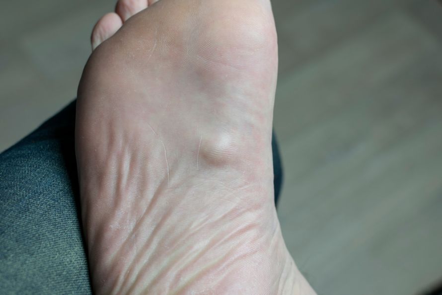
In Ledderhose disease, harmless but bothersome nodules form on the sole of the foot @ Farantsa /AdobeStock
Diagnosis: How is Ledderhose disease diagnosed?
Ledderhose disease is usually suspected based on the description of symptoms and a physical examination of the foot. A doctor will determine the exact location of the nodules and assess the degree of tissue hardening.
To confirm the diagnosis and rule out other conditions (such as malignant tumors or cysts), doctors use imaging techniques.
-
Ultrasound (sonography): This technique clearly visualizes the nodules in the plantar aponeurosis.
-
MRI (magnetic resonance imaging): An MRI provides precise images of the extent of the condition in depth. This is particularly important when surgery is planned, to determine how deeply the fibromatosis extends into the foot muscles.
For an accurate diagnosis, it is often advisable to visit a clinic specializing in foot surgery or radiology. There, procedures such as ultrasound or MRI can provide clarity.
Treatment of Ledderhose disease: Conservative
Treatment depends on the patient’s level of discomfort. As long as the nodules are small and painless, it is often possible to wait and see. However, if symptoms arise, conservative treatment is the first step.
Orthopedic Insoles The most important aid is orthopedic insoles. These are custom-fitted and feature a recessed area (soft padding) at the site of the nodule. This reduces pressure on the nodules while walking. Although orthopedic insoles cannot stop the growth, they can often significantly relieve the pain associated with Ledderhose disease.
Radiation Therapy In the early stages, when the nodules are still actively growing, low-dose X-ray radiation can slow cell growth and reduce the size of the nodules. You can find more information on this in our Radiation Therapy section.
Physical Therapy: Stretching exercises for the calf muscles and the plantar fascia can help relieve tension on the tendon plate and alleviate symptoms.
Surgical Treatment: When Is Surgery Necessary?
If conservative measures such as orthotics and physical therapy are no longer sufficient and the patient suffers from chronic pain, surgical treatment is considered. Surgery is also recommended if the nodules are so large that no shoes fit anymore.
There are various surgical approaches for treating Ledderhose disease:
-
Removal of the nodules: In this procedure, only the visible nodules are excised. The disadvantage is that the recurrence rate (return of the condition) is very high. New cases of Ledderhose disease can quickly develop from the remaining plantar fascia tissue.
-
Partial or total fasciotomy: To prevent recurrence, specialists often remove the entire affected tendon plate (plantar aponeurosis). This is a more extensive procedure, but it significantly reduces the risk of the condition returning.
Surgical removal is challenging because wound healing on the sole of the foot is often difficult and the skin is under tension. After surgery, the foot usually cannot bear full weight for several weeks.
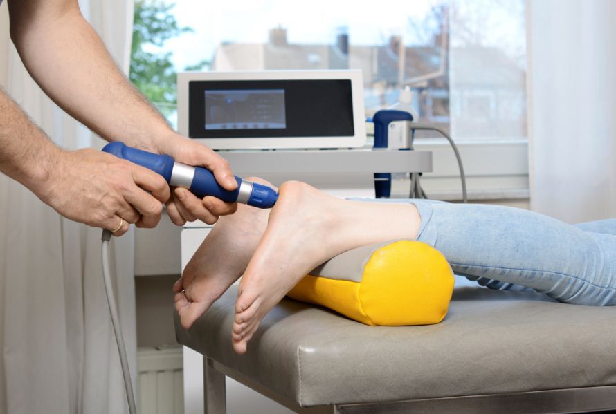
Shockwave therapy for Ledderhose disease @ Dan Race /AdobeStock
Prognosis and Living with the Condition
Ledderhose disease is a chronic condition. This means that even after successful treatment, patients must remain vigilant. New nodules can form at the edges of the removed fascia even after surgery.
Nevertheless, the condition can usually be managed well. With soft footwear, orthotics, and early treatment, most people affected can live pain-free. Since Ledderhose disease is a benign condition, overall life expectancy is not affected. Regular checkups are important to detect any regrowth of the nodules at an early stage.
FAQ: The 8 Most Important Questions About Ledderhose Disease
What is Ledderhose disease?
Ledderhose disease is a benign condition of the connective tissue on the sole of the foot (plantar fibromatosis). It involves the formation of firm nodules and strands in the plantar fascia, which can cause pain when walking.
Is Ledderhose disease dangerous?
No, Ledderhose disease is a benign condition. The nodules are not malignant tumors and do not metastasize. The main issue is the pain and the resulting limitation in walking ability.
Do the nodules go away on their own?
As a rule, the nodules do not resolve on their own. Ledderhose disease is a chronic condition that often progresses in episodes. Without treatment, the nodules may grow larger or increase in number.
Do orthotic insoles help with Ledderhose disease?
Yes, orthopedic insoles are the gold standard in conservative treatment. A targeted recess (hole or soft padding) at the site of the nodule relieves pressure when stepping, which significantly alleviates the pain.
When is surgery necessary?
Surgical removal of the nodules or the entire tendon plate is recommended if conservative measures such as orthotic insoles no longer provide relief, if the pain while walking becomes unbearable, or if the nodules are so large that they make it impossible to wear shoes.
What is the difference between this and Dupuytren’s disease?
Both conditions are types of fibromatosis and often occur together. Dupuytren’s disease affects the palm of the hand and leads to curling of the fingers. Ledderhose disease affects the sole of the foot and primarily causes pressure pain; curling of the toes is less common.
Which doctor treats Ledderhose disease?
The first point of contact is often an orthopedic surgeon or foot surgeon. Radiation therapists may also be involved in treatment during the early stages. A radiologist is responsible for diagnosis via MRI.
Can Ledderhose disease be prevented?
Since the exact cause is unknown and genetic factors play a major role, there is no sure way to prevent it. However, avoiding risk factors such as excessive alcohol consumption and smoking can reduce the risk of fibromatosis in general.
Share this article

Sources
flexikon.doccheck.com/de/Morbus_Ledderhose
Heyd R et al., Radiation therapy for early stages of morbus Ledderhose. Strahlenther Onkol 2010; 186(1): 24–29. doi: 10.1007/s00066-009-2049-x
medlexi.de/Morbus_Ledderhose
Seegenschmiedt RH & Attassi M. Strahlentherapie beim Morbus Ledderhose—Indikation und klinische Ergebnisse. Strahlentherapie und Onkologie 2003; 179: 847–853