An angioma is a vascular malformation that is usually benign and can take various forms. Among the best-known types are cavernomas and arteriovenous malformations, in which a direct shunt forms between an artery and a vein. Many angiomas go unnoticed for a long time and are discovered incidentally. Depending on their location, however, neurologically significant symptoms such as headaches, seizures, or neurological deficits may occur.
An angioma can be congenital and become noticeable over the course of a person’s life. Diagnosis is made using modern imaging techniques and neurological examinations. If an angioma is diagnosed, various treatment options—such as embolization, radiation therapy, or surgery—may be considered. Treatment depends on the size, location, and individual risk of bleeding.
What is an angioma?
An angioma is essentially a malformation of the blood vessels, the exact cause of which is not yet fully understood. However, angiomas are often present from birth.
Angiomas consist of a cluster of blood vessels comprising an artery and a vein, through which the artery directly delivers blood to the vein. A distinction is made between developmental angiomas and tumor-like angiomas.
Developmental angiomas usually occur where blood flows directly from an artery into a vein. These include, for example, aneurysms, varicose veins, cavernomas, and arteriovenous malformations. On the other hand, there are tumor-like angiomas, such as hemangiomas and lymphangiomas.
Where do angiomas occur?
Angiomas—commonly known as “blood blisters”—often appear on the skin of the face, on the eyelids, nose, lips, neck, or scattered across the torso. On the skin, angiomas are usually visible as small red nodules—flat or slightly raised—and are harmless.
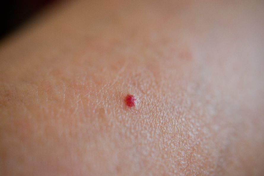
A small angioma on the skin © waranyu #221240842 | AdobeStock
When such skin angiomas appear after the age of 40, they are usually senile angiomas (angioma senile), which can develop sporadically or in large numbers as a result of aging. If “blood clotting” (thrombosis) occurs inside these clusters of blood vessels, senile angiomas can turn an almost black color and harden. In such cases, you should definitely consult a dermatologist to definitively distinguish the angioma from a nodular, malignant melanoma (“black skin cancer”).
In principle, however, angiomas can also occur in all major organs of the body. When found within organs such as the liver or heart, doctors refer to them as cavernous hemangiomas, or cavernomas for short. In the brain, these vascular anomalies can even have serious health consequences for patients.
What symptoms occur?
In the majority of cases, skin angiomas are asymptomatic. Although they can grow to be a few millimeters to a few centimeters in size, they usually regress during childhood. Removal of skin angiomas is generally indicated only if they cause repeated mechanical irritation in problematic areas of the body.
However, in about 2–4% of brain angiomas (= cerebral angiomas), bleeding can occur, leading to neurological deficits, loss of consciousness, and/or acute headache. Depending on the severity of such bleeding episodes, the condition can even be life-threatening.
Furthermore, cases have been described in which angiomas have caused seizures. This may be caused purely mechanically by the proximity of the seizing structures to the angioma, or it may be due to oxygen deprivation caused by the angioma.
Cerebral angiomas also cause circulatory disturbances in the brain, so that, depending on their location, a variety of other symptoms may occur, such as visual disturbances, paralysis, speech disorders, and personality changes.
How are angiomas diagnosed?
While skin angiomas can be identified fairly easily through visual examination due to their shape and red color, organ-based angiomas—such as cerebral angiomas in the brain—can only be diagnosed using specialized imaging techniques.
Traditionally, angiomas in the body’s organs are visualized using angiography, in which a contrast agent is injected to flow through all blood vessels and make the blood flow visible. Nowadays, however, angiography is increasingly being replaced by a more modern technique: magnetic resonance imaging (MRI). This allows for high-resolution images of, for example, the interior of the head.
Nevertheless, cerebral angiomas and cavernomas are often incidental findings as long as no bleeding has occurred that is accompanied by neurological deficits. In the latter case, an emergency CT scan—a so-called computed tomography scan—is usually performed at the hospital, which can detect cerebral hemorrhages very quickly.
Is there a treatment for angiomas?
If an asymptomatic cerebral angioma is detected, there is still a 50% risk that bleeding could occur at some point in a person’s life. Therefore, doctors generally recommend treatment on a case-by-case basis.
There are currently three different treatment options for cerebral angiomas:
-
In embolization, a catheter is guided into the brain’s blood vessels to inject an adhesive that seals off the angioma’s vessels. However, there is a risk that the adhesive could enter other small brain vessels and cause further damage to blood flow there.
-
If the angioma is easily accessible, however, it should be surgically removed. This reduces the size of the angioma and lowers the risk of bleeding. On the other hand, this surgical method also carries risks; for example, the blood vessel could be injured or blood clots could be released, which in turn could damage other areas of the brain.
-
Cerebral angiomas that are difficult to access surgically are treated with stereotactic radiation therapy. In this process, the radiation triggers growth in the vascular cells, which very slowly—over the course of years—grow into the vessel lumen, thereby closing off the angioma’s vascular mass from the inside. After three years, approximately 80% of the cerebral angiomas treated in this way are occluded.
Which doctors treat angiomas?
Angiomas are vascular malformations and, as such, are primarily treated by specialists in angiology (angiologists). Angiology is a subspecialty of internal medicine that focuses on the diagnosis and treatment of vascular diseases.
FAQ
What is an angioma?
An angioma is a vascular malformation—usually benign—that results from abnormalities in blood vessels. Types of angiomas include cavernomas, hemangiomas, and arteriovenous malformations. In an arteriovenous malformation, blood flows directly from an artery into a vein without passing through the normal capillary system. This results in a tangled mass of blood vessels with an increased risk of complications.
What symptoms can occur with an angioma?
Many angiomas cause no symptoms and are discovered by chance. However, symptomatic angiomas can cause headaches, seizures, epileptic fits, neurological symptoms, or neurological deficits. If a cerebral hemorrhage occurs, paralysis or other serious neurological symptoms may also develop.
How is an angioma diagnosed?
Diagnosis is made using modern imaging techniques such as MRI, CT, and angiography. Imaging procedures allow for the precise visualization of blood vessels, arteries, and veins. Neurosurgeons, neurologists, neuroradiologists, and neurosurgery specialists work closely together, particularly when a cerebral arteriovenous malformation or cavernoma is suspected.
How are angiomas treated?
The treatment of angiomas depends on their size, location, and risk of bleeding. Possible procedures include catheter-based embolization, surgical removal of the angioma, or radiosurgery using a specialized radiation device. In embolization, coils, glue, or other materials are used to occlude the affected vessels.
When is treatment necessary?
Not every patient with an angioma requires immediate treatment. For asymptomatic angiomas, initial monitoring may be sufficient. Treatment is often necessary in cases of bleeding, neurological symptoms, seizures, or an increased risk due to blood supply. The decision regarding treatment is made on an individual basis, taking into account various treatment options and current scientific findings, such as those from the ARUBA study.
Share this article

Sources
- https://diggefa.de/
- www.altmeyers.org/de/dermatologie/angiom-seniles-458
- https://flexikon.doccheck.com/de/Angiom
- Piek J et al., Neurovaskuläre Erkrankungen: Klinik und Therapie zerebraler Kavernome. Dtsch Arztebl 2001; 98(25): A-1690 / B-1432 / C-1343. www.aerzteblatt.de/archiv/neurovaskulaere-erkrankungen-klinik-und-therapie-zerebraler-kavernome-5ca89d10-41f4-42eb-b310-0cfb80f6af9a [zuletzt aufgerufen am 07.09.2020]