About 30–40% of patients with brain tumors have a glioma. About 60% of patients are men, and 40% are women. The peak age for gliomas is between 40 and 65 years, but gliomas also occur quite frequently in children.
Low-grade gliomas arise from degenerated supporting cells of the nervous tissue and usually grow only slowly. They are of low malignancy, hence the name. In English, they are accordingly referred to as “low-grade gliomas.” Treating low-grade gliomas is not easy and therefore requires, among other things, specialized neurosurgery.
Below you will find further information as well as a selection of specialists for low-grade gliomas.
Definition: Low-grade gliomas
Low-grade gliomas are solid tumors that arise from abnormal cells in the brain or spinal cord. They originate directly in the central nervous system (CNS), which is why low-grade gliomas are also referred to as primary CNS tumors.
In principle, such low-grade gliomas can develop anywhere in the CNS. However, they tend to occur more frequently in certain locations:
- in children, primarily in the cerebellum or in the central regions of the cerebrum;
- in adults, predominantly in the cerebrum.
Causes of Low-Grade Gliomas
The causes of low-grade gliomas remain largely unknown.
Tumor development originates in the supporting cells of the CNS, known as glial cells. The most common glial cells in the CNS are called astrocytes and oligodendrocytes. If the glioma results from cellular changes in these cells, it is therefore referred to as an astrocytoma, oligodendroglioma, or oligodendrocytoma.
Only about 5% of cases of cellular changes in adults that ultimately lead to a tumor are attributable to hereditary genetic mutations.
In addition, certain congenital malformations appear to increase the risk of low-grade gliomas. These include, for example,
- neurofibromatosis type 1 or
- tuberous sclerosis.
For example, one in five children with congenital neurofibromatosis type 1 develops low-grade gliomas early in life.
A history of cancer can also be a risk factor for low-grade gliomas. For example, radiation therapy administered to childhood leukemia patients increases their later risk of developing such a brain tumor.
Classification of Low-Grade Gliomas
The World Health Organization (WHO) classifies gliomas into four risk categories (Grades I–IV). Low-grade gliomas fall into risk categories I and II. Grades III and IV, on the other hand, are reserved for high-grade gliomas.
Grade I gliomas are benign CNS tumors that grow slowly and usually remain well-localized. Although Grade I gliomas can grow in a mass-forming manner, they do not destroy layers of tissue or spread into deeper tissues.
In contrast, Grade II gliomas grow diffusely and also invade surrounding tissues.
The transition from a Grade II to a Grade III glioma (= dedifferentiation) is gradual in adults—in children, however, this occurs only rarely.
What symptoms does a low-grade glioma cause?
The symptoms of a low-grade glioma depend primarily on the tumor’s location in the CNS and its extent of spread. A distinction is made between general and specific symptoms.
The general symptoms of a low-grade glioma are nonspecific and therefore not very characteristic. This means they are also found in other diseases. These general symptoms include:
- Headaches
- Back pain
- Stiff neck
- Dizziness
- Nausea and vomiting
- Weight loss
- Difficulty concentrating
- Hydrocephalus resulting from impaired drainage of cerebrospinal fluid (CSF)
Low-grade gliomas often develop gradually. They frequently grow over several years. Typically, symptoms may appear during this time and manifest as follows, depending on the affected area of the central nervous system (CNS):
- If the glioma is located in the cerebellum—as is more commonly observed in children—it leads to balance, gait, and movement disorders.
- A low-grade glioma in the cerebrum, on the other hand, frequently leads to seizures, paralysis, or speech disorders.
- If the tumor is located in the spinal cord, however, various types of paralysis, sensory disturbances, and pain may occur.
How is a low-grade glioma diagnosed?
The diagnosis of a low-grade glioma is often an incidental finding. Initial indications of a potentially malignant tumor in the CNS arise from
- the patient’s medical history as well as
- the physical and neurological examination.
Deficits, paralysis, or gait disturbances are usually also noticeable during these examinations. Various imaging techniques are available for further evaluation of a suspected tumor, such as
- magnetic resonance imaging (MRI),
- computed tomography (CT), and
- positron emission tomography (PET).
Using these methods, the physician can detect a low-grade glioma and simultaneously determine its size and location within the CNS. PET imaging allows for the spatial visualization of particularly active tumor centers, known as “hot spots.”
Unlike higher-grade gliomas, low-grade gliomas do not take up contrast agent. As a result, they appear on MRI as a diffuse cloud. This is because the blood-brain barrier is usually intact, and the contrast agent cannot yet cross into the tumor. In principle, this is a positive prognostic factor.
A tissue sample from the low-grade glioma is used for the final pathological characterization of the tumor. In this process, the tumor is characterized histologically and through molecular biology. The following factors play a particularly important role:
- the IDH mutation status,
- the MGMT promoter status, and
- the 1p/19q codeletion.
In low-grade gliomas, particular attention is paid to the IDH mutation status. This refers to isocitrate dehydrogenase, whose gene mutates relatively frequently in this type of tumor. On the one hand, this increases the likelihood that the glioma will dedifferentiate into a higher-grade tumor over time. On the other hand, this tumor group responds better to chemotherapy, allowing for more targeted treatment
The 1p/19q codeletion provides information about the structure of the chromosomes in the mutated tumor cells. Human DNA is coiled up into what are known as chromosomes. In oligodendrogliomas in particular, a segmental loss occurs on chromosomes 1 and 19. If this deletion is present, however, the tumors respond well to chemotherapy. This treatment would follow surgical removal of the tumor.
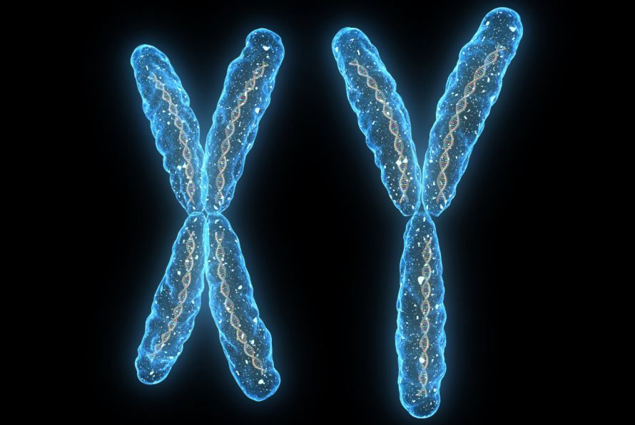
X and Y chromosomes contain the actual DNA strands © SciePro | AdobeStock
In healthy cells, the MGMT promoter is responsible for repairing damaged breaks in the DNA. Every day, a wide variety of DNA segments on the chromosomes are read and replicated, a process during which coding errors can occur. The MGMT promoter searches for these damages and repairs them.
In gliomas, especially in glioblastomas, this promoter is often methylated. This means that carbon and hydrogen atoms have been incorrectly attached to it. As a result, the enzyme becomes inactive and DNA damage can no longer be repaired. For this reason, tumors with a methylated MGMT promoter respond better to combination therapy consisting of chemotherapy and radiation.
What treatment options are available for low-grade gliomas?
Low-grade gliomas are primarily treated surgically. The goal is the complete surgical removal of the tumor.
In some cases, the tumor cannot be completely removed, for example,
- due to its location,
- if there are multiple tumor foci, or
- due to its growth pattern, which can sometimes infiltrate over a wide area.
Further surgery would risk causing neurological damage to the brain.
In such cases, the functionally active mixed tissue consisting of brain and tumor tissue is left in place. Depending on the histological and molecular biological analysis (see above),
- follow-up observation or
- treatment with radiation therapy and/or chemotherapy.
Awake craniotomy: A specialized surgical procedure for adult glioma patients
Wakeful craniotomy is a specialized surgical procedure for tumor resection in the CNS. It is used for
- localized low-grade gliomas with infiltrative growth and multiple tumor foci, as well as
- higher-grade tumors
.
Its major advantage is that the patient is briefly responsive during the operation. This makes it possible to operate very close to healthy brain tissue.
At the start of the surgery, patients are under general anesthesia. The surgeon then opens the skull and creates an access route to the tumor. Once the target area is reached, the patient is gently awakened. Dissection within the brain does not cause pain, as brain tissue lacks pain receptors.
Neuropsychologists and neurologists monitor the awake surgery. They conduct various tests with the patients before the surgery and repeat them during the procedure.
Brain functions in the surgical area are simultaneously visualized through “brain mapping” using brief microelectrical stimulations. This provides the doctors with a functional map of the brain. Using this map, the doctors determine which areas allow for the safe removal of tumor tissue without causing
- motor function,
- speech, or
- other sensory functions
. If various types of fine electrical stimulation indicate deficits, the patient feels them immediately while awake during the operation. The surgeons then adjust the tumor resection accordingly and refine it.
Intraoperative MRI is often performed
- intraoperative MRI,
- high-resolution ultrasound,
- tumor fluorescence, and
- 3D navigation
are used.
Prognosis for low-grade glioma
Low-grade glioma has a good prognosis when the tumor is completely resected. Approximately 90% of patients survive long-term.
Studies have shown that the extent of tumor resection is one of the most important factors for the best possible prognosis. Therefore, all available technical resources should be utilized. This allows surgeons to strike a balance between maximal tumor resection and the risk of surgery-related neurological deficits.
Even if residual tumor remains, the prognosis can still be improved. To achieve this, oncologists must determine the appropriate, personalized radiation therapy and/or chemotherapy regimen.
This is always carried out by an interdisciplinary team of specialists from the fields of
- neuro-oncology,
- radio-oncology, and
- radiosurgery.
Share this article