Anatomy and Function of the Intestine
Anatomically, the human intestine is divided into four sections:
- Duodenum: The duodenum begins immediately after the stomach. It surrounds the front part of the pancreas and plays a crucial role in the digestion of food. It mixes the acidic chyme with bile and pancreatic juice.
- Small Intestine (Jejunum and Ileum): The small intestine is between 5 and 7 meters long. This is where the majority of digestion takes place through the absorption of proteins, fats, and carbohydrates.
- Large intestine (colon): In the large intestine, which is approximately 1.5 meters long, the stool is primarily thickened through the removal of water.
- Rectum: The rectum collects the intestinal contents in a reservoir. Working in close coordination with the sphincter muscles, it empties the intestinal contents.
Which intestinal diseases require the removal of a section of the intestine?
The following conditions can occur in the intestine:
- Tumors: Benign and malignant
- Diverticular disease (diverticulitis)
- Chronic inflammatory bowel diseases (Crohn’s disease, ulcerative colitis)
- Adhesions/intestinal obstruction (adhesions, paralytic ileus)
- Below you will find more information about these conditions.
- Benign Tumors in the Intestine
In the vast majority of cases, benign tumors in the bowel are polyps (adenomas) of the colon. These benign tumors can be detected during a colonoscopy. Doctors can remove them during the colonoscopy without surgery.
Since benign tumors can become malignant over time, doctors must always remove them. Medical professionals estimate that it takes approximately 10 years for a benign tumor to turn into a malignant one.
Therefore, you should undergo a colonoscopy every 10 years as a preventive measure to avoid colon cancer.
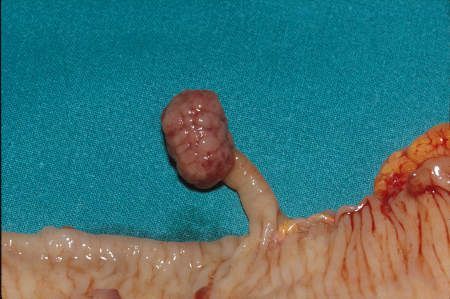
Figure 1: Adenoma in the colon
Malignant Tumors in the Colon
Malignant tumors are also found almost exclusively in the large intestine. These are tumors that initially develop from benign tumors of cells in the large intestine’s mucous membrane. They progress to colorectal cancer. Colon cancer begins to grow from the inside of the bowel. It can narrow the bowel, which can lead to a bowel obstruction.
Over time, the tumor invades the wall of the bowel. It can also spread outside the bowel into other organs. A general characteristic of malignant tumors is their ability to spread tumor cells and form secondary tumors (metastases).
In colorectal cancer, tumor cells can spread to the lymph nodes of the intestine. They then form metastases and exhibit malignant growth themselves.
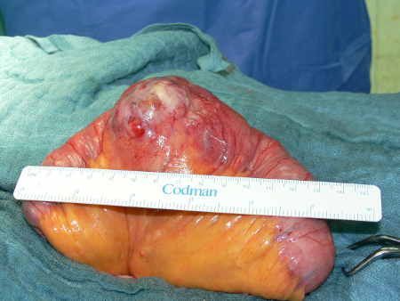
Figure 2: Malignant tumor in the colon
Since lymphatic vessels and lymph nodes are located near the colon, surgeons must remove the corresponding lymphatic drainage area during colon surgery. Otherwise, additional tumors may develop from any lymph nodes left behind.
Another mechanism by which colorectal cancer can spread is the dissemination of cancer cells via the bloodstream. The cancer cells then preferentially settle in the liver or the lungs, where they form metastases.
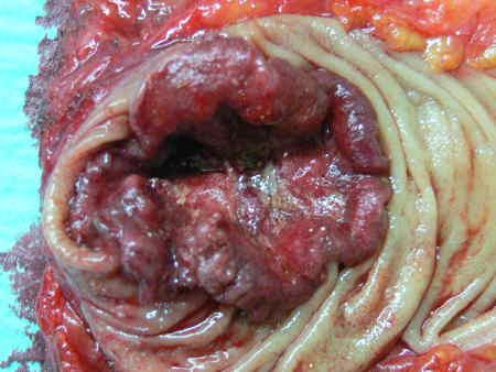
Figure 3: Malignant tumor in the colon
Diverticular Disease and the Resulting Diverticulitis
Diverticula are outward protrusions of the intestinal wall. They are therefore always benign lesions. These diverticula occasionally occur in the small intestine, but are much more common in the large intestine.
Diverticula may be congenital, but in most cases they develop in middle age. The protrusions are usually caused by increased pressure in the large intestine, and a congenital weakness of the connective tissue also appears to play a decisive role.
Diverticula are present in approximately 70 percent of people in industrialized countries as they age. They do not in themselves constitute a disease.
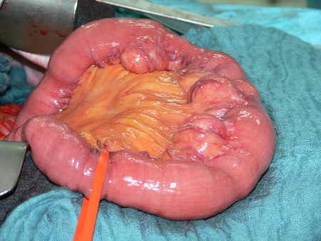
Figure 4: Diverticula in the small intestine
Through mechanisms that are not yet fully understood, diverticula can cause painful inflammation of the intestine (diverticulitis). If the inflammation progresses, it can lead to a perforated bowel and life-threatening peritonitis.
Diverticula frequently occur throughout the entire large intestine. However, they are most common in the S-shaped section of the intestine just before the rectum (sigma). Inflammation often occurs in episodes and can lead to narrowing of the intestine over the course of years. In addition to inflammation, severe bleeding can also occur as a result of diverticula.
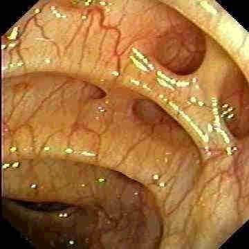
Figure 5: Diverticular openings in the large intestine
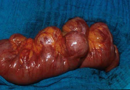
Figure 6: Diverticula in the large intestine
Chronic Inflammatory Bowel Diseases
This condition, the cause of which is not yet fully understood, leads to severe inflammatory flare-ups that persist over many years or decades. These flare-ups occur primarily in the lower small intestine, but also in the large intestine or rectum.
The disease most commonly develops in younger people. Women are affected more frequently.
Treatment for this intestinal inflammation primarily involves medication. If severe complications such as bowel perforations, strictures, or fistulas occur, intestinal surgery is necessary. During the procedure, doctors remove the affected section of the intestine as sparingly as possible.
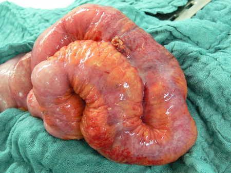
Figure 7: Chronic Inflammatory Bowel Disease (Crohn’s Disease)
Adhesions and Intestinal Obstruction
Adhesions in the abdominal cavity occur following inflammation or surgery. They usually cause no problems. Occasionally, however, they can lead to symptoms if sections of the intestine are fused together or if adhesions form between the intestine and the abdominal wall.
These relatively extensive adhesions can prevent the intestine from moving freely. It may also be unable to pass through narrowings or kinks.
Intestinal surgery is necessary if the symptoms are clearly attributable to these adhesions.
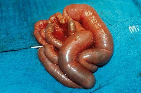
Figure 8: Intestinal obstruction (ileus) in the small intestine
Furthermore, cord-like adhesions (brides) can form after surgery. The intestine can become entangled around these, which can lead to acute intestinal obstruction (ileus).
Blood flow to the intestine is then no longer guaranteed. This results in damage to the intestine. If left untreated, this leads to the death of that section of the intestine. In this situation, only timely intestinal surgery can prevent damage to and loss of the intestine.
Medical Specialties
Surgeries on the intestine are performed by specialists in visceral surgery or colorectal surgery. The field of proctology specializes in diseases of the intestine.
Coloproctology centers of excellence bring together experts from various disciplines who focus specifically on diseases and treatments of the rectum.
Open Bowel Surgery or Minimally Invasive Procedure?
For a bowel resection, there are generally two surgical techniques to choose from:
- open surgery
- laparoscopic surgery
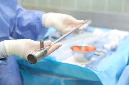
Figure 9: Laparoscopic surgery (laparoscopy)
The type of surgical procedure used for bowel resection depends on the underlying condition. In cases of malignant disease, doctors must remove a larger portion of the bowel than in cases of benign disease.
Open procedures require a larger abdominal incision. Laparoscopic procedures (minimally invasive technique, “keyhole surgery”) do not require a large incision or direct visualization of the body.
The two procedures differ not in the extent of bowel resection, but in the method of accessing the abdominal cavity.
Partial Bowel Resection via Open Surgery
Open abdominal surgery is the most commonly performed surgical procedure in Germany.
In this procedure, the surgeon opens the abdominal cavity through an incision. This incision is usually made along the midline of the abdomen, above and/or below the navel. The procedure is performed under general anesthesia.
During a partial bowel resection, doctors free the bowel from natural adhesions to prepare it for removal.
They ligate the blood vessels in the section of the intestine and resect that portion. The two sections of the intestine are then connected using sutures or mechanical staplers. Finally, the abdominal wall is closed with sutures.
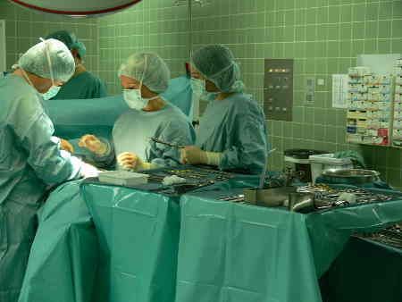
Figure 10: Open Surgery
Partial bowel resection via laparoscopic surgery
In principle, laparoscopic surgeries do not differ from open surgeries in terms of the extent of bowel resection. The main difference lies in the small access point to the abdominal cavity.
Special tubes (trocars) allow for small incisions (between 5 and 10 millimeters) in the abdominal wall. Through these trocars, surgeons can insert specialized instruments into the abdominal cavity (endoscopic camera). A monitor displays the images from the abdominal cavity.
The procedure requires space inside the body. To create this space, doctors pump harmless carbon dioxide gas into the abdominal cavity to expand it.
All surgical steps can be performed using special instruments and staples, just as in open surgery. The only time an incision of a few centimeters is needed in the abdominal wall is to remove the excised intestinal tissue from the abdominal cavity.
The advantage of this minimally invasive technique is that patients experience less pain after surgery. They recover more quickly from the procedure. Additional benefits include a shorter hospital stay and smaller scars.
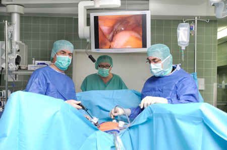
Figure 11: Laparoscopic Surgery
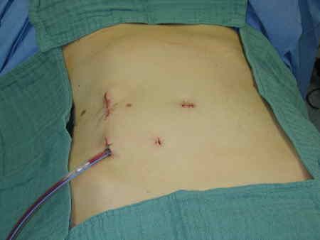
Figure 12: Incisions after laparoscopic surgery
Surgical Procedures for Intestinal Resection
A partial bowel resection can be performed using various surgical techniques:
- Wedge resection
- Segmental resection
- Right hemicolectomy
- Transverse resection
- Left hemicolectomy
- Sigmoid resection
- Anterior rectal resection
- Rectal amputation
- Total colectomy
- TEM (Transanal Endoscopic Microsurgery)
Partial bowel resection with wedge resection
In this procedure, doctors remove a portion of the intestinal wall. This is done, for example, in cases of a broad-based benign polyp that cannot be removed endoscopically.
The continuity of the bowel is preserved. The resection site is closed either with sutures or using a stapler.
Partial bowel resection with segmental resection
In cases of benign conditions affecting the small or large intestine, the affected section of the intestine is removed in a minimally invasive manner. This is the standard surgical approach, particularly for chronic inflammatory bowel diseases.
Surgeons reconnect the sections of the intestine before and after the removed segment using sutures or a stapler.
Partial bowel resection with right hemicolectomy
This refers to the removal of the right-sided portion of the large intestine (ascending colon) up to the transverse colon. This procedure is performed for tumors of the right-sided large intestine, including the removal of the supplying blood vessels and lymphatic vessels.
After removal, doctors reconnect the end of the small intestine (ileum) to the transverse colon (transversum).
Partial bowel resection with transverse colon resection
For tumors located exactly in the middle of the transverse colon, doctors remove the entire transverse colon. They then connect the right side of the colon (ascending colon) to the left side of the colon (descending colon).
Partial bowel resection with left hemicolectomy
This refers to the removal of the left-sided portion of the large intestine (descending colon), beginning at the transverse colon. This procedure is performed for tumors of the left-sided large intestine and involves the removal of the supplying blood vessels and lymphatic vessels.
After removal, surgeons reconnect the end of the transverse colon to the remaining large intestine.
Partial bowel resection with sigmoid resection
Doctors can treat tumors in this S-shaped section of the colon by removing the affected portion of the colon (along with the supplying blood vessels and lymph vessels).
Diverticular disease frequently occurs in this section of the colon. If diverticulitis occurs frequently or if an inflammatory flare-up is severe, removal of this section of the bowel is necessary. Today, these procedures are performed in most hospitals using laparoscopic surgery, i.e., a minimally invasive technique.
Partial Bowel Resection with Anterior Rectal Resection
In cases of cancer in the rectum (the lower 16 centimeters of the bowel), doctors must remove part or all of the rectum. The surrounding fatty tissue around the rectum, which contains the lymph nodes, is also removed.
The difficulty with this intestinal surgery is that the rectum lies deep within the pelvis and is therefore more difficult to access. After removing the rectum, doctors reconnect the remaining rectal stump, along with the sphincter muscle, to the colon above it.
Nowadays, with the appropriate experience, this surgery can also be performed using minimally invasive techniques that are gentle on the patient.
In principle, thanks to improved surgical techniques, the sphincter can now be preserved even in cases of tumors located near it.
However, the rate of anastomotic leaks is higher in these surgeries compared to surgeries in other areas of the large intestine. Therefore, as a safety measure, doctors create a colostomy, which they can later reverse without complications.
Partial Bowel Resection with Rectal Amputation
If the tumor is located at the sphincter, a rectal resection involving removal of the sphincter is necessary.
As with rectal surgery, the goal of the procedure is to preserve the sphincter muscle. It is preferably performed using a minimally invasive approach.
After removal of the sphincter muscle, an artificial bowel opening (stoma) is created. An appropriate ostomy care system ensures that patients can go about their daily activities without difficulty.
Partial Bowel Resection with Total Colectomy
Removal of the entire large intestine (colon) may be necessary in cases of chronic inflammatory bowel disease such as ulcerative colitis, especially when the entire large intestine is affected.
In this procedure, doctors remove the entire large intestine and create a reservoir (pouch) from the lower portion of the small intestine. They connect this pouch directly to the lower rectum above the sphincter muscle (ileoanal pouch).
In younger patients in particular, this surgery is performed almost exclusively using minimally invasive techniques.
Partial Bowel Resection with TEM (Transanal Endoscopic Microsurgery)
Benign tumors are located in the rectum between the sphincter and the junction between the rectum and the lower colon (sigma). They are difficult to reach from the abdominal cavity. Therefore, local removal is not possible.
However, local removal of benign adenomas from the rectal wall is possible via the anus. A specially designed rigid endoscope enables minimally invasive removal of the tumor.
This procedure is particularly useful for tumors located between 8 and 12 centimeters from the sphincter. Otherwise, this area would be inaccessible via the anus. Doctors can easily remove benign tumors located closer to the sphincter from the rectal wall under direct visualization.

About the medical author
Sabine Schneider
Editor-in-Chief
Sabine Schneider – medical author: Explore expert articles and medical expertise in the Leading Medicine Guide.
More about the medical author →