Macular degeneration is a common disease of the retina that primarily affects older adults and can significantly impair vision. Age-related macular degeneration, in particular, affects the macula—the area of the eye responsible for the sharpest vision. There are two forms: dry and wet, with wet age-related macular degeneration often progressing more rapidly. In wet AMD, new blood vessels form that can damage the retina and lead to a decline in visual acuity.
In advanced cases, in addition to injections and medication, surgical procedures such as retinal rotation are also used. This specialized surgery relocates the affected macula to utilize healthy photoreceptor cells. The goal is to stabilize or improve vision. Early diagnosis by an ophthalmologist is crucial for slowing the progression of the disease.
What is retinal rotation?
Retinal rotation can restore near-optimal vision for patients in many stages of age-related macular degeneration (AMD). Currently, there is still no way to completely cure AMD. At best, all treatment methods can halt or slow the progression of the disease. In this context, retinal rotation has come to play a significant role.
During retinal rotation, an incision is made in the affected retina, which is then detached and rotated by approximately 30 degrees. The goal is to reposition the macula—the eye’s visual center—to a different location within the pigment epithelium. In the best-case scenario, this allows it to reattach to an intact pigment epithelium cell.
If this is successful, the patient can see more clearly again after a short healing and recovery period. An optimal surgical outcome can restore vision to an average of 70 percent for five to ten years. Further improvement in visual acuity can be achieved through additional medication and the use of special visual aids.
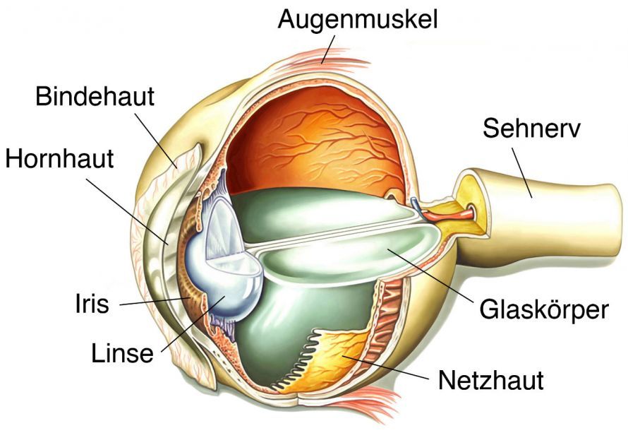
However, an optimal result cannot be guaranteed with retinal rotation. The success of the treatment depends
- on the condition of the retina and
- the course of the surgery
. The retinal rotation itself is also followed by surgery on the eye muscles. Optimal treatment success is only possible if both surgical steps are successful.
In addition to AMD, retinal rotation can also be used to treat other eye conditions, such as
- other types of growths on the retina or
- damage to the center of vision (macula) caused by external factors (e.g., accidents or physical trauma)
Background and Development of Retinal Rotation
The first 20 documented retinal rotations were performed in 1997–1998. To determine the ideal procedure, surgeons tested various techniques, such as rotating the retina by different angles ranging from 20 to 45°.
After the recovery phase, visual acuity had improved in more than half of the patients. Just under half of the treated patients were able to read without difficulty again after retinal rotation. The procedure has since become more refined, and success rates have increased further. However, in some cases, statutory health insurance may not cover the entire cost of treatment.
Necessary Preparation and Preoperative Examinations Before Retinal Rotation
Surgical intervention is always preceded by thorough ophthalmological examinations. The ophthalmologist determines the severity of macular degeneration and, if possible, its cause. Retinal rotation is not equally suitable for all patients. The doctor must weigh whether the expected success of the surgery is commensurate with the potential risks.
The patient must also be willing to cooperate with the treatment. They must largely eliminate the risk factors that contribute to the progression of macular degeneration. These include
- Diabetes: Should be optimally controlled.
- High blood pressure: A healthy lifestyle lowers blood pressure.
- Tobacco: Smoking should be completely stopped.
Follow-up care after a successful surgery is also planned in advance as much as possible. While the final details can only be determined after evaluating the surgical outcome, you still need to know in advance which options are feasible for you, both personally and financially.
Performing the Procedure
Retinal rotation is a fairly complex procedure consisting of two surgical stages:
- the actual retinal surgery and
- the subsequent eye muscle surgery.
Retinal Rotation
First, the surgeon removes the lens and the vitreous humor. Only then can he make an incision in the retina and detach it. To remove new blood vessels, the optic nerve must be folded back. The surgeon then rotates the retina along with the macula and positions it over healthy tissue. The retina is then seamlessly reattached to the eye’s tissue using a laser.
The empty cavity in the eye created by the vitrectomy is filled with silicone oil or gas to prevent it from collapsing. This filler is removed from the eye two to three months after the procedure.
Eye Muscle Surgery
During a second procedure, the eyeball must be adjusted to the macula’s new position. Without this second step, the patient’s vision would be severely distorted after the procedure. This is also the case if the surgeon does not correctly adjust the macula’s new position.
Complications and Risks
Retinal rotation is associated with several risks:
- Insufficient repositioning: For physical or surgical reasons, the macula may not be able to be repositioned far enough away from the deformed tissue area.
- The body’s immune response to the silicone oil used as a vitreous substitute (relatively rare).
- Retinal tears.
- Deterioration of vision, even days or weeks after a successful procedure.
- In some cases, visual distortions due to a loss of the ability to optimally coordinate both eyes.
- Retinal detachment following the procedure.
A retinal detachment is an ophthalmic emergency. If the patient notices any deterioration or changes in vision, they should seek medical attention immediately.
Follow-up Care for Retinal Rotation
The healing phase lasts about three months, even though vision is restored much more quickly.
The following can be detrimental to the healing process:
- Excessive alcohol consumption,
- unhealthy eating,
- nicotine, especially for the delicate blood vessels of the retina,
- spending too much time watching TV or using a computer,
- excessive physical exertion,
- and excessive exposure to bright sunlight.
Long-term protection of the eyes from glaring sunlight must be ensured. Smoking—even if resumed later—can also lead to the surgery’s benefits lasting for a shorter period.
The carotenoid lutein, often called the “eye vitamin,” aids in the healing of the retinal blood vessels. It is found in
- yellow and red vegetables (such as bell peppers and corn),
- peas, and
- broccoli
.
You should also be sure to keep all your follow-up appointments with your eye doctor.
FAQ
What is macular degeneration?
Macular degeneration is a disease of the retina in which the macula is damaged. This region is responsible for sharp vision and contains many sensory cells and photoreceptors. The condition is usually age-related and can lead to a loss of visual acuity.
What is the difference between dry and wet age-related macular degeneration?
The dry form develops slowly and is characterized by degeneration of the sensory cells. The wet form, on the other hand, is caused by new, abnormal blood vessels that can lead to swelling and scarring. Wet AMD progresses more rapidly and, if left untreated, can lead to blindness.
When is retinal rotation performed?
Retinal rotation is used in severe cases of wet age-related macular degeneration when other treatments, such as injections or anti-VEGF therapy, are not sufficiently effective. In this procedure, the retina is surgically repositioned to relieve pressure on the damaged macula.
What treatment options are available?
The most important therapies include intravitreal injections of medications such as ranibizumab, bevacizumab, or aflibercept. These VEGF inhibitors prevent the growth of new blood vessels. Laser therapy or surgical procedures are also used as complementary treatments.
How is the condition diagnosed?
Diagnosis is performed by an ophthalmologist using vision tests and imaging techniques such as OCT. The retina is examined in detail, particularly the macular region. This allows changes to be detected early and the appropriate treatment to be initiated.