
Aortic valve stenosis is one of the most common heart valve diseases and is among the leading causes of reduced cardiac output. The narrowing between the left ventricle and the aorta impedes blood flow, which—depending on the severity of the condition—leads to increasing strain on the heart muscle. Many patients with aortic valve stenosis initially show few symptoms; however, calcific or congenital aortic valve stenosis often progresses if left untreated. Modern diagnostic procedures such as echocardiography, transesophageal ultrasound, or cardiac catheterization allow for an accurate assessment of the severity. The choice of the best possible treatment depends on clinical guidelines, the causes and severity of aortic valve stenosis, as well as any comorbidities.
What is aortic valve stenosis?
Let’s first take a look at the anatomy of the heart. The aortic valve forms the passageway from the left ventricle to the aorta (Fig. 1).
Blood enriched with oxygen in the lungs is pumped from the left ventricle into the body at high pressure. In a healthy aortic valve, it flows evenly through the valve.
After each ejection (systole)—which can be felt as a pulse wave—the aortic valve closes again (diastole).
The aortic valve consists of three delicate, sail-shaped leaflets (Fig. 2). The opening area of a healthy aortic valve is 3–4 cm². In an adult at rest—depending on height and weight—approximately 4–5 L/minute flow through the aortic valve.
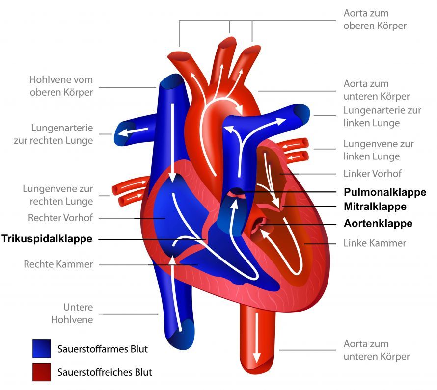
Fig. 1: Diagram of the human heart and heart valves © bilderzwerg | AdobeStock
When the leaflets of the aortic valve no longer open fully, this is referred to as aortic valve stenosis. It is a result of stiffening and calcification of the leaflets (Fig. 3). The left ventricle must generate increasingly higher pressure to pump the same volume of blood through the narrowing.
Aortic valve stenosis is considered severe if the remaining opening area is less than 1 cm². The smaller the remaining opening area, the higher the blood flow velocity and the pressure difference before and after the aortic valve (mean pressure gradient).
The heart initially responds to the pressure load by thickening the heart muscle (hypertrophy). If aortic valve stenosis persists for a long time, left ventricular enlargement (dilatation) may develop in addition to hypertrophy.
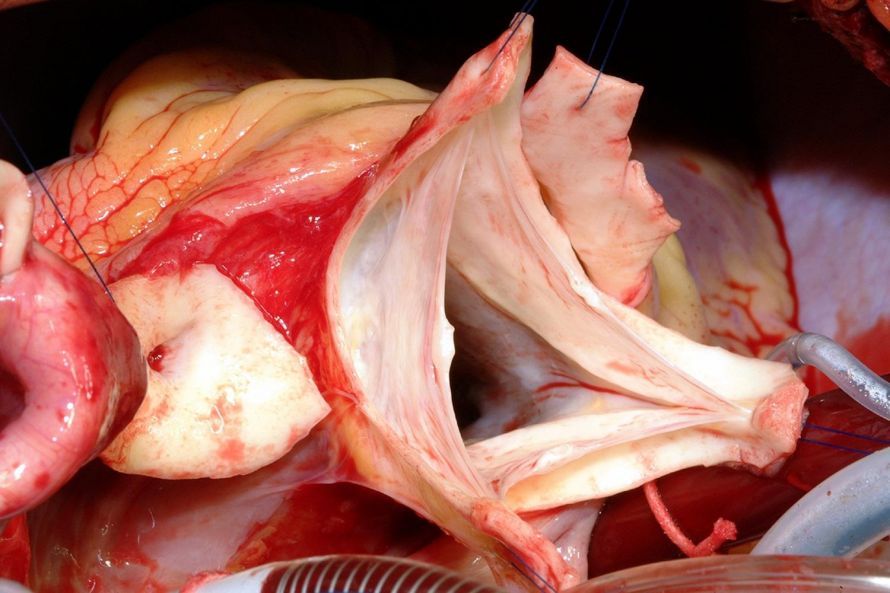
Fig. 2: Aortic valve with the three delicate leaflets, in the open position (image taken by Prof. Dr. med. Alexander Albert during a DAVID aortic valve reconstruction)
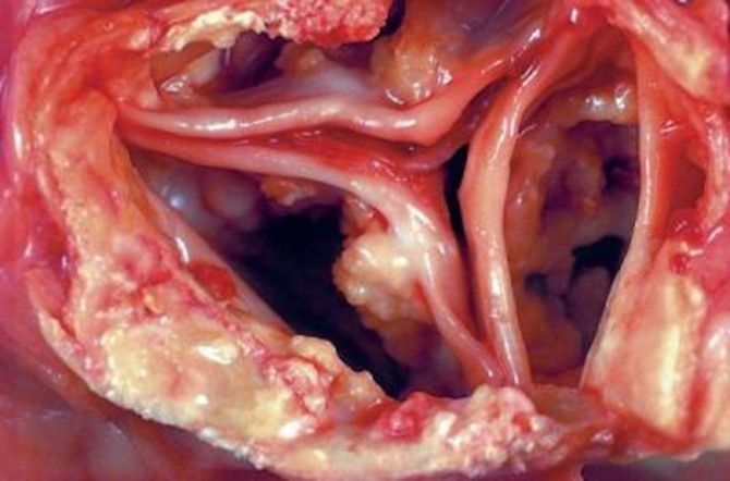
Fig. 3: Stenotic aortic valve with three leaflets that have become stiff due to calcium deposits and thickening and can no longer open properly.
What causes aortic valve stenosis?
Aortic valve stenosis can have various causes. First, a distinction is made between congenital aortic valve stenosis and that acquired over the course of a person’s life. Only 10 percent of all cases of aortic valve stenosis are present at birth. In most cases, the heart condition develops later in life.
Aortic valve stenosis most commonly occurs in older adults over the age of 60. Various factors contribute to age-related wear and tear. These lead to connective tissue remodeling and calcification of the valve leaflets. Factors that contribute to the accelerated progression of this not fully understood process include:
- High blood pressure
- Diabetes
- Smoking
- Kidney failure
- Excessively high calcium levels in the blood
- Genetic factors
What are the symptoms of aortic valve stenosis?
Aortic valve stenosis develops gradually and is usually asymptomatic in the early stages. Therefore, it is not uncommon for aortic valve stenosis to go unnoticed for years and only be detected during routine examinations.
In the early stages, symptoms occur only during physical exertion. During more strenuous activity, the heart is no longer able to pump enough blood through the stenosis. If too little blood reaches the brain, this manifests as dizziness and lightheadedness. This can even lead to a brief loss of consciousness (syncope).
The progressive thickening of the heart muscle leads to muscular and connective tissue remodeling of the heart (negative remodeling). Once the muscle reaches a certain thickness, it can no longer be adequately supplied with oxygen. Similar to coronary heart disease, this leads to
- a feeling of pressure in the chest,
- tightness, or
- severe pain (angina pectoris).
In addition, the heart becomes stiffer and less elastic (reduced compliance).
As a result, the atrium must generate increasingly higher pressure to distend and fill the left ventricle. This leads to pathological enlargement (dilatation) as well as muscular and connective tissue remodeling of the atrium.
This triggers cardiac arrhythmias—particularly atrial fibrillation. The remodeling of the entire heart also increases the risk of ventricular (malignant) arrhythmias. These, in turn, can cause loss of consciousness.
In later stages, the entire heart enlarges and cardiac output decreases. Blood then backs up into the lungs, leading to shortness of breath and, in severe cases, pulmonary edema. When left untreated, shortness of breath as a symptom of heart failure is associated with a very poor prognosis.
How is aortic valve stenosis diagnosed?
Aortic valve stenosis produces a characteristic heart murmur. Doctors can hear this murmur during a routine examination with a stethoscope and make a preliminary diagnosis.
The diagnosis is confirmed by an ultrasound examination of the heart (echocardiography).
The severity of aortic valve stenosis is classified as follows:
| Aortic valve stenosis | Mild stenosis | Moderate stenosis | Severe stenosis | |
| Max. flow velocity, m/sec | <2.5 | 2.5–3 | 3–4 | >4 |
| Average pressure gradient, mmHg | No gradient | <20 | 20–40 | >40 |
| Aortic valve orifice area | 3–4 cm² | >1.5 | 1–1.5 | <1 |
Cardiac function is also important for the prognosis (left ventricular ejection fraction—EF):
- Normal ≥ 55%
- Moderately impaired 30–55%,
- Severely impaired < 30%
Surgery for Aortic Valve Stenosis
The standard treatment for aortic valve stenosis is surgery.
Heart valve insufficiencies (leaks) can often be repaired. In cases of stenosis, the valve leaflets or pockets are typically destroyed by inflammation and degeneration. They no longer open properly.
Consequently, aortic valve replacement is always necessary in cases of (acquired) aortic valve stenosis.
Timing of Heart Valve Replacement
Heart valve replacement is necessary in cases of severe aortic valve stenosis accompanied by typical symptoms. These symptoms may also only occur during physical exertion.
Sometimes the patient has few or no symptoms, but the heart already shows significant damage as a result of aortic valve stenosis (EF <50%). In such cases, surgery is also necessary.
Sometimes, the severity of aortic valve stenosis is difficult to determine. This is the case, for example, when heart function is significantly impaired. In such cases, specialized tests by a cardiologist are necessary to determine the severity of the aortic valve stenosis.
Possible procedures: Open-heart surgery, minimally invasive surgery, or TAVI?
Traditionally, aortic valve replacement is performed via open-heart surgery.
Access to the heart is gained through a longitudinal incision in the sternum [Fig. 6, sternotomy]. The patient is connected to a heart-lung machine. This machine takes over the functions of the heart and lungs during the surgery. In this way, the heart is “shut down,” allowing the surgeon to work on it safely.
The surgeon then removes the old, diseased parts of the aortic valve and sutures a prosthesis into place.
The advantage of open-heart surgery is that the surgeon has a direct and clear view of the aortic valve.
The same procedure can also be performed using a minimally invasive approach at specialized centers. This involves either a partial incision of the sternum [Fig. 7, partial sternotomy] or lateral access by spreading the ribs.
Nowadays, aortic valve replacement is increasingly performed using minimally invasive techniques, but without a heart-lung machine. Of great importance here is the so-called catheter-based procedure (TAVI). It is not a surgical procedure in the traditional sense.
In this procedure, the new prosthetic heart valve is implanted via the groin [Fig. 9, transfemoral TAVI] or between the ribs through the apex of the heart [Fig. 10, transapical TAVI].
Aortic valve replacement via transfemoral TAVI can be performed while the patient is awake.
When to use TAVI, and when not to?
TAVI is currently indicated for
- older patients (> 75 years) and/or
- patients with multiple comorbidities and/or
- patients at increased surgical risk
.
There is still insufficient experience with younger patients. In addition, the physician will consider various technical factors that may support or argue against TAVI.
Factors in favor of TAVI:
- Previous heart surgeries
- calcified vessels
Factors against TAVI include certain anatomical characteristics
- of the aortic valve,
- the aortic root, and
- the coronary arteries.
TAVI prostheses are biological prostheses. For this reason, patients under the age of 50 to 60 will receive a mechanical prosthesis or undergo the Ross procedure.
Biological or mechanical prostheses?
For non-TAVI procedures, the patient can choose between biological and mechanical prostheses (see Figs. 4 and 5). TAVI prostheses are always biological prostheses. Biological prostheses are made from the heart valves or pericardium of pigs or cattle. The material is modified so that it is accepted by the body’s immune system and integrates very well.
The disadvantage of these prostheses is that they wear out very quickly, especially in younger patients. They are ideal for patients over 70 years of age, where they are expected to last more than 20 years.
However, there are other options available today in cases of wear and tear or the development of new aortic valve stenosis. Using the so-called “valve-in-valve” procedure, TAVI can be performed without the need for another surgery.
With the goal of avoiding repeat surgery, biological prostheses are now even recommended for patients between the ages of 50 and 60. However, with each new TAVI procedure, the valve orifice becomes smaller and smaller. As a result, this valve-in-valve procedure can generally only be performed once.
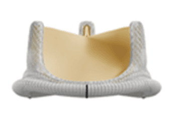
Fig. 4: Biological heart valve made from bovine material (courtesy of Edwards)
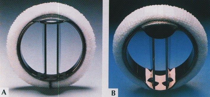
Fig. 5: Mechanical heart valve (courtesy of Medtronic)
Mechanical prostheses are recommended for patients between the ages of 50 and 65. These are made of robust carbon-containing material and, in theory, last forever. However, blood clots can easily form on this artificial surface, which can lead to blockages of the prosthesis and strokes.
Therefore, patients must take blood-thinning medication—usually Marcumar or warfarin—for the rest of their lives. The problem with Marcumar therapy is that even minor injuries can lead to severe bleeding. Cerebral hemorrhages are particularly feared in this context.
The risk of severe bleeding is not particularly high, at 1–2% per year. However, especially in younger patients, the risk accumulates over the decades. Particularly for athletic patients or those with physically demanding jobs, this Marcumar therapy can lead to significant limitations in quality of life.
With the appropriate experience, mechanical prostheses can also be implanted using a minimally invasive approach through the ribs (antero-lateral minithoracotomy). Understandably, this is never done as a TAVI procedure because the prostheses cannot be folded.
A (truly) biological solution for young people: the Ross procedure?
For younger patients, this means that neither the biological nor the mechanical prosthesis represents a truly good solution.
The Ross procedure offers an alternative. In this procedure, the aortic valve is replaced with the body’s own heart valve, the pulmonary valve. This valve is located between the right side of the heart and the pulmonary circulation. In this low-pressure circuit, it is exposed to significantly less blood pressure and stress than the aortic valve and is therefore somewhat more delicate. Otherwise, its anatomical structure is similar to that of the aortic valve.
It is harvested during surgery and sutured into place in place of the diseased aortic valve. The pulmonary valve (pulmonary autograft) possesses all the characteristics of a natural heart valve. It therefore does not require blood-thinning medication and is not subject to wear and tear. It is thus a true biological prosthesis.
The surgery is also performed on children with aortic valve stenosis, in which case the valve actually grows along with the child.
A so-called homograft is implanted at the site where the pulmonary valve was harvested. This homograft comes from a deceased donor and is provided by specialized homograft banks prior to surgery.
This homograft is subjected to less stress in the pulmonary circulation. As a result, it lasts significantly longer there than if it were implanted directly in the aortic valve position.
The Ross procedure is quite complex and is performed only by a few surgeons with years of experience in this technique.
In Detail: The Classic (Complete) Sternotomy
Complete sternotomy is the standard procedure for cases that are not suitable for TAVI. These cases involve younger patients without a significantly increased surgical risk.
In most cases, these patients would also be eligible for a minimally invasive procedure known as an anterolateral mini-thoracotomy (see below).
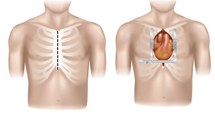
Fig. 6: Conventional sternotomy, image source [https://adultct.surgery.ucsf.edu/conditions--procedures/minimally-invasive-aortic-valve-surgery.aspx]: “Minimally-invasive Cardiac Surgery” by Tobias Deuse, M.D.
In Detail: Minimally Invasive Surgical Procedures
Partial sternotomy
For which patients is this procedure suitable?
Many surgeons no longer perform this procedure. Because the sternum is opened, there is a risk of instability and complications in this area.
As a truly minimally invasive alternative to surgery via sternotomy, surgeons prefer the anterolateral mini-thoracotomy, which does not involve opening the sternum at all (see below).
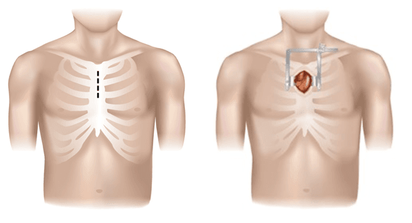
Fig. 7: Partial sternotomy, image source [https://adultct.surgery.ucsf.edu/conditions--procedures/minimally-invasive-aortic-valve-surgery.aspx]: “Minimally-invasive Cardiac Surgery” by Tobias Deuse, M.D.
The anterolateral mini-thoracotomy
The anterolateral mini-thoracotomy involves opening the chest near the sternum between the second and third ribs.
During MIC surgery, the surgeon accesses the heart through a small incision between the ribs. The sternum is not opened.
The heart-lung machine is connected via the femoral vessels (leg arteries and veins) through a puncture (without an incision).
For which patients is this procedure suitable?
In the hands of an experienced surgeon, nearly all aortic valve surgeries can be performed using this approach. It is used when a TAVI procedure is not an option (see below).
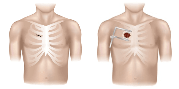
Fig. 8: The anterolateral mini-thoracotomy, image source [https://adultct.surgery.ucsf.edu/conditions--procedures/minimally-invasive-aortic-valve-surgery.aspx]: “Minimally-invasive Cardiac Surgery” by Tobias Deuse, M.D.
Transcatheter Aortic Valve Implantation (TAVI)
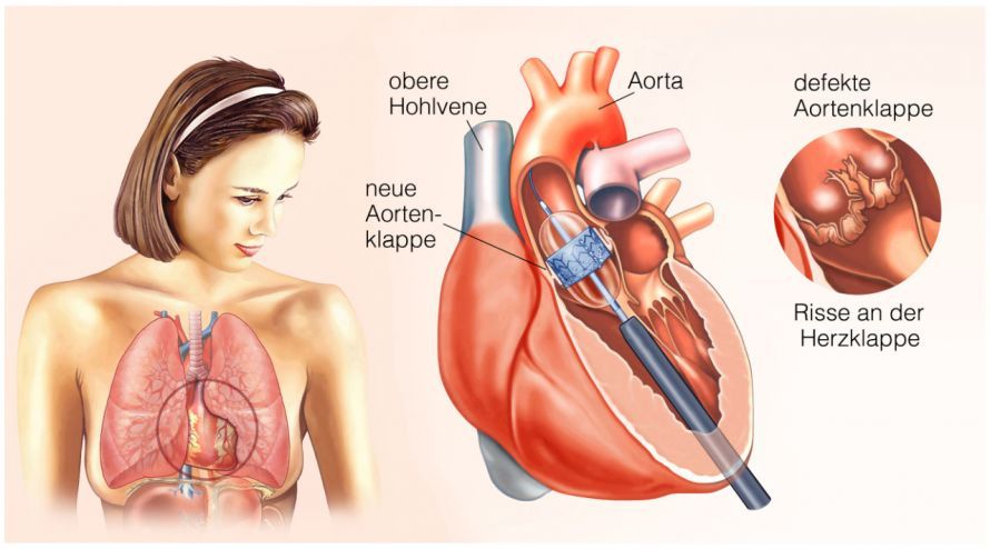
Transfemoral TAVI
In a TAVI procedure, a biological aortic valve is attached to the tip of a catheter. The surgeon then advances it to the heart and secures it in place at the aortic valve position by expanding it.
The TAVI procedure was developed for the following patients:
- severe aortic valve stenosis
- Need for heart valve replacement
- The risk of conventional aortic valve replacement is too high
The procedure is performed in a so-called hybrid operating room. It combines the capabilities of a cardiac catheterization lab (mobile X-ray system) with the equipment of an operating room.
With this TAVI procedure, it is not necessary to cut open the sternum or use a heart-lung machine. The catheter is usually advanced into the heart via the femoral artery (transfemoral).
Transfemoral TAVI has now become the gold standard for treating aortic valve stenosis in
- older patients or
- those at increased surgical risk.
The procedure can be performed under general anesthesia or with the patient awake.
Transapical TAVI
Transapical means that the valve is inserted through the apex of the heart. This method is an option when the femoral arteries are too small or the aorta is severely calcified.
A skin incision a few centimeters long is made on the left side of the chest wall, below the nipple. The catheter is then inserted directly through the apex of the heart.
A folded biological heart valve prosthesis is advanced through the catheter to the site of the diseased aortic valve and expanded. The diseased heart valve is thereby pushed into the wall of the aorta and replaced.
This procedure has now become the standard for aortic valve replacement. In certain cases, it is also possible to replace the mitral valve in this manner (TMVI).
Transapical aortic valve replacement is an option for all patients who are older or have an increased surgical risk.
Transfemoral TAVI is even less invasive than transapical TAVI. It can also be performed while the patient is awake. For this reason, transfemoral TAVI is the preferred method today. Exceptions include patients with
- severe atherosclerotic changes in the leg vessels or
- very small vessel diameters through which the TAVI prostheses cannot be advanced.
In cases of coronary artery disease, a combination of TAVI and a single coronary bypass to the anterior wall of the heart is also performed.
It is also used as a hybrid procedure followed by PCI (stent placement) on the lateral and/or posterior walls of the heart.
Ross procedure
The Ross procedure is suitable for
- younger, active patients between the ages of 11 and 55,
- people at high risk of injury in their line of work who therefore do not wish to take Marcumar, and
- women who wish to have children.
Certain anatomical criteria must be met. These can usually be determined preoperatively through appropriate imaging tests such as CT or MRI. In particular, the sizes of the pulmonary and aortic valves should not differ too greatly from one another.
Aortic Valve Stenosis and Sports: What Physical Activity Is Permitted—and When Caution Is Advised
For patients with aortic valve stenosis, physical activity is possible, but the extent always depends on the individual severity of the condition. In cases of mild valvular aortic stenosis without significant symptoms, moderate endurance sports can often still be pursued—provided that exercise tolerance has been assessed by a cardiologist. However, as the valve narrows and blood flow decreases, the risk of dizziness, chest pain, or brief loss of consciousness increases, especially if symptoms occur during exertion.
Patients with moderate or severe aortic valve stenosis should continue athletic activities only after medical evaluation. Guidelines recommend, in these cases, a thorough assessment of cardiac output and symptoms, as well as auscultation and, if necessary, supplemental stress echocardiography. Sports involving high intensity, rapidly changing levels of exertion, or potential risks of falls and collisions (e.g., soccer, martial arts, intense strength training) are considered unsuitable because the circulatory system has a limited ability to respond when the aortic valve is narrowed.
After the implantation of a new heart valve—for example, through surgery or transcatheter aortic valve replacement (TAVR/TAVI)—physical activity can generally be gradually increased. Structured follow-up care is crucial, as approximately 10 to 20 percent of patients still require adjustments to their medication regimen. It is particularly important to note that if you experience any form of chest pain, shortness of breath, or arrhythmia, you must stop exercising immediately and seek medical evaluation.
FAQ on Aortic Valve Stenosis
What are the typical symptoms of aortic valve stenosis?
Symptoms such as shortness of breath, chest pain, dizziness, or intolerance to physical exertion often do not appear until the disease has reached an advanced stage. These symptoms occur because aortic valve stenosis places an increasing strain on the heart and restricts blood flow.
How does calcific or valvular aortic valve stenosis develop?
The most common form is calcific aortic valve stenosis in the elderly, caused by calcification and structural changes in the aortic valve. Other causes include rheumatic fever or congenital abnormalities. The course of the disease varies significantly depending on the cause and treatment.
How is the severity of aortic valve stenosis determined?
The severity is assessed using ultrasound, transesophageal echocardiography, or sometimes cardiac catheterization. During these procedures, blood flow, pressure gradients, and the valve’s opening area are measured. The heart’s pumping capacity also plays a role.
When is treatment necessary?
Treatment is necessary when symptoms appear, when cardiac output decreases, or when the aortic valve is severely narrowed. Depending on the severity of the condition, treatment options may include medication, surgical procedures, or catheter-based procedures such as transcatheter aortic valve implantation.
Can aortic valve stenosis affect life expectancy?
Yes. If left untreated, aortic valve stenosis can significantly weaken the heart and, in the long term, lead to heart failure, arrhythmias, or endocarditis. Timely diagnosis and treatment significantly improve life expectancy.
What role does medication play?
Medications cannot cure the condition, but they can alleviate symptoms and stabilize comorbidities such as high blood pressure or arrhythmias. They are part of the overall treatment plan for aortic valve stenosis, but they do not replace surgery in cases of severe stenosis.
Which diagnostic method is most important for diagnosis?
Aortic valve stenosis is primarily diagnosed using echocardiography. Auscultation, transesophageal echocardiography, or cardiac catheterization may also be necessary, especially when decisions regarding surgical treatment must be made.
Share this article
Sources
- S2k-Leitlinie „Kongenitale Aortenklappenstenose“ (AWMF-Register-Nr. 023-047), Stand 21.01.2022: register.awmf.org/de/leitlinien/detail/023-047