Urinary stones are among the most common disorders of the urinary tract and affect the kidneys, ureters, or bladder. Kidney stones and ureteral stones form due to the deposition of mineral salts, oxalic acid, or other components in the urine. Depending on their composition, stones can vary in size and cause severe discomfort. Typical symptoms include cramping pain, renal colic, blood in the urine, or discomfort in the lower abdomen.
Small stones are often passed through the urethra, while larger stones require treatment in a urology clinic. Modern procedures such as ESWL, ureterorenoscopy, or extracorporeal shock wave lithotripsy allow for the gentle fragmentation of urinary stones. In specialized centers or hospitals, kidney and ureteral stones are碎石 or surgically removed. Drinking enough fluids and taking preventive measures help prevent the recurrence of stone formation.
What are urinary stones?
Urinary stones are small, stone-like formations (concrements) that form from mineral salts and can block the urinary tract.
In Germany, urinary stones are among the most common conditions. Over the past few decades, the rate of urinary stone diagnoses has tripled. This is due to changes in dietary habits and lifestyle, as well as improved diagnostic capabilities.
Urinary stones are given different names depending on where they are located:
- Bladder: Bladder stones (cystolithiasis). Bladder stones can form directly in the bladder (primary bladder stones). However, they often form in the kidney and are flushed into the bladder with the urine (secondary bladder stones).
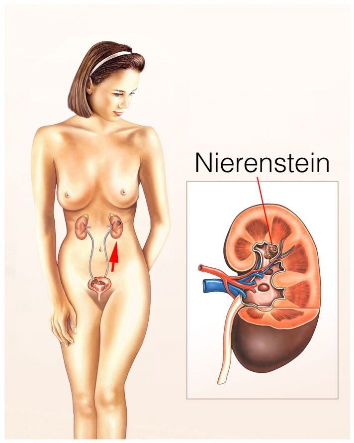
- Kidneys: Kidney stones (nephrolithiasis),
- Ureter: Ureteral stones (ureterolithiasis) and
- Urethra (condition: urethralithiasis).
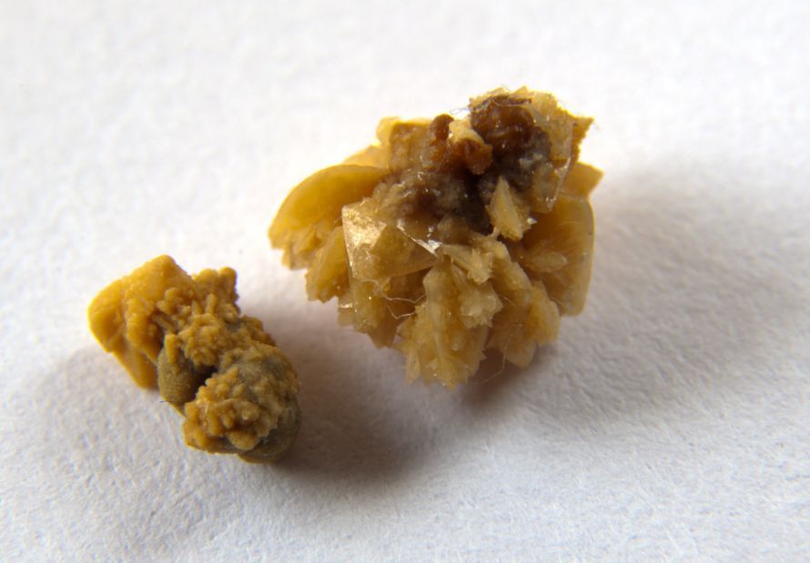
Two kidney stones. The crystalline structure of the stone is clearly visible on the right kidney stone © remik44992 | AdobeStock
Most urinary stones are very small, so the body can pass them in the urine. As a result, many patients do not even realize that they have or have had urinary stones. However, if the stones reach a certain size, they can become lodged in the urinary tract. This can cause severe, cramp-like pain (colic). In such cases, they must be removed.
It is estimated that one in ten people will develop urinary stones at some point in their lives. One in four of those affected will even develop them multiple times.
How do urinary stones form?
If the urine contains high concentrations of certain mineral salts, these salts precipitate when the urine is sufficiently acidic. This means they are no longer soluble. They then form small crystals (grit), which aggregate to form larger structures. In extreme cases, a urinary stone can be so large that it fills the entire renal pelvis (pelvic stone).
Urinary stones differ based on their main component.
- Three-quarters of all urinary stones are calcium oxalate stones (also known as whewellite or weddellite).
- About one in ten urinary stones consists of magnesium ammonium phosphate. Stones of this type are called struvite.
- 5% of all urinary stones contain uric acid (urate stones).
- One in twenty stones is a calcium phosphate stone.
- Xanthine and cystine stones are even rarer.
Known risk factors for the development of urinary stones include
- lack of exercise,
- being overweight,
- diabetes mellitus,
- advanced age, and
- being male.
In older men, bladder stones often occur in conjunction with benign prostatic hyperplasia. Benign prostatic hyperplasia is associated with impaired bladder contraction, which makes it difficult to pass urine.
Inflammation of the urinary tract promotes the formation of stones. Certain neurological disorders (paraplegia, multiple sclerosis) also restrict urine flow. As a result, they often lead to the formation of urinary stones as well.
Struvite stones often develop as a result of urinary tract infections. An unhealthy diet high in stone-forming salts also contributes to the development of stones. The body cannot excrete excessive amounts of these chemical compounds in sufficient quantities through the urine. These stone-forming salts include
- calcium,
- ammonium compounds,
- phosphates,
- oxalates,
- uric acid.
What are the symptoms of urinary stones?
Whether symptoms occur depends on the size of the urinary stones and their exact location. Bladder grit and small stones usually do not cause any problems.
However, if urinary stones restrict urine flow, symptoms may occur. Sharp-edged urinary stones rub against the organ’s wall, causing
- mucosal irritation,
- minor bleeding, and
- in some cases, even damage to the affected organ due to tears. The resulting scarring then causes further symptoms.
If urine can no longer drain, it backs up all the way to the kidney. The resulting urinary retention (ischuria) requires urgent emergency medical care, as it can otherwise lead to sepsis. Insufficient drainage of urine can lead to permanent kidney damage.
During urination, sudden, extreme pain in the lower abdomen (colic) may occur. Sometimes the urine stream simply stops. Irritation of the mucous membrane leads to blood in the urine. Other patients complain of a constant urge to urinate but can only pass a few drops of urine (pollakiuria).
If the kidney stones cause pain, the patient may try to find relief by changing their body position. They may lie down, stand upright, or walk around. Sometimes the symptoms are so severe that the patient feels nauseous and vomits.
If you notice any of these symptoms, please see a urologist as soon as possible.
How are urinary stones diagnosed?
During the physical examination, the urologist first listens to the patient’s lower abdomen and then gently palpates it. If the urologist suspects the presence of a urinary stone, they will take a urine and blood sample. The urine sample shows whether the patient’s urine contains bacteria, blood, or crystals. The blood sample provides information about uric acid levels, kidney function, and a possible urinary tract infection.
The most important imaging techniques are ultrasound (sonography) and computed tomography (CT).
Ultrasound is easy to perform and does not involve radiation. On an ultrasound image, kidney stones can be detected in the kidney once they reach a size of at least 5 mm. Stones of this size and larger increasingly cause problems. Computed tomography is primarily used to detect stones in the ureters. It can also reveal urinary obstruction and very small stones that are not visible on ultrasound.
Occasionally, as an alternative to computed tomography, an X-ray of the urinary tract without contrast medium is performed. However, this examination is not as sensitive as a CT scan.
If urinary diversion is necessary, a procedure called ureteropyelography is performed beforehand. In this procedure, a contrast agent is administered to the patient via a catheter, which allows for very clear visualization of the ureters and renal pelvis.
How are urinary stones treated?
Various medications can help relieve renal colic. These are usually non-opioids, with metamizole considered the first-line treatment. Indomethacin and diclofenac can also alleviate symptoms. Heat therapy, such as hot baths, may also provide pain relief in some cases.
The urologist does not need to treat urinary stones that do not cause symptoms. Instead, the urologist simply monitors them at regular intervals. Urinary stones that are 5 mm or smaller often pass on their own. Alternatively, medications that help flush them out can be used. Uric acid stones may also be dissolved with medication under certain circumstances.
It is always important for the patient to drink plenty of fluids. Regular exercise can also help dislodge the urinary stone. Urinary drainage is necessary if the urinary tract is so blocked that urine can no longer be excreted (urinary retention). In this procedure, a thin plastic tube (ureteral stent) is advanced past the blocked area and into the kidney. Urine can then drain out through this tube. This protects the kidney and reduces the risk of further kidney damage.
Urinary stones that block the flow of urine must be removed. The same applies to stones that continue to cause pain despite treatment or that do not pass on their own. In most cases, surgical removal is now performed using minimally invasive techniques. Ureteral stones and smaller kidney stones are treated using ureterorenoscopy. In this procedure, an endoscope is advanced through the urethra, the bladder, and the ureters into the renal pelvis. The stones are then removed using mini-forceps; larger stones are first碎ed with a laser.
Large kidney stones, including outlet stones, are removed using minimally invasive or open percutaneous (through the skin) stone surgery. This procedure is known as percutaneous nephrolitholapaxy. However, open surgery is rarely performed these days.
In extracorporeal shock wave lithotripsy (ESWL), energy waves are focused from outside the body onto a ureteral or kidney stone, shattering it. The fragments are then passed in the urine.
However, large urinary stones can produce larger fragments, which in turn can block the urinary tract. In this case, further procedures such as ureterorenoscopy or the placement of a ureteral stent would be necessary.
If the kidney stone developed in connection with another medical condition, the specialist must also treat that condition.
Once the kidney stone has been successfully passed, the specialist must determine its underlying cause. This is the only way to prevent the formation of additional kidney stones in the future.
Can Kidney Stones Be Prevented?
Many patients who have had a kidney stone removed go on to develop kidney stones again. To prevent this, you should drink at least 2.5 to 3 liters of fluid daily. This dilutes the urine, preventing mineral salts from precipitating and forming crystals.
Sugar-sweetened soft drinks increase the risk of stone formation. Therefore, you should avoid these beverages.
In addition, make sure to maintain a balanced diet. People with uric acid stones should limit their intake of purine-rich foods, as the body breaks down purines into uric acid. Foods high in purines include
- meat
- offal
- sausage
Oxalate stones can be prevented by avoiding the consumption of
- rhubarb
- spinach
- nuts
- coffee
.
FAQ
What are urinary stones?
Urinary stones are solid concretions or stone deposits in the kidneys, ureters, bladder, or urinary tract. Stone formation results from mineral salts, oxalic acid, uric acid, or other components in the urine. Depending on their location, they are classified as kidney stones, ureteral stones, or bladder stones.
What symptoms do urinary stones cause?
Typical symptoms include cramping pain, colic, renal colic, and blood in the urine. Many patients also experience nausea and vomiting or pain in the lower abdomen. If a stone blocks the ureter, severe discomfort and difficulty urinating may occur.
How are urinary stones diagnosed?
Diagnostic methods include ultrasound, CT scans, cystoscopy, or X-rays. In urology, the composition, location, and size of the stones are also assessed. In complicated cases, ureteroscopy or ureterorenoscopy is performed.
How are kidney stones and ureteral stones treated?
For small stones, drinking more fluids often helps flush them out. Larger stones are broken up using shock waves through ESWL (extracorporeal shock wave lithotripsy). Alternatively, stones can be removed endoscopically, percutaneously via PCNL, or surgically under anesthesia.
How can urinary stones be prevented?
To prevent urinary stones, experts recommend drinking enough fluids and following a balanced diet. Good follow-up care and monitoring the composition of the stones reduce the risk of stone recurrence. People with a history of urinary stones or struvite stones, in particular, should have their kidneys and urinary tract checked regularly.
Share this article

About the medical author
Dr. Claus Puhlmann
Medical Author · Medical journalist
View full expert profile →Sources
- aerzteblatt.de (2017) Urolithiasis: Diagnostik zu oft mit Computertomographen statt Ultraschall. Deutsches Ärzteblatt vom 27. April 2017
- Deutsche Gesellschaft für Urologie et al. (2015) S2k-Leitlinie zur Diagnostik, Therapie und Metaphylaxe der Urolithiasis. awmf-Registernummer 043-025
- Fisang C et al. (2015) Urolithiasis – interdisziplinäre Herausforderung in Diagnostik, Therapie und Metaphylaxe. Dtsch Arztebl Int 112(6):83-91. DOI: 10.3238/arztebl.2015.0083
- Knoll T et al. (2015) Urolithiasis: Worauf zu achten ist. Dtsch Arztebl 112(37). DOI: 10.3238/PersUro.2015.0911.02