
Tinea cruris is a fungal infection of the lower leg. It usually develops from an untreated case of athlete's foot. Women are affected more often because they spread the fungal pathogens when shaving their legs. Severe itching and brownish-reddish, scaly nodules are the result and the first signs of this condition.
Here you will find further information as well as a selection of tinea cruris specialists.
Definition: What is tinea cruris?
The term tinea cruris refers to a fungal infection of the lower leg. “Tinea” is the medical term for infections caused by dermatophytes. Dermatophytes are filamentous fungi and common causes of fungal skin infections. In anatomy, “crus” is the name for the lower leg, or calf.
Other forms of tinea include, among others
- tinea pedis (athlete’s foot),
- tinea capitis (a fungal infection of the scalp, which most commonly affects children),
- tinea manus (hand fungus), and
- tinea inguinalis (jock itch).
In the Anglo-American world, tinea cruris is often mistakenly equated with tinea inguinalis. The latter, however, refers to jock itch. Due to the warm, moist environment there, skin fungi can easily take hold. However, this article will not focus on tinea inguinalis.
Dermatophytes as the causative agents of tinea cruris
Dermatophytes are filamentous fungi that specialize in infecting the skin of humans and animals.
Fungi are found everywhere in nature. They can colonize the soil, plants, animals, and humans. Depending on the species, they thrive in the prevailing environment. About a quarter of the Earth’s total biomass consists of fungi! Many fungal species live in symbiosis with trees and shrubs, enabling their growth.
From a human medical perspective, in addition to dermatophytes, there are also
- yeasts,
- ascomycetes, and
- molds.
Skin fungi feed on human keratin, an essential component of the stratum corneum. They therefore exclusively infect
- skin,
- hair, and
- nails.
We usually contract them through direct contact or via inanimate objects:
- tiles in a swimming pool,
- a damp public shower, or
- gym mats at the fitness center.
All of these are common routes of transmission for dermatophytes. Transmission from animal fur to humans is also possible. This is particularly common in children (for example, in the case of tinea capitis).
Factors Contributing to a Fungal Infection
For a mycosis—that is, a fungal infection—to develop, the pathogen must first penetrate the skin. Once there, it must evade the host’s immune defenses.
Most fungi are facultatively pathogenic. This means that they can only cause disease when the skin barrier is compromised or when a person’s immune system is impaired. This is referred to as an “opportunistic infection.”
Factors that favor such opportunistic infections can include, for example, circulatory disorders of the skin. These include, for instance, pre-existing conditions such as
- atherosclerosis or
- peripheral arterial disease (PAD).
Diabetes mellitus—the “diabetes” that is very common today—also weakens the immune system.
Many other underlying conditions and modern medications weaken the immune system.
However, you don’t have to be seriously ill to contract a fungal infection: Young children often have an immature immune system and are relatively prone to developing a skin fungus.
Development of Tinea Cruris
Tinea cruris is usually not a primary infection. Typically, the affected person has already been suffering from athlete’s foot for some time. If the infection spreads upward, tinea cruris develops.
Athlete’s foot is the most common dermatophyte infection. It usually affects the sweaty spaces between the toes, where microorganisms find their ideal living conditions.
Over a long period of time, the athlete’s foot infection can go unnoticed. As a result, people may not even realize that there is a risk of the infection spreading. The fungus can be “smeared” by hand and thus spread toward the ankle region and lower leg.
The razor often serves as the entry point for tinea cruris: When shaving the legs, it’s impossible to avoid causing minor skin abrasions. The dermatophytes inevitably picked up by the razor blade thus enter the skin directly and colonize primarily the torn hair follicles.
Symptoms of Tinea Cruris
In general, fungal infections usually manifest as
- itching,
- skin redness,
- scaling
.
Sometimes the skin oozes or cracks. This is because the fungi cause inflammation in the outermost layers of the skin (tinea superficialis). This infection can also extend deeper and cause pustules and inflammatory infiltrates (tinea profunda). However, this only occurs in cases of severely compromised immunity or with particularly aggressive fungi (especially those dermatophytes found in animals).
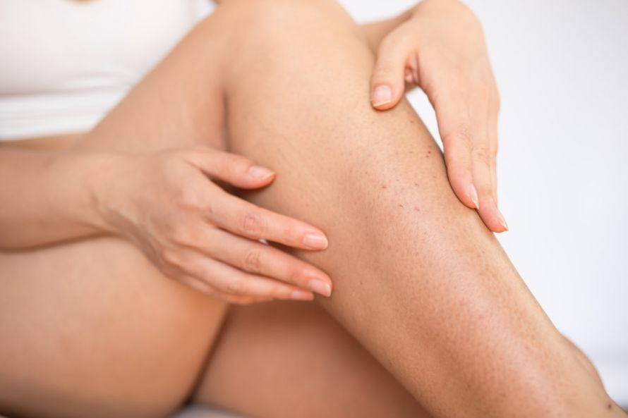
In tinea cruris, the inflammation can often be clearly traced to the hair follicles. They appear as brown or bluish-red, pea-sized nodules and usually have a scaly border. Severe itching in the affected areas is a common symptom. The outer sides of the lower legs are particularly affected.
If you notice these symptoms, you should see a dermatologist (or your primary care physician first). They can diagnose the problem.
Diagnosis of Tinea Cruris
For the doctor, tinea cruris is often a visual diagnosis. It is based on
- the typical external appearance and
- the patient’s medical history (e.g., women, leg shaving, concurrent athlete’s foot).
If there is any doubt, the diagnosis must be confirmed microscopically.
However, this does not yet reveal exactly which pathogen caused the fungal infection. A fungal culture is necessary for this. It is grown from the tissue sample in a Petri dish over the course of several weeks. This allows for conclusions to be drawn about the exact fungal species and possible resistance to antifungal medications. Fast-growing dermatophytes can be identified after one week at the earliest.
The specimens used for diagnosing tinea cruris are
- skin flakes,
- nail clippings, or
- plucked hairs,
which are collected from the peripheral areas of the affected regions. In the case of the lower leg, a vigorous swab is usually sufficient.
Another method for diagnosing fungal infections is the Wood’s lamp. A Wood’s lamp emits UV light. This allows certain species of dermatophytes to be identified as fluorescent areas on the skin in a darkened room. However, this method is of only secondary importance, since fungal microscopy and culture are usually performed anyway.
Treatment of Tinea Cruris with Antifungals
The treatment of fungal skin infections such as tinea cruris generally follows a two-step approach:
- topical therapy and
- systemic therapy.
Topical therapy
The first line of treatment is topical therapy, which should always be attempted whenever possible. “Topical” means that the antifungal agent is applied “locally.” It is therefore a local treatment. Solutions, creams, or ointments are available for this purpose.
The active ingredients used here are primarily
- azole antifungals,
- tolnaftate, or
- terbinafine.
For very superficial cases of tinea cruris, application over a period of a few days to four weeks may be sufficient.
Side effects are rare, as the active ingredient does not enter the bloodstream in significant amounts. As a result, it cannot spread throughout the body.
A disadvantage of topical therapy is that treatment often takes quite a long time.
Individual fungal cells can persist for a long time as spores in the horny layers of the epidermis. Spores are very robust and are difficult to eliminate. Although antifungal medications prevent the pathogens from growing and multiplying, they do not kill them. Therefore, the goal of treatment is to continue it until all current skin cells—including the spores they contain—have been shed.
If itching and redness subside after a few days, the inflammation has already been successfully treated. However, up to four weeks of topical therapy are sometimes necessary to completely clear the skin.
A second problem with topical therapy is that it often acts only very superficially. Deeper-seated fungi are not affected, as the skin’s defensive barrier also acts against the medication.
In addition, reinfection can easily occur.
Systemic Therapy
Persistent or widespread fungal skin infections such as tinea cruris are therefore often treated “systemically” from the outset. Systemic means that the active ingredients are not applied locally but act throughout the entire body.
This is done in the form of tablets. Since the entire body is affected, side effects may occur.
Systemic antifungals can damage the liver. Therefore, good liver function is a prerequisite for most antifungal agents.
The following are used:
- griseofulvin (the only approved medication for young children),
- itraconazole,
- fluconazole, or
- terbinafine.
Systemic administration leads to higher concentrations in the cornea and better cure rates. As a result, the treatment duration can usually be shortened by about half. Two weeks is typical in most cases. Combination with topical therapy is also possible.
Summary of Tinea Cruris
Tinea cruris is a serious and persistent condition. It requires decisive treatment. Although the fungi pose no immediate danger, as they thrive only on the skin, However, the infection is a major cosmetic problem and, as it progresses, leads to skin inflammation and almost unbearable itching.
The risks of a two-week course of treatment, on the other hand, are quite manageable. Liver function tests may be necessary. Tinea cruris is, however, usually very treatable.
Share this article