
An astrocytoma is a tumor of the central nervous system that originates from astrocytes. Astrocytomas are a type of glioma and are among the most common brain tumors in children and young adults. Astrocytomas are typically diagnosed using MRI and additional molecular tests. Depending on the WHO grade, the tumor may be benign or malignant and may grow at varying rates. Treatment often involves surgery, followed by radiation therapy or chemotherapy.
The prognosis depends, among other factors, on the tumor’s location, grade, and molecular profile. Modern neurosurgical techniques allow for the safest possible tumor removal while preserving important brain functions. For those affected, early diagnosis and individualized treatment are crucial.
What is an astrocytoma, and what are the different grades?
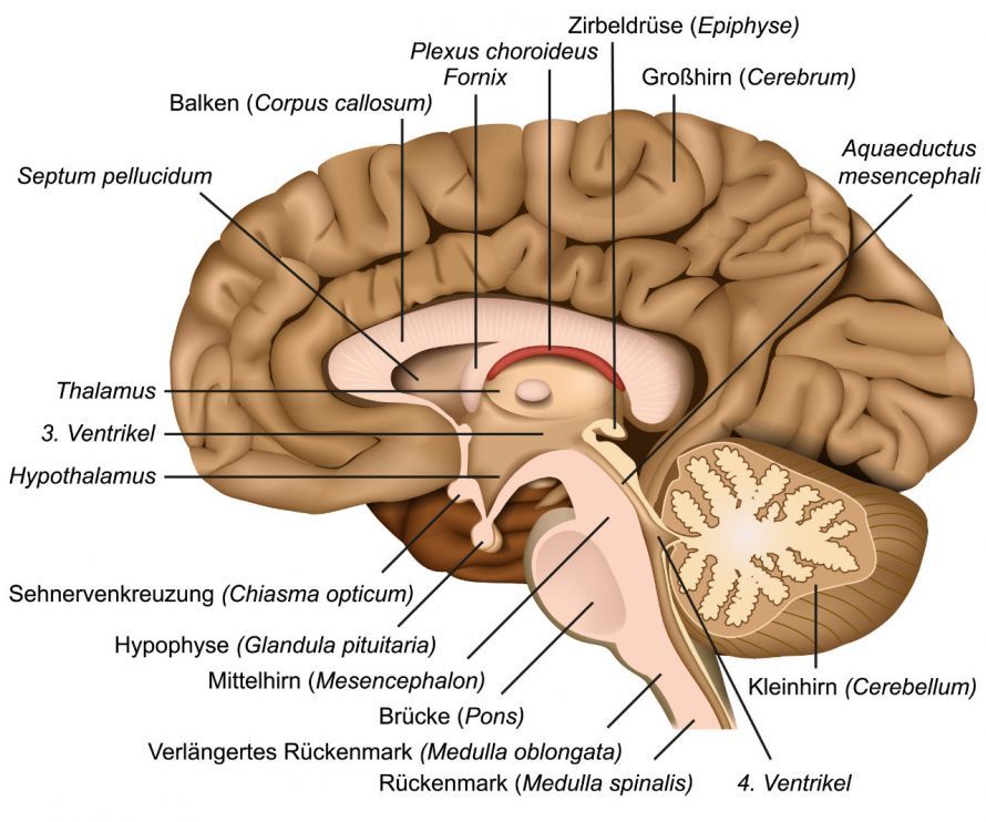
Pilocytic astrocytoma (Grade 1) most commonly occurs in children and young adults. Pilocytic astrocytoma is the most common brain tumor in children.
It primarily affects
- the cerebellum,
- the basal ganglia,
- the thalamus,
- the brainstem, and
- the optic nerve (nervus opticus; known as an optic glioma).
A Grade 1 brain tumor grows only slowly and is clearly demarcated from the surrounding brain tissue in terms of its growth characteristics. In 99 percent of cases, it is benign and can be cured with complete surgical removal.
Diffuse astrocytoma (Grade 2) most commonly affects middle-aged individuals (ages 30 to 40). In most cases, it is located in the white matter of the cerebrum, frequently in the frontal lobe. Diffuse astrocytoma is also frequently found in the thalamus, midbrain, and pons. Less commonly, this tumor occurs in the temporal or parietal lobes. Grade 2 astrocytoma grows very slowly. However, it also invades healthy surrounding tissue (“diffuse growth”). If left untreated, it progresses to a malignant form in about 10 percent of cases per year.
An anaplastic astrocytoma (Grade 3) occurs in people between the ages of 35 and 45. The tumor grows faster than Grade 2 astrocytomas. It is located in the cerebrum and can also progress to glioblastoma. Glioblastoma (Grade 4) typically occurs in people over the age of 50. It is the most malignant glioma and, with 3 new cases per 100,000 people, the most common.
The tumor primarily originates in the white matter of the cerebrum. It grows infiltratively, meaning it invades healthy brain tissue.
Symptoms
In general, astrocytoma causes symptoms similar to those of other brain tumors. However, the exact symptoms depend primarily on the tumor’s location. Astrocytomas located near the optic tract impair vision. This leads to protrusion of the eyeball (known as exophthalmos).
If the tumor grows in the cerebellum, the patient may experience
- trembling of the hands,
- impaired coordination of the limbs, and
- other functions controlled by this region of the brain.
Other possible symptoms of a brain tumor include
- dizziness,
- nausea and vomiting,
- headaches,
- fatigue,
- personality changes,
- Symptoms of paralysis,
- Unsteady gait and tendency to fall, as well as
- seizures.
Causes and Risk Factors
This type of tumor develops from astrocytes. This cell type belongs to the group of supporting cells (glial cells) in the brain. Supporting cells are responsible for isolating nerve cells from blood vessels and the surface of the brain.
Astrocytomas arise from genetic changes that disrupt the growth control of astrocytes. This leads to uncontrolled cell division and the development of a tumor.
Various factors are discussed as possible causes of such genetic changes. It is well established that brain tumors can develop as a result of radiation therapy.
However, the medical community generally assumes that astrocytomas are not usually hereditary. That said, there are certain genetic disorders in which astrocytomas occur more frequently. These include, for example, neurofibromatosis types 1 and 2 (a rare skin disorder).
Examinations and Diagnosis
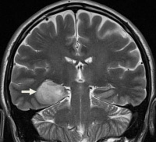
The most important diagnostic method when an astrocytoma is suspected is magnetic resonance imaging (MRI, also known as nuclear magnetic resonance imaging; Fig. 1). With the help of a contrast agent, neurologists can identify the astrocytoma as either a well-defined or diffuse area on the MRI scan.
In a Grade 1 astrocytoma, the affected region of the brain appears distinctly bright on the scan because the tumor is sharply defined.
Grade 2 and 3 tumors are visible only as more or less dark areas. Glioblastoma appears as a ring-shaped structure.
A biopsy is often used to confirm the diagnosis. During this procedure, the neurosurgeon surgically removes a tissue sample from the affected region of the brain. This sample is examined in the laboratory for malignant cells. The results of this histological examination are crucial: they allow physicians to determine the tumor’s grade of malignancy and plan treatment accordingly.
In selected cases, tests are conducted to detect the presence of specific molecular markers. Their presence or absence can influence the prognosis and treatment decisions.
Another diagnostic method is cerebrospinal fluid (CSF) analysis. This involves the collection and examination of fluid from the spinal canal. It plays a particularly important role in distinguishing astrocytomas from other diseases.
Treatment of Astrocytomas
When deciding on a treatment, the treating physician must weigh the risks and benefits. Factors considered include
- the patient’s age,
- the patient’s general condition, and
- neurological status
into the treatment plan.
In addition to these factors that cannot be influenced, there are also factors that can be influenced. These, too, determine the success of a treatment. They include, among other things,
- the time until surgery (the shorter, the better),
- the extent of tumor resection (partial resection vs. complete tumor resection), as well as
- the use of chemotherapy or radiation therapy for inoperable astrocytomas.
In general, the therapeutic options for astrocytoma are therefore
- surgery,
- radiation therapy, and
- chemotherapy
are available.
Surgery for Astrocytoma
The modern strategy of very early surgery for astrocytoma is based on relatively recent observations. A study compared the survival times of patients from two universities using two different but consistently applied strategies. It demonstrated a clear survival advantage for patients who underwent surgery.
In contrast, long-term observation over several years, or a biopsy alone followed by chemotherapy or radiation without surgery, more frequently led to malignant transformation and an associated accelerated progression of the disease.
In the case of pilocytic astrocytoma (WHO Grade 1), surgery is usually sufficient. If the tumor is located in the cerebellum, it can be completely removed in most cases. This results in a very good prognosis, and symptoms improve rapidly.
Pilocytic astrocytomas
- in the basal ganglia,
- in the thalamus, and
- in the brainstem
are removed using microsurgical techniques. However, complete removal is often not possible, so radiation therapy usually follows.
Smaller diffuse astrocytomas (WHO Grade 2), in particular, can be surgically removed. Depending on the results of the histopathological examination, the next step is
- either a wait-and-see approach is taken to see if tumor regrowth occurs after brain tumor surgery, or
- radiation therapy is initiated.
The goal of surgery is normally the complete removal of the tumor tissue. However, the tumor also grows into healthy brain tissue. Therefore, there is a risk of neurological deficits if healthy brain tissue is removed along with the tumor tissue. For this reason, larger tumors generally cannot be completely removed. In these cases, radiation therapy is important. Chemotherapy is used only for very extensive tumors and in cases of tumor regrowth following radiation therapy.
Anaplastic astrocytoma (WHO Grade 3) also invades healthy tissue. Therefore, complete surgical removal, as is possible with other gliomas, is not feasible. Rather, the goal of surgery is to reduce the size of the tumor. This increases the chances of success for subsequent radiation therapy or chemotherapy.
Course and Prognosis of Astrocytomas
Following a diagnosis of astrocytoma, lifelong follow-up monitoring via MRI is typically performed. However, the imaging interval is adjusted on an individual basis. The following intervals are currently considered standard:
- every 3 months: after surgery, for WHO Grade III tumors, in cases of visible residual tumor, for IDH wild-type tumors, and for PET-active residual tumors,
- every 3–12 months: depending on tumor type, molecular profile, extent of resection, and the duration of previous tumor-free or progression-free intervals.
According to the Bern OPTIMISST protocol, most patients should resume normal activities as early as the day after surgery. Patients with larger tumors or more complex locations should plan for about one to two months of recovery time after surgery. It is important to create optimal and stress-free conditions for the surgery within the family and with the employer.
In cases of particularly difficult surgeries, deficits are to be expected. However, in 95% of cases, these deficits are temporary. In such cases, intensive neurorehabilitation should be scheduled. The histological diagnosis is available no later than one week after surgery. The molecular diagnosis follows another week later. Afterward, the medical team plans the next steps during a tumor board meeting. The determined grade of the tumor has a major impact on the patient’s future quality of life and the course of the disease.
Grade 1 astrocytomas have a favorable prognosis. Five years after the tumor was diagnosed, 9 out of 10 patients were still alive. Most patients are also considered “cured” after surgery if the tumor can be completely removed.
For patients with Grade 2 tumors, additional surgery is often necessary after 2–8 years. In cases of small tumors with favorable characteristics, surgery can remove more than 99.99% of the tumor cells. Only a few cells remain, and not all of these cells will regrow afterward. Consequently, there are patients in whom the tumor does not recur for decades or even during their lifetime. It is therefore more accurate to speak of “long-term control” rather than a cure.
With Grade 3 tumors, the tumor tissue regrows more rapidly. However, depending on the exact characteristics of the cells, it is also possible to remain tumor-free for several years. Life expectancy for patients with Grade 3 astrocytoma is much lower than for patients with Grade 1 astrocytoma. Grade 4 astrocytoma (“glioblastoma”) has the poorest prognosis: five years after diagnosis, only 5 out of 100 patients were still alive. However, modern chemotherapy can help extend survival times, particularly in younger patients.
FAQ
What is an astrocytoma?
An astrocytoma is a tumor that arises from astrocytes and belongs to the group of gliomas. Astrocytomas develop in the brain and are among the most common brain tumors. Depending on the WHO grade, these tumors can be benign or malignant. Diffuse astrocytoma and anaplastic astrocytoma are among the most common forms.
What symptoms does an astrocytoma cause?
The symptoms of an astrocytoma depend on the tumor’s location in the brain. Headaches, nausea, and vomiting, as well as neurological deficits or epileptic seizures, are common. If the tumor grows near the optic tract or other important brain regions, additional symptoms may arise. Diagnosis is therefore made as early as possible using magnetic resonance imaging (MRI).
How is an astrocytoma diagnosed?
An astrocytoma is typically diagnosed using MRI (magnetic resonance imaging). Biopsies, molecular analyses, and additional clinical and neurological examinations may also be necessary. The World Health Organization classifies astrocytomas according to WHO Grade 1 through WHO Grade 4. This classification is crucial for prognosis and treatment.
How is an astrocytoma treated?
Treatment depends on the tumor’s location, WHO grade, and the patient’s overall health. In most cases, the primary treatment is surgery to remove the tumor. Depending on the findings, radiation therapy, radiotherapy, or chemotherapy may follow. For astrocytomas with an IDH mutation, treatment approaches and prognosis may differ in some respects from those of other tumor types.
What is the prognosis and life expectancy for an astrocytoma?
The prognosis and life expectancy depend heavily on the tumor grade. A pilocytic astrocytoma (WHO Grade 1) often has a favorable prognosis, whereas an anaplastic astrocytoma (Grade 3) or a glioblastoma (Grade 4) has a significantly more aggressive course. Factors such as tumor growth, molecular markers, complete tumor removal, and potential recurrences also influence the prognosis. Malignant astrocytomas often have a significantly poorer prognosis than low-grade tumors.
Share this article
Sources
- Back M et al., Influence of molecular classification in anaplastic glioma for determining outcome and future approach to management. J Med Imaging Radiat Oncol 2019; doi:10.1111/1754-9485.12850
- Feiden S & Feiden W. WHO-Klassifikation der ZNS-Tumoren. Pathologe 2008; 29, 411–421
- Jakola AS et al., Comparison of a strategy favoring early surgical resection vs a strategy favoring watchful waiting in low-grade gliomas. JAMA 2012; 308(18): 1881–1888
- Jones DT et al., Recurrent somatic alterations of FGFR1 and NTRK2 in pilocytic astrocytoma. Nat Genet 2013; 45: 927–932
- krebshilfe.de/infomaterial/Blaue_Ratgeber/Gehirntumoren_BlaueRatgeber_DeutscheKrebshilfe.pdf
- Leitlinien der Deutschen Gesellschaft für Neurologie. AWMF-Registernummer: 030/099 [derzeit in Überarbeitung bis voraussichtl. Ende 2019]
- Weller M (2014) Gliome - Leitlinien für Diagnostik und Therapie in der Neurologie. Herausgegeben von der Kommission
- Wick W., Astrozytäre Tumoren (Gliome). In: Hacke W (Hrsg) Neurologie. 2015. Springer, Heidelberg