A urinary tract tumor is a malignant lesion that can develop in various parts of the urinary tract—namely, the kidney, renal pelvis, ureter, bladder, or urethra.
The most common type is urothelial carcinoma, which arises from the lining (urothelium) of the urinary tract. Men are affected more frequently than women, and the peak incidence occurs between the ages of 60 and 70.
A typical initial symptom is painless blood in the urine. In the early stages, a urinary tract tumor often causes no symptoms. Early urological diagnosis is therefore crucial for detecting the disease in time and treating it successfully.
What is a urinary tract tumor, and how does it develop?
A urinary tract tumor is a malignant growth (malignant tumor) in the urinary tract. This includes the kidney, the renal pelvis, the ureter, the bladder, and the urethra.
The most common type is urothelial carcinoma of the upper urinary tract—that is, a tumor that originates in the lining (urothelium) of the renal pelvis or ureter.
Such a malignant urinary tract tumor may remain localized or invade neighboring structures via the urinary tract.
When detected early, tumors of the urinary tract are usually treatable—especially when diagnosis and treatment are performed by experienced urologists.
Depending on the location, the following distinctions are made:
| Area | Typical Tumor | Proportion / Frequency |
|---|---|---|
| Renal pelvis | Urothelial carcinoma of the renal pelvis | Approx. 5–10% of all urothelial carcinomas |
| Ureter | Ureteral carcinoma | Very rare (< 1%) |
| Bladder | Bladder carcinoma | Most common form, over 90% of all cases |
| Urethra | Urothelial carcinoma of the urethra | Rare |
Urothelial carcinoma of the upper urinary tract (i.e., the renal pelvis and ureter) accounts for about 5–10% of all urothelial carcinomas.
Since the entire urinary tract is lined with the same mucosa, multiple tumors can develop simultaneously or sequentially. Tumors of the upper urinary tract—specifically in the renal pelvis and ureter—often present with symptoms associated with a renal pelvis or ureteral tumor, such as painless blood in the urine.
Together with bladder cancer, these conditions are among the most significant malignant changes affecting the urinary tract.
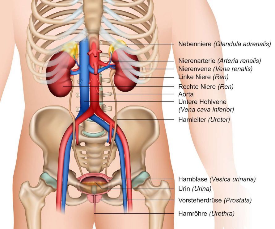
The ureters transport urine from the kidneys to the bladder @ bilderzwerg /AdobeStock
Causes and Risk Factors for a Urinary Tract Tumor
The causes of urinary tract tumors are varied and not fully understood.
Medical professionals believe that genetic factors, environmental influences, and chronic irritation of the urinary tract may contribute to the development of a urinary tract tumor.
People with a long history of tobacco use, exposure to aromatic amines, or chronic urinary tract infections are at particularly high risk.
These factors promote changes in the mucous membrane (urothelium) and can increase the likelihood of developing urothelial carcinomas of the upper urinary tract.
- Tobacco smoking (the most significant risk factor; up to 50% of all cases)
- Exposure to aromatic amines (e.g., in paints, varnishes, and the rubber industry)
- Chronic urinary tract infections or urinary retention
- Analgesic abuse (long-term use of pain relievers, e.g., phenacetin)
- Arsenic exposure in drinking water
- Genetic factors and familial clustering
- Previous radiation or chemotherapy in the pelvic area
In patients with ureteral or renal pelvis tumors, tumors may later develop in the bladder—and vice versa.
Symptoms and early signs of a urinary tract tumor
A urinary tract tumor often causes no symptoms for a long time.
The first sign is usually painless blood in the urine, which occurs at some point in nearly all patients.
Other possible symptoms include:
- Flank pain or a dull, pressing sensation in the back
- Frequent urge to urinate or a burning sensation when urinating
- Difficulty passing urine, possibly with hydronephrosis
- Tenderness in the kidney area
- General symptoms such as fatigue, weight loss, and night sweats
Important: Blood in the urine should always be evaluated by a urologist, even if there is no pain.
Diagnosis of a urinary tract tumor: Tests and procedures
Diagnosis is performed by experienced urology specialists.
It combines physical examination, laboratory tests, endoscopy, and imaging:
Laboratory Diagnostics
- Urine cytology: Detection of tumor cells (urothelial carcinoma cells) in the urine.
- Tumor markers in the blood can aid in monitoring the course of the disease.
Imaging Diagnostics for Urinary Tract Tumors (CT, Ultrasound, Cystoscopy)
- Ultrasound examination of the bladder and kidneys to detect urinary obstruction.
- CT urography or MRI urography with contrast medium to assess tumor size, infiltration, and lymph nodes.
- Retrograde pyelography: Visualization of the renal pelvis using a contrast agent.
- Cystoscopy (bladder examination) and ureterorenoscopy for direct endoscopic evaluation and tissue sampling (biopsy).
During endoscopic examination of the urinary tract, urologists pay particular attention to the openings of the ureters into the bladder, as tumor cells or bleeding may be present there.
During transurethral resection (TUR), smaller tumors in the bladder can be removed and examined histologically.
This allows for the early detection of potential metastases or tumor infiltration.
This diagnostic procedure enables precise staging according to the TNM classification and helps determine the appropriate course of treatment.
Treatment and Management of Urinary Tract Tumors
Treatment of urinary tract tumors depends on the tumor stage, location, and the patient’s overall health. Treatment covers the entire spectrum of urology—from endoscopic examination to minimally invasive procedures and radical surgery.
In early stages, transurethral resection (TUR) is often sufficient to remove the tumor.
The goal is to completely remove the urinary tract tumor while preserving the function of the upper urinary tract.
Endoscopic and Minimally Invasive Removal of Urinary Tract Tumors
Smaller tumors in the renal pelvis or ureter can be removed minimally invasively using ureterorenoscopy or laser ablation.
This preserves the organ. This is often followed by local irrigation therapy with mitomycin C to prevent recurrence.
Surgical removal (nephroureterectomy) and other forms of treatment
For larger or infiltrating tumors, a nephroureterectomy (surgical removal of the kidney and ureter) is required.
The ureteropelvic junction of the affected ureter is also removed to prevent recurrence.
Chemotherapy and immunotherapy for urinary tract tumors
In advanced stages, systemic chemotherapy may be necessary (e.g., with cisplatin).
For inoperable or metastatic urinary tract tumors, immunotherapies using checkpoint inhibitors (e.g., pembrolizumab, atezolizumab) are increasingly being used. By combining various forms of therapy, physicians can make full use of the entire spectrum of modern urological oncology.
In individual cases, additional treatment of the prostate is performed if tumor cells are present there or if there are drainage problems.
Follow-up Care and Monitoring
Regular follow-up care is crucial for the early detection of recurrences or secondary tumors. Particular attention is paid to the upper urinary tract.
Both the upper urinary tract and the bladder are regularly monitored via endoscopy or ultrasound to rule out tumor regrowth.
A tumor of the ureter or renal pelvis may also develop later, which is why comprehensive monitoring of the entire urinary tract is recommended.
This includes ultrasound, CT, cystoscopy, and urine cytology at specified intervals (every 3–6 months in the first year, then annually).
Today, treatment of urinary tract tumors is usually performed using minimally invasive or endoscopic methods, provided the tumor is still localized.
For larger tumors, surgical removal via nephro-ureterectomy remains the treatment of choice.
Specialized urology clinics, such as the University Hospital of Cologne, offer all modern diagnostic and therapeutic procedures.
Prognosis
The prognosis for a urinary tract tumor depends heavily on the tumor stage, the depth of infiltration, and lymph node involvement.
If the tumor is detected early and completely removed, the chances of a cure are very good.
In advanced stages with metastases, the disease is often chronic but can frequently be controlled over the long term with modern therapies.
FAQs on Urinary Tract Tumors – Diagnosis, Treatment & Prognosis
What tests are necessary to diagnose a urinary tract tumor?
To diagnose a urinary tract tumor, urologists use several imaging and diagnostic procedures.
In addition to ultrasound examination of the bladder and kidneys, computed tomography (CT) with contrast dye is considered the method of choice for determining the extent of the tumor and identifying potential metastases.
In addition, a cystoscopy or ureterorenoscopy is often performed, during which the lining of the urinary tract is directly examined and a tissue sample is taken.
These procedures allow for a diagnostically reliable staging of the tumor.
German Cancer Society – Bladder Cancer 2024; Onkopedia 2024.
How is a urinary tract tumor surgically removed?
Surgical removal depends on the tumor stage and location.
Smaller tumors can often be treated by endoscopic removal—for example, using a laser during a minimally invasive ureterorenoscopy.
In cases of advanced disease, nephroureterectomy—that is, the complete removal of the kidney and ureter—is the treatment of choice.
Following the procedure, local mitomycin C irrigation or additional therapy such as chemotherapy or immunotherapy may be administered to prevent recurrence.
What is the difference between ureteral carcinoma and a renal pelvis tumor?
Both are classified as urothelial carcinomas of the upper urinary tract.
In ureteral carcinoma, the tumor is located in the mucosa of the ureter and can obstruct urine flow.
A renal pelvis tumor, on the other hand, is located at the junction between the kidney and the ureter.
Since the entire urinary tract is lined with the same mucosa, both types of tumors can develop simultaneously or sequentially.
Therefore, guidelines recommend regular monitoring of the entire urinary tract following an initial diagnosis.
How is the tumor stage determined for a urinary tract tumor?
Tumor staging describes how deeply the urinary tract tumor has invaded the mucosa and surrounding tissue, and whether lymph nodes or other organs already show signs of metastasis.
Staging is performed according to the TNM classification using computed tomography (CT), contrast-enhanced imaging, and endoscopic biopsy.
The tumor stage is crucial for treatment planning and the patient’s prognosis.
Where is a urinary tract tumor treated, and according to which guidelines?
Treatment usually takes place in a urology clinic, often at specialized centers such as the University Hospital of Cologne or comparable university medical centers.
Here, experts work in an interdisciplinary manner according to the current guidelines for the diagnosis and treatment of urothelial carcinomas of the upper urinary tract.
Depending on the tumor stage, these guidelines recommend endoscopic, minimally invasive, or radical surgery, combined with chemotherapy or immunotherapy if necessary.
Share this article

Sources
- krebsinformationsdienst.de/tumorarten/harnblasenkrebs/index.php
- S3-Leitlinie Früherkennung, Diagnose, Therapie und Nachsorge des Harnblasenkarzinoms, Langversion 1.1 – November 2016; AWMF-Registernummer: 032/038OL
- S3-Leitlinie Diagnostik, Therapie und Nachsorge des Nierenzellkarzinoms, Langversion 2.0 - August 2020; AWMF-Registernummer: 043/017OL
- urologielehrbuch.de/harnleiterkarzinom_02.html
- urologie.uk-koeln.de/erkrankungen-therapien/urothelkarzinom-oberer-harntrakt/