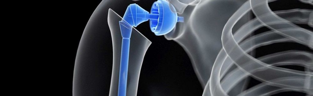
Shoulder arthroplasty refers to the complete or partial replacement of the shoulder joint with artificial materials. If the joint is completely replaced—that is, the glenoid cavity of the scapula and the head of the humerus—this is referred to as a total shoulder replacement (TSR); if only one component of the joint is replaced, it is called a hemiprosthesis.
Certification as an Endoprosthetics Center (EPZ)
 Physicians who hold this seal are affiliated with medical facilities that are certified by endocert as an Endoprosthetics Center (EPZ) or as a Maximum Care Endoprosthetics Center (EPZmax) and have thus demonstrated through an audit that they meet the established requirements. Recertification is required every three years.
You can find the criteria, requirements, and differences between EPZ and EPZmax here.
What is shoulder endoprosthetics?
The shoulder joint is formed by the head of the humerus and the glenoid cavity of the scapula. It is the most mobile joint in the human body. Since there is minimal bone-to-bone contact, the ligaments and, above all, the muscles play a significant role in stabilizing the joint. The tendons of the stabilizing muscles are arranged like a cuff around the shoulder joint and are therefore collectively referred to as the rotator cuff.
Due to the joint’s high range of motion and the relatively limited soft-tissue stabilization—particularly in the forward-downward direction—shoulder dislocations can occur relatively easily. In addition, the bony components of the shoulder joint can fracture due to external forces, and the muscles and tendons of the rotator cuff can tear.
Similar to the hip and knee joints, joint wear (osteoarthritis) can also occur in the shoulder joint. Depending on the severity of the joint wear, a complete or partial replacement of the shoulder joint with artificial materials—that is, a shoulder prosthesis—may be necessary. When both the glenoid cavity and the humeral head are replaced, the procedure is referred to as a total shoulder arthroplasty (TSA); when only the humeral head is partially replaced, it is called a shoulder hemiarthroplasty.
Shoulder prostheses, which are very well tolerated by the body, are usually made of a titanium or chromium-chromium alloy and are anchored in the bone—depending on the condition of the bone and the patient’s age—with or without bone cement (a special plastic).
What conditions do specialists in shoulder arthroplasty treat?
Shoulder injuries—usually associated with previous bone fractures, rheumatic diseases (rheumatoid arthritis), or other degenerative (wear-and-tear-related) conditions can lead to osteoarthritis—that is, joint wear and tear in the shoulder joint—in which the cartilage is initially degraded, and in advanced stages, the bone itself is also degraded.
Other conditions treated by experts in shoulder arthroplasty include, for example:
- Extensive rotator cuff defects (defect arthropathy)
- Avascular necrosis of the humeral head (humeral head necrosis), meaning that bone tissue dies as a result of an accident or a disease
- Tumors of the upper part of the humerus (humerus tumors)
- Pseudarthrosis (false, artificial, or pseudo-joint) at the junction of the humeral head and shaft (non-union of a bone fracture)
- Recurrent fracture of a previously healed fracture
- Impacted shoulder dislocation (dislocated shoulder joint)
- Omarthrosis resulting from an infection
What diagnostic procedures do specialists in shoulder arthroplasty use?
The patient interview (medical history)—for example, regarding the nature and duration of symptoms—as well as a physical and clinical examination form the basis of the diagnosis. In many cases, this alone is sufficient to narrow down the possible causes of the patient’s symptoms.
However, imaging studies are indispensable. X-ray examinations (radiographic diagnostics) play a key role in this process. They can be used to detect fractures and bone changes. Computed tomography (CT) allows specialists to assess bone structure, among other things.
Soft tissues, such as the rotator cuff, bursae, and other muscles, can be visualized using magnetic resonance imaging (MRI). To better visualize the joint space, a contrast agent is occasionally injected directly into the shoulder joint, followed by an MRI (known as MR arthrography).
Ultrasound (sonography) is a widely used diagnostic method for evaluating soft tissue structures.
What treatment methods are included in the scope of services provided by a specialist in shoulder arthroplasty?
The range of services offered by a specialist in shoulder arthroplasty includes, among others, the following methods:
- Implantation of anatomical shoulder prostheses—that is, replacement of the humeral head and the glenoid cavity with anatomical prosthetic components
- Implantation of reverse shoulder prostheses—that is, the joint components are swapped so that the spherical component is implanted on the glenoid side and the concave component on the humeral head side. The goal is to alter the joint’s pivot point in such a way that the deltoid muscle is preloaded
- Inverse shoulder prostheses are used in cases of rotator cuff damage, following trauma, or when an anatomical shoulder prosthesis has become loose
- Shaftless shoulder prostheses are increasingly being implanted on the humeral side
- Revision (correction) and replacement of shoulder prostheses
- Treatment of complex fractures of the humeral head
- Treatment of long-term complications of humeral head fractures
What sets specialists in shoulder arthroplasty apart?
Experts in shoulder arthroplasty are typically specialists in orthopedics, trauma surgery, sports medicine, or orthopedic surgery who have specific experience in the implantation of shoulder prostheses.
When deciding whether or not to proceed with surgery, they weigh the benefits against the risks and consider possible alternatives to joint replacement. Through certifications, they ensure a high standard of surgical care.

About the medical author
Prof. Felix Zeifang
Medical Author · Manager
Prof. Dr. med. Felix Zeifang – Author: Discover expert medical articles at Leading Medicine Guide.
More about the medical author →Sources
- Deutsche Gesellschaft für Orthopädie und Orthopädische Chirurgie(DGOOC) (2017) S2e-Leitlinie „Rotatorenmanschette“. AWMF-Leitlinien-Register Nr. 033/041
- Deutsche Gesellschaft für Unfallchirurgie e.V. (DGU) et al. (2017) Oberarmkopffraktur. Leitlinien Unfallchirurgie. AWMF-Leitlinien-Register Nr. 012/023
- Garving C, Jakob S, Bauer I et al. (2017) Impingementsyndrom der Schulter. Dtsch Arztebl Int 114(45): 765-76. doi: 10.3238/arztebl.2017.0765
- Jerosch J, Heisel J (2003) Schulterendoprothetik: eine Standortbestimmung – Implantation bei Omarthrosen und Frakturen des proximalen Humerus. Dtsch Arztebl 100(37): A-2366 / B-1978 / C-1865
- Schünke M et al. (2018) Prometheus. Allgemeine Anatomie und Bewegungssystem: LernAtlas der Anatomie. Thieme, Stuttgart