Brain aneurysm surgery is the neurosurgical treatment of a dilated blood vessel in the brain (brain aneurysm). There are two procedures—clipping and coiling—from which the doctor and patient choose the one that is most likely to be successful in each individual case.
Learn more here about the procedure for brain aneurysm surgery and find selected brain aneurysm surgeons.
What is brain aneurysm surgery?
An aneurysm generally refers to a bulge in the wall of a blood vessel. This means that individual layers or all layers of a blood vessel expand outward.
There are many causes for such a defect. Common risk factors include
- high blood pressure,
- atherosclerosis (hardening of the arteries),
- smoking,
- alcohol consumption, and
- injuries.
Brain aneurysms can also be congenital, resulting from a weakness in the arterial wall. If a blood vessel in the brain is affected, it is referred to as a cerebral aneurysm. Cerebral aneurysms are usually located at vessel bifurcations.
Cerebral aneurysm surgery is the neurosurgical procedure used to remove or occlude this bulge in the brain.
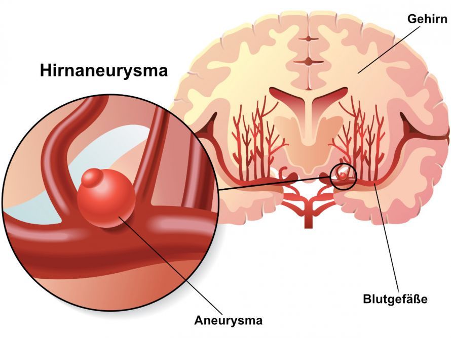
A cerebral aneurysm can lead to life-threatening brain hemorrhages if it ruptures © rob3000 | AdobeStock
There are various procedures from which the doctor can choose in consultation with the patient. The procedure selected depends on the individual
- risk factors,
- family history, and
- severity
.
Why should an aneurysm be treated?
A brain aneurysm usually does not cause any symptoms on its own and is often discovered incidentally during an examination. Sometimes
- headaches,
- episodes of dizziness, or
- visual disturbances
may indicate the presence of an aneurysm.
However, as the aneurysm grows larger, the risk of it rupturing increases. This can lead to a dangerous brain hemorrhage, known as a subarachnoid hemorrhage, which must be avoided at all costs. The rupture of an aneurysm can be triggered by physical exertion or occur spontaneously while at rest.
A hemorrhage must be treated as quickly as possible, as it can cause numerous long-term complications. In 30 percent of cases, it is fatal. Possible complications include, for example,
- hydrocephalus (a drainage disorder of the brain’s blood vessels),
- areas of the brain with reduced blood flow and resulting symptoms,
- epileptic seizures,
- electrolyte imbalances,
- vitreous hemorrhages, or
- cardiac complications such as arrhythmias.
The aneurysm should be surgically removed if the risk of surgery is lower than the risk of rupture. The risk of rupture is determined based on several factors. These include, among others,
- the size of the aneurysm,
- high blood pressure,
- location,
- shape,
- family history,
- alcohol consumption,
- previous bleeding, and
- other genetic factors.
What surgical methods are available for brain aneurysm surgery?
There are two different neurosurgical procedures for the surgical treatment of brain aneurysms:
- microsurgical clipping
- coiling
Microsurgical clipping
In microsurgical clipping, the surgeon closes off the aneurysm sac with a clip. The clip is precisely tailored to the size and shape of the aneurysm and isolates the aneurysm sac within the blood vessel from the blood flow. The clip is made of titanium or other metals.
For this brain aneurysm surgery, the surgeon makes a small incision in the skin and opens the skull. This provides a minimally invasive access route to the aneurysm. For larger aneurysms, a bypass may need to be created to bypass the aneurysm. The advantages of this technique are
- the reliable occlusion of the aneurysm with a low risk of rupture,
- effectiveness against aneurysms with a wide neck,
- good intraoperative management of complications, and
- the ability to treat concomitant cerebral hemorrhages.
Significant disadvantages of this cerebral aneurysm surgery are
- its invasiveness, which increases the overall surgical risk,
- the increased risk of intraoperative ruptures, and
- the inaccessibility of certain locations.
Coiling
The second treatment option is coiling. This cerebral aneurysm surgery is performed via an endovascular approach, that is, through the bloodstream. Unlike clipping, it is a neuroradiological procedure performed under continuous X-ray guidance.
A specialist punctures an artery in the thigh. Through this opening, the specialist advances a thin catheter along the blood vessels upward to the aneurysm. Then, another thin catheter is advanced through the first one and guided into the aneurysm.
Through this catheter, the surgeon inserts thin platinum coils into the aneurysm. These coils cause a blood clot to form inside the aneurysm, thereby blocking it. If the neck of the aneurysm is too wide, a balloon or stent may need to be inserted to dilate the adjacent blood vessel. This also prevents the balloon or stent from being washed out of the aneurysm.
The advantages of this technique include its minimally invasive nature and, according to studies, better long-term outcomes.
The disadvantages are the slightly higher risk of rupture and incomplete occlusion, which occurs in 15 percent of cases.
Which procedure is the right one?
The choice depends on individual factors. When both options are equally suitable, the surgeon generally prefers coiling because it carries a lower surgical risk.
Clipping is more suitable
- for younger patients,
- for patients without significant comorbidities,
- for aneurysms that are more centrally located,
- for aneurysms with complex shapes, and
- in cases of concomitant bleeding.
Coiling is the preferred treatment
- for older patients with comorbidities,
- for aneurysms in the posterior circulation,
- for simple aneurysms with a thin neck, and
- in the absence of bleeding.
The experience of the treating neurosurgeons and neuroradiologists is also a decisive factor in the choice of procedure.
What does follow-up care look like after brain aneurysm surgery?
The hospital stay lasts about three to four days. After a postoperative follow-up MRI and the completion of all examinations, the patient is discharged.
Follow-up appointments are scheduled on-site. It is also determined whether these appointments will take place at the radiologist’s office or at the hospital. It is important that you attend these appointments to ensure optimal monitoring of the operated aneurysm.
If the aneurysm is completely occluded, it can no longer cause complications. For this reason, follow-up visits are rarely necessary after clipping.
Following endovascular therapy, follow-up MRI scans must be performed at six months and two years. With this procedure, recanalization occurs more frequently, often requiring additional treatment.
If occlusion is insufficient, physicians may change the treatment strategy as needed. Following treatment with coiling in combination with a stent, patients should undergo four weeks of therapy with two different blood-thinning medications, followed by monotherapy.
Untreated cerebral aneurysms should be monitored annually via MRI, even if their size remains stable. Patients with acquired aneurysms should minimize risk factors as much as possible:
- Maintain a balanced diet,
- keep blood lipid levels low,
- control your blood pressure,
- and avoid nicotine and alcohol.
What are the risks and complications? What are the chances of success?
In general, serious complications are very rare in surgery for unruptured aneurysms smaller than 10 millimeters. For more complex aneurysms, such as giant aneurysms or more complicated types, the treatment risk is higher.
Treatment with clipping may lead to
- wound infections,
- postoperative bleeding,
- epileptic seizures, or
- vascular occlusion leading to a stroke
. With coiling, there is a risk of
- perforation of the aneurysm, leading to bleeding,
- an allergic reaction to contrast dye, as well as
- vascular occlusion
.
However, the probability of vascular occlusion is less than two percent for both procedures. The overall prognosis for both procedures is very good. With coiling, however, more frequent follow-up visits are necessary.