Capsule endoscopy is an innovative procedure in gastroenterology used to visualize the digestive tract. During capsule endoscopy, the patient swallows a small capsule that captures images as it passes through the intestines. This procedure is particularly well-suited for examining the small intestine, which is only partially accessible using conventional methods such as colonoscopy.
After the capsule is swallowed, the video capsule travels autonomously through the small intestine and later through the large intestine. The images captured are transmitted to a receiver attached to a portable data recorder. Capsule endoscopy is a purely diagnostic procedure for visualizing the mucosa of the digestive tract. It is particularly useful in cases of unexplained bleeding or suspected Crohn’s disease.
Applications of Capsule Endoscopy
Capsule endoscopy has been in use for nearly 20 years. It has been CE-certified and FDA-approved since 2001.
The procedure provides over 50,000 high-quality photos directly from the intestine in a gentle, non-invasive manner. The photos are later compiled into a video.
A gastroenterologist can then evaluate the video and use it to make an accurate diagnosis of the patient’s symptoms. Live evaluation of the transmitted images is now also possible.

This is particularly
- in cases of unexplained iron-deficiency anemia, where causes in the stomach and large intestine have been ruled out, or
- in cases of suspected chronic inflammatory bowel disease of the small intestine, such as Crohn’s disease,
capsule endoscopy is used.
Diseases of the small intestine can be examined more thoroughly using capsule endoscopy than with a conventional magnetic resonance imaging (MRI) scan. An MRI often lacks sufficient resolution. Capsule endoscopy, on the other hand, can reveal even small lesions.
As it travels through the gastrointestinal tract, the camera capsule passes through all sections of the intestine. It can therefore also be used for colorectal cancer screening. There are special capsules for the small intestine and the large intestine. The colon capsule has two cameras, one at each end. This allows images to be captured on both sides. The small-bowel capsule, on the other hand, is equipped with only one camera.
The Capsule Endoscopy Procedure
Capsule endoscopy is significantly less uncomfortable than a colonoscopy. The camera capsule is swallowed with a little water, just like a regular vitamin capsule. Neither a hospital stay nor a sedative is necessary.
The capsule endoscopy procedure is fully automated. However, some preparatory steps are necessary beforehand to ensure high-quality images of the intestine:
- Consultation with the doctor to determine whether the swallowing reflex is functioning properly or if there are any known narrowings of the intestine. A self-dissolving preliminary examination capsule can be used for this purpose.
- To ensure that the intestinal lining is clearly visible, the patient should fast for 12 hours before taking the capsule, meaning they should refrain from eating or drinking.
- The doctor may prescribe a laxative beforehand, which should be taken prior to the examination.
- Two hours after swallowing the capsule, patients may resume drinking fluids. However, they should avoid red-colored or dairy-based beverages so as not to interfere with the images. Light meals are permitted again after about four hours.
Before swallowing the camera capsule, the patient is given a recording device to wear on their belt. They usually take the recording device home with them and return it the next day.
As the capsule passes through the intestines, it continuously transmits images from the digestive tract. The recording device stores the transmitted images for later analysis. This process can result in up to 60,000 images.
The capsule is now propelled by natural bowel movements (peristalsis). Its position can be monitored via the recording device.
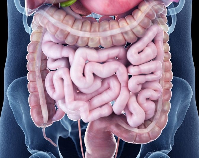
During the recording phase of capsule endoscopy, there are no special rules to follow other than a slightly reduced diet. The patient can behave and move completely naturally.
Completion and Evaluation of Capsule Endoscopy
After the camera capsule has passed through the gastrointestinal tract, it is naturally excreted in the bathroom. This usually occurs after about 9–14 hours, but in some cases it may take several days.
At the doctor’s office or clinic, a video is created on a computer from the thousands of individual photos. The specialist then evaluates this video. If bleeding is present, the footage can show the location of the bleeding. Capsule endoscopy can be a valuable tool for diagnosing Crohn’s disease, as well as tumors and polyps.
Risk factors for which capsule endoscopy is not used
Capsule endoscopy is a diagnostic procedure that offers many advantages for the patient. The only prerequisites for a successful examination are
- a functioning swallowing reflex and
- bowel segments that are completely patent and free of narrowings where the capsule could become stuck.
However, these conditions are thoroughly assessed before capsule endoscopy.
Risk factors that preclude capsule endoscopy therefore include
- severe swallowing difficulties,
- severe constipation (for example, caused by certain medications or diabetes),
- intestinal obstruction (mechanical ileus),
- adhesions in the intestine that narrow the bowel,
- deep diverticula in the small intestine.
Advantages and Disadvantages of Capsule Endoscopy
Capsule endoscopy is a very minimally invasive procedure. It benefits patients who have unexplained abdominal symptoms or who are reluctant to undergo a conventional colonoscopy.
As a precise diagnostic tool, capsule endoscopy provides very good insights into the individual course of the disease. The advantages of capsule endoscopy at a glance:
- non-invasive, painless, and gentle examination procedure,
- no radiation exposure,
- no hospital stay required,
- no sedatives or tranquilizers,
- no contrast agents,
- Examination of the entire small intestine is only possible with capsule endoscopy,
- Cancer screening with a low barrier to entry.
Capsule endoscopy is an innovative yet now well-established diagnostic tool for the digestive tract. It provides a unique view of the small intestine and is comparable to colonoscopy for examining the large intestine.
One disadvantage of capsule endoscopy is that it serves a purely diagnostic purpose. Unlike with colonoscopy, doctors cannot perform any procedures on suspicious tissue changes during the examination.
During a colonoscopy, on the other hand, doctors can remove colon polyps and take tissue samples while the procedure is still in progress. Capsule endoscopy can detect suspicious areas, but a conventional colonoscopy is then necessary to perform any intervention.
Costs and Conclusions Regarding Capsule Endoscopy
The issue of cost coverage by health insurance providers has not yet been definitively resolved.
In cases of unexplained intestinal bleeding (“unexplained mid-intestinal bleeding”), health insurance companies may cover the costs. However, certain criteria must be met if the capsule endoscopy is performed on an outpatient basis.
Patients should therefore discuss reimbursement with their doctor before undergoing a capsule endoscopy. In individual cases, reimbursement may be obtained from the insurance provider upon prior application. Capsule endoscopies performed as part of a hospital stay are billed by the hospital using flat-rate fees.
In summary, capsule endoscopy is an excellent way to thoroughly examine the entire intestinal tract, particularly the small intestine. Inflammatory bowel diseases or causes of bleeding in the small intestine can thus be diagnosed almost imperceptibly.
Capsule endoscopy should also be regarded as a milestone in medical technology for colorectal cancer screening.
FAQ
How does capsule endoscopy work?
During a capsule endoscopy, the patient swallows a small capsule containing built-in control and transmission electronics as well as a camera. As the capsule travels through the gastrointestinal tract, it automatically captures images—two per second—and transmits them to an external receiving and storage unit. The capsule travels through the digestive tract on its own until it is excreted naturally.
When is a capsule endoscopy recommended?
The most important indication, however, is the investigation of unexplained bleeding in the small intestine. It is also indicated when a small intestine disorder is suspected, such as Crohn’s disease, or when pathological changes—such as polyps or strictures—are suspected. Small-bowel capsule endoscopy allows for the examination of the entire small intestine, which is not possible with conventional endoscopy or colonoscopy.
What are the advantages of capsule endoscopy?
A major advantage of capsule endoscopy is that it is a non-invasive procedure that does not require sedation. The procedure is generally well tolerated by patients and does not require any surgical intervention, unlike a conventional colonoscopy. Capsule endoscopy also allows for the examination of hard-to-reach sections of the small intestine.
Are there any risks or contraindications?
The most important contraindication for capsule endoscopy is a known narrowing in the intestine. Narrowings or an increased risk of bleeding can cause the capsule to get stuck. In such cases, the feasibility of performing a capsule endoscopy must be carefully evaluated.
How is the procedure performed?
A thorough bowel preparation is necessary before a capsule endoscopy can begin. After swallowing the capsule, the patient can usually resume their normal daily activities. The capsule reaches the large intestine after passing through the small intestine and is then excreted. The results of the examination are later evaluated by a gastroenterologist using the stored images.
Be sure to read our fascinating expert interview with Prof. Dr. med. Robert Ehehalt on the Leading Medicine Guide blog!

About the medical author
Prof. Robert Ehehalt
Medical Author · Internist,Gastroenterologist · Heidelberg
More about the medical author →