The knee joint is the largest joint in the human body and plays a central role in movement and stability. The anatomy of the knee joint involves a complex interplay of bones, cartilage, menisci, and ligamentous structures. The knee joint connects the femur to the tibia and allows for flexion and extension of the joint.
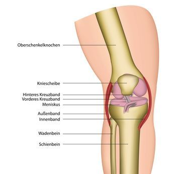
The kneecap, also known as the patella, protects the knee joint and aids in force transmission via the tendon. The cruciate ligaments and collateral ligaments stabilize the knee joint and ensure proper alignment of the articular surfaces. The menisci act as fibrous cartilage and distribute the load evenly throughout the knee joint. Due to the high load it bears, the knee joint is susceptible to injuries and conditions such as osteoarthritis or gonarthrosis.
The Knee Joint
The knee joint allows the leg to bend and straighten, as well as to rotate moderately inward and outward (internal and external rotation) when bent. These movements are guided and stabilized by the menisci, ligaments, tendons, and muscles. Important blood vessels and nerves run through the popliteal fossa at the back of the knee joint.
The Articular Cartilage
In a healthy person, the contact surfaces of the bones in the knee joint are covered by a whitish layer of cartilage several millimeters thick. This articular cartilage enables smooth and pain-free movement of the knee joint. It consists of living cartilage cells (chondrocytes) embedded in a matrix tissue.
Due to natural aging processes and improper weight-bearing, as well as a lack of nourishment to the cartilage caused by a sedentary lifestyle, the cartilage can become dry and brittle. This increases the risk of injury and accelerates wear and tear. In such cases, cartilage surgery may be considered as a treatment option.
The Menisci (Articular Discs)
Between the rounded ends of the thigh bone (femoral condyles) and the nearly straight head of the shin bone (tibial plateau) lies a joint space containing the two menisci(plural of meniscus):
- the medial meniscus (meniscus medialis) and
- the lateral meniscus (meniscus lateralis).
These crescent-shaped cartilage discs can adapt to movement, thereby distributing weight and pressure and acting as shock absorbers and sliding bearings. They also assist the ligaments in stabilizing the knee joint.
Since the medial meniscus is firmly fused to the medial collateral ligament and the joint capsule and therefore has less mobility than the lateral meniscus, it is more prone to injury (meniscus tear).
The Ligaments and Tendons of the Knee Joint
The ligaments of the knee play an important role. Without them, the femur and tibia would not be connected to one another. Stabilization of the knee joint in the anterior and posterior directions is provided by the two cruciate ligaments (anterior and posterior cruciate ligaments).
The medial and lateral collateral ligaments provide lateral stability to the knee joint and the joint capsule. The patellar ligament complements the patellar tendon. The patellar tendon is the most important tendon of the knee joint. The kneecap (patella) is embedded in it.
The Joint Capsule
The knee joint is enclosed by a strong joint capsule. This capsule produces a fluid that nourishes the articular cartilage and reduces friction between the cartilage surfaces.
Diseases and Injuries of the Knee Joint
The knee joint is particularly susceptible to degenerative changes (knee osteoarthritis or gonarthrosis—wear and tear of the knee joint) and ligament injuries (especially cruciate ligament tears). Knee osteoarthritis can result from, among other things, leg misalignments (e.g., bowlegs or knock-knees), improper weight-bearing, or injuries.
Cartilage damage in the knee does not necessarily lead to knee osteoarthritis, as there are a variety of treatment options available for knee cartilage damage. In cases of knee osteoarthritis, a total knee replacement (knee prosthesis) can significantly improve quality of life.
Sports injuries frequently affect the knee. In addition to a torn cruciate ligament (cruciate ligament rupture), injuries may include tears of the collateral ligaments, meniscal tears, and the avulsion of a piece of cartilage or bone.
Other conditions and injuries affecting the knee include arthrofibrosis, patella fractures, and Baker’s cysts. Find your specialist in sports trauma.
Conclusion
The knee joint is the largest joint in the human body and plays a central functional role in stability and movement. In the anatomy of the knee joint, the articulatio is a complex hinge joint between the femur and the tibia.
The condyles of the femur are in close contact with the articular surfaces of the tibia, while the joint capsule encloses and protects the joint. The structure and function of the knee joint are further supported by important blood vessels and nerves, as well as stabilizing structures. Clinically, injuries to the knee joint and conditions such as knee osteoarthritis are of great significance.
Damage can severely limit mobility and, in the long term, affect the entire musculoskeletal system. Overall, human anatomy and general anatomy make it clear that the knee joint is the largest and, at the same time, one of the most vulnerable joints in the body.
FAQ
What is the knee joint?
The knee joint is a large joint consisting of the femur, tibia, and patella. It enables flexion and extension and is crucial for mobility in the leg.
What is the function of the menisci in the knee joint?
The menisci are crescent-shaped pieces of fibrocartilage in the knee joint that act as shock absorbers. The medial and lateral menisci distribute the load across the joint surfaces.
What role do the cruciate ligaments play?
The cruciate ligaments stabilize the knee joint and prevent the femur and tibia from slipping out of place. The anterior cruciate ligament and the posterior cruciate ligament are particularly important in this regard.
What conditions can affect the knee joint?
Common conditions affecting the knee joint include osteoarthritis, knee osteoarthritis, or meniscus damage similar to a herniated disc. Injuries to the knee joint also frequently affect the ligaments.
What is the anatomy of the knee joint?
The anatomy of the knee joint includes bones such as the femur and tibia, the patella, menisci, cartilage, the joint capsule, as well as the cruciate ligaments and collateral ligaments. These structures work together to ensure stability and mobility.