A patellar fracture is a break in the kneecap and is one of the most common injuries to the knee joint. A patella fracture is usually caused by a fall onto the knee or by a traffic accident involving a direct impact to the patella. Depending on the type of fracture, the displacement of the fracture fragments, and the stability of the extensor mechanism, a patella fracture can be treated conservatively or surgically.
Typical symptoms include swelling, pain on the front of the knee joint, and an inability to lift the leg while keeping it straight. Diagnosis involves clinical examinations, X-rays, CT scans, MRI, and sometimes ultrasound. For complex patella fractures, surgical procedures such as tension band osteosynthesis, cerclage, or plate osteosynthesis are used.
Treatment of a patellar fracture also includes physical therapy, an orthosis, a splint, and a gradual return to full weight-bearing on the leg. Early management of a patellar fracture significantly improves the prognosis, healing, and mobility of the knee joint.
The Patella and Causes of a Patella Fracture
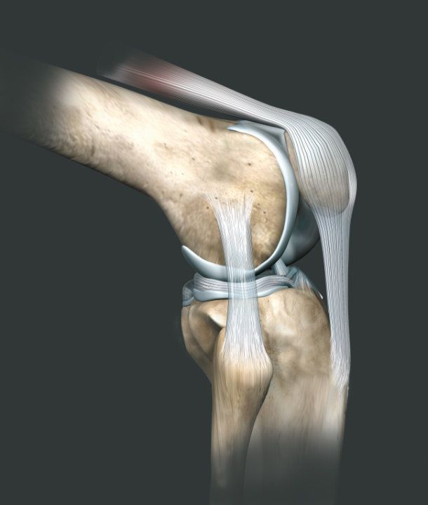
The kneecap (patella) is part of the knee’s extensor mechanism. At the level of the knee joint, it acts as a pulley for the extensor muscles of the lower leg. It is therefore involved in the active extension of the knee joint.
The patella prevents the tendons of the thigh extensor muscles from rubbing directly against the knee joint. It glides along a corresponding groove in the femur.
The mechanism of injury in a patella fracture is usually a fall onto the kneecap or a severe impact trauma.
Symptoms and Diagnosis of a Patella Fracture
A patella fracture is accompanied by severe pain on the front of the knee joint and directly over the patella.
Significant swelling of the knee joint usually occurs as well. Typically, the patient is unable to actively extend the lower leg or keep the knee joint extended. A definitive diagnosis can be made using X-rays.
Treatment of a Patellar Fracture
Treatment depends on:
- Type of patella fracture
- Number of fragments
- Alignment of the fracture line and
- the degree of displacement of the fragments
In principle, doctors can treat non-displaced, stable fractures conservatively. Surgical treatment is recommended for displaced, unstable fractures.
Splinting and immobilization of the affected knee joint are always necessary. An increased risk of a blood clot (thrombosis) requires the prophylactic administration of anticoagulants.
As part of surgical treatment, doctors secure the fracture fragments using drill wires or screws. A special bandage absorbs the tensile forces (cerclage). Once the patella fracture has healed, it is usually necessary to remove the implants in a subsequent surgery.
Follow-up Care for a Patella Fracture
Initially, the knee joint is immobilized using special splints. The patient should not put weight on the leg and should avoid actively extending the knee.
The patient can begin follow-up physical therapy early on:
- pain-managed rehabilitation with full axial weight-bearing in the extended position (Mecron splint)
- X-ray follow-up after weight-bearing and after 14 days to rule out secondary dislocation
- Range of motion restrictions: Weeks 1–3: Ext/Flex 0–0–30; Weeks 4–6: Ext/Flex 0–0–60
Mobilization following surgical treatment depends on the extent of the injury and the surgical procedure. Osteosynthesis with screws and tension straps can usually also be mobilized according to the following schedule:
- Weeks 1–3: Extension/Flexion 0–0–30
- Weeks 4–6: Extension/flexion 0–0–60
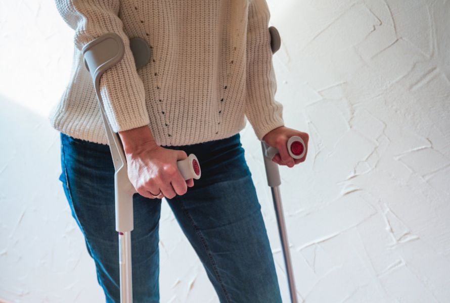
The use of a continuous passive motion (CPM) device is helpful. When bearing full axial weight, the patient must use forearm crutches for gait training. This allows the patient to relieve pressure on the extensor mechanism and reduce retropatellar compression.
Prognosis Following a Patellar Fracture
The prognosis for an uncomplicated patella fracture is usually very good. The patient can use the affected leg without restriction.
Occasionally, residual symptoms such as a tendency to swell or sensitivity to weather changes may occur and can persist for several months.
A long-term complication may be premature joint wear in the patellar glide surface. This occurs when, following comminuted fractures, an irregularity in the cartilage on the back of the patella persists despite surgery.
Medications for a Patella Fracture
Initial and postoperative local and systemic analgesic therapy should be administered.
Conclusion
If a patellar fracture is suspected, a clinical examination should be performed promptly to identify a fracture of the patella and possible injuries to the quadriceps tendon or extensor mechanism in a timely manner. The exact characterization and classification of the injury is determined using X-rays or computed tomography (CT), particularly if displacement of the fracture fragments or involvement of the articular surface is suspected.
Treatment of a patellar fracture can be conservative or surgical; however, minimally displaced distal pole fractures often heal without surgery. For unstable fractures, procedures such as tension band osteosynthesis or cerclage are used as part of surgical management to prevent the fragments from diverging. Early treatment following a fall on the knee or a traffic accident is crucial for the healing and management of a patellar fracture, so that long-term issues such as pain on the front of the knee, difficulty climbing stairs, a bent knee joint, or joint wear can be avoided, and the knee does not remain actively restricted.
FAQ
What is a patellar fracture?
A patella fracture is a fracture or break of the kneecap. The patella connects the quadriceps, the patellar tendon, and the knee joint and is important for knee extension. A patella fracture is often caused by a direct fall onto the knee or by traffic accidents involving an impact to the kneecap.
What are the symptoms of a patella fracture?
Typical symptoms of a patella fracture include severe pain on the front of the knee joint, swelling, and limited mobility. With a patella fracture, the knee can no longer be actively extended. Other symptoms include bruising, an unstable leg, or pain when climbing stairs.
How is a patella fracture diagnosed?
If a patella fracture is suspected, a clinical examination of the knee joint is performed first. X-rays, CT scans, MRI, or ultrasound are used to identify the type of fracture, the joint surface involved, and any possible associated injuries. The classification of patellar fractures is important for planning further treatment.
When is a patella fracture treated surgically?
A patellar fracture is treated surgically if the fracture fragments are displaced or if the extensor mechanism has been injured. Tension band osteosynthesis, cerclage, or plate osteosynthesis are frequently used. The goal of surgical treatment is to ensure stable healing of the fracture and to restore function to the knee joint.
Can a patella fracture be treated conservatively?
Yes, minimally displaced distal pole fractures or stable longitudinal fractures can be treated conservatively. Conservative treatment usually involves wearing a brace or splint while keeping the leg extended. Physical therapy, full weight-bearing after medical clearance, and thrombosis prophylaxis also aid in rehabilitation following a patella fracture.
Share this article
Sources
- S2e-Leitlinie „Patellafraktur“ (AWMF-Register-Nr. 187-021), Stand 04.02.2021: register.awmf.org/de/leitlinien/detail/187-021