Whole-body MRI—or whole-body MRI for short—is an imaging technique used to diagnose various diseases. MRI stands for magnetic resonance imaging, but the terms “nuclear magnetic resonance imaging” and “nuclear magnetic resonance” are also widely used. Below, you’ll learn when a whole-body MRI is used, how the procedure works, and what you as a patient should keep in mind. You’ll also find specialists in whole-body MRI here.
What is an MRI, and how does it work?
X-rays and computed tomography (CT) involve a certain amount of radiation exposure for the patient. Magnetic resonance imaging (MRI), on the other hand, does not use any radiation at all.
It uses strong magnetic fields that set the hydrogen atoms in the human body in motion. These movements are analyzed and converted into cross-sectional images. The more hydrogen a tissue contains, the more detailed the image can be with magnetic resonance imaging.
The following are particularly well visualized in an MRI:
- soft tissue,
- the entire vascular system, arteries, veins, and
- most organs
For examining bones, X-rays are still considered the best diagnostic method. For examining the lungs and heart specifically, computed tomography (CT) is either the preferred method or is used as a supplement.
A whole-body MRI does not take much time: As a rule, the entire body is examined in a maximum of 60 minutes.
How does a whole-body MRI work?
Before the whole-body MRI begins, the patient removes all metallic objects such as
- rings,
- necklaces,
- piercings, and
- glasses
. In some cases, a contrast agent is also administered. This is used to make the arteries and veins appear even more clearly. To do this, the doctor inserts a cannula into a vein in the crook of the arm.
For the actual whole-body MRI, the patient lies down on an examination table. They are given headphones to protect their ears from the loud knocking sounds of the MRI machine. The table slowly moves through the MRI coil, starting at the head or the feet.
It takes about 40 to 60 minutes for the entire body to be scanned. The patient remains in contact with the radiology technician via an intercom. The patient is also given an emergency button to press if the exam becomes too loud or uncomfortable. The MRI scan is then stopped immediately.
After the whole-body MRI, a radiologist evaluates the data.
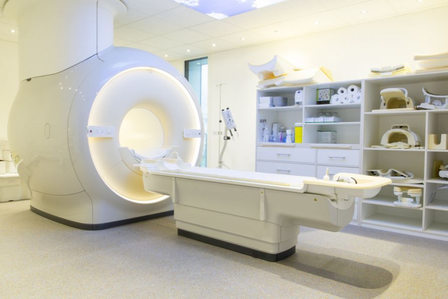
Lying in front of an MRI machine © digitale-fotografien | AdobeStock
Applications: When is an MRI used?
A whole-body magnetic resonance imaging scan is used for the early detection of various diseases. Cardiovascular diseases, in particular, are often asymptomatic in their early stages. However, they should be detected early to reduce the risk of a heart attack or stroke.
If a doctor’s suspicion is not confirmed by a physical examination and blood tests, they may additionally order a whole-body MRI. This makes calcifications and narrowing of blood vessels (such as arterial stenosis or atherosclerosis) visible. This allows for timely initiation of appropriate treatment.
Magnetic resonance imaging is also used for rheumatic inflammatory diseases. Such diseases often affect not only the joints but also other parts of the body.
A whole-body MRI offers the advantage of scanning the entire body in a single session. This often eliminates the need for additional diagnostic procedures, which are associated with high costs and significant stress for the patient.
Another area of application for whole-body MRI is cancer diagnosis, specifically the detection of metastases. Benign bone growths (exostoses), whose development must be monitored regularly, can also be quickly examined using MRI.
Patients can undergo a whole-body MRI as part of a preventive health screening. However, health insurance companies will only cover the costs if the doctor has a reasonable suspicion of a specific disease.
Anyone who wishes to cover the cost of the examination themselves should request a cost estimate in advance and seek comprehensive advice.
Which specialists use MRI?
A full-body MRI is primarily required and ordered by the following specialists for diagnostic purposes:
Since not every doctor’s office has an MRI machine, patients are usually referred to
- a radiologist,
- a neuroradiologist, or
- a hospital with a radiology department.
What should patients be aware of before an MRI?
A full-body MRI is not suitable for all groups of people. For example, pregnant women should avoid an MRI during the first three months of pregnancy, as the potential effects on the unborn child have not yet been sufficiently studied.
Furthermore, a full-body MRI is only possible to a limited extent and with special precautions for the following individuals:
- Patients with electronic implants (pacemakers, subcutaneous insulin pumps, neurostimulators)
- Patients with artificial heart valves
- Patients with metallic joint implants (endoprostheses)
- Patients with middle ear implants that are not made of gold or Teflon
- Patients with cochlear implants
If you have an implant in your body, be sure to talk to your doctor. Depending on the material used, a full-body MRI is still possible in many cases. This is especially true for newer implants, which are often made of gold, Teflon, or other non-magnetic materials.
If there is a possibility that metal fragments are present in your body, you should also discuss this with your doctor. This is because any metal parts will be set in motion by the MRI machine’s strong magnets, which carries a high risk of injury.
Tattoos can cause burns and image distortion. In such cases, a careful assessment must be made as to whether the examination should be performed.
Are there any risks or complications?
A full-body MRI carries no risks whatsoever. The exam involves no radiation at all and is completely painless.
However, many patients complain about the high noise level inside the MRI machine, which can be very distressing even when wearing headphones. Nowadays, however, there are more modern MRI machines that operate more quietly and are also less sensitive to movement.
Patients who are prone to panic attacks in confined spaces can be given a mild sedative before the exam begins. Alternatively, many hospitals and radiology practices now offer open MRI machines, which are less confining.
What are the advantages and disadvantages of whole-body MRI?
Despite its numerous advantages, some medical professionals view whole-body magnetic resonance imaging with skepticism. Their main criticism is that this diagnostic procedure often reveals abnormalities that are not indicative of disease. However, these findings can still cause anxiety for patients. This is the case in about 40 to 50 percent of examinations.
A detailed discussion with the patient after the data has been evaluated is therefore essential. The doctor should provide the patient with comprehensive information and make it clear that not every abnormality is a cause for concern.

About the medical author
Sabine Schneider
Editor-in-Chief
Sabine Schneider – medical author: Explore expert articles and medical expertise in the Leading Medicine Guide.
More about the medical author →Sources
- https://www.aerzteblatt.de/archiv/moeglichkeiten-und-grenzen-der-ganzkoerper-mrt-303115e9-adcb-4428-a91b-59f873eb1f04
- https://www.dkfz.de/radiologie