Recommended specialists
Article overview
Radiology - Further information
In the search for specialists in radiology you will find medical experts in imaging diagnostics. You would like to have preventive screening carried out for the early detection of cancer, such as breast cancer, cardiovascular disease or osteoporosis? Or you would like to contact a specialist in radiology for a second medical opinion? Here you will find selected specialists in radiology.
Specialist radiology/radiologist
Radiology is a speciality that uses medical imaging to diagnose and treat diseases within the body. Visual representations of the body’s interior are used to support the process of clinical analysis and medical intervention. Such medical imaging techniques can also be used to capture the detailed functioning of some organs or tissues (physiology), and to create a database of normal anatomy and physiology, making it possible to detect and identify abnormalities much faster and more accurately.
As a broad discipline within the overall field of biological imaging, radiology utilises modern imaging techniques, such as X-ray radiography, ultrasound, CT (computed tomography), nuclear medicine and MRI (magnetic resonance imaging), to create data that informs medical investigations and treatments.
The process of capturing medical images is usually undertaken by a radiographer, also known as a radiologic technologist. Raw visual data is then interpreted by a diagnostic radiologist who sends the images and a report back to the clinician who requested the procedure. Data and images collected during investigations are then digitally archived and stored so they can be quickly and easily accessed by appropriate healthcare professionals.
Radiologists are medical doctors (MDs) or doctors of osteopathic medicine (DOs) who specialise in diagnosing and treating diseases and injuries using medical imaging techniques. Interventional radiology is a further extension of this discipline, which involves the performance of (usually minimally invasive) medical procedures, guided by a variety of imaging technologies.

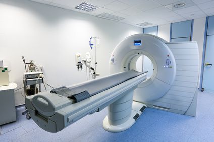
CT scanner © zlikovec / Fotolia
Which illnesses do radiology specialists treat?
While radiology specialists are now called upon to investigate most diseases and conditions for diagnostic purposes, they also play a vital role in preventive screening and interventional procedures.
Some medical areas where preventive screening can be used to promote continued wellness include:
- breast cancer
- cardiac
- carotid artery
- colorectal cancer
- lung cancer
- osteoporosis
- repair of narrowing arteries (angioplasty)
- peripheral vascular disease
- renal artery stenosis
- gastrostomy tube placements
Such peripheral and minimally invasive interventions minimise the patient’s physical trauma, while also reducing infection rates and recovery times, as well as the duration of hospital stays.
What treatment methods are used by radiology specialists?
Even though the boundaries are becoming harder to define, the technologies deployed by radiology specialists can be subdivided into those that are mainly used for investigative and diagnostic purposes, and others in which the intervention has a clear treatment goal.
Diagnostic imaging modalities include:
- Projection (plain) radiography, which was the original X-ray imaging technique used to support medical diagnosis. Even though much more detailed and powerful imaging methods are now available, projection radiography is still the default diagnostic mode for some applications, such as breast cancer mammography.
- Fluoroscopy and angiography, which are special adaptations of X-ray imaging technology that use swallowed or injected radio-contrast agents. These liquids are used in conjunction with X-rays and display screens to highlight the detail of internal processes in real time. This technique facilitates investigations of blood vessel functions, and much more.
- CT (computed tomography) imaging technology uses computing power to create three-dimensional body images from X-ray data. In recent times, powerful digital data processing has enabled CT scans to produce sophisticated internal detail, which healthcare professionals need to make accurate evaluations of major health problems, such as cerebral haemorrhage and pulmonary embolism (clots in the lung arteries).
- Ultrasound employs high-frequency sound waves to generate images of internal soft tissue in real time. This technique is considered safer because it uses no radiation, and has thus been extensively used to track pregnancies. More sophisticated ultrasound using sound-filtering techniques can also be used to conduct vascular investigations and find blood clots in deep veins.
- MRI (magnetic resonance imaging) creates strong magnetic fields to influence the behaviour of hydrogen protons within body tissue, and uses the resultant micro activity to generate internal images. Because of the relative ease with which MRI can construct images in any chosen plane of movement, the technique is greatly favoured in many medical areas, including, for example, neuroradiology.
- Nuclear medicine has developed radiopharmaceutical substances capable of seeking out certain kinds of body tissue and tracking its function. This allows targeted monitoring of functions such as blood flow to heart muscle. The use of biologically active substances can also track cell growth, which is very helpful in the diagnosis of certain cancers.
Interventional radiology is the logical extension of minimally invasive diagnostics to include treatment interventions that can be monitored in real time. Some techniques can be performed while the patient is wide-awake and require minimal sedation, if any.
What additional qualifications are required by radiology specialists?
Radiology is an expanding and very competitive medical field. Diagnostic radiologists must complete four years at medical school to earn a medical degree, then one year of internship, followed by a four-year training residency. To become an interventionalist, radiologists may then opt for a further one or two years of speciality fellowship training.
Medical spectrum
Therapies
- Embolization
- Interventional radiology
- Laser(-induced) thermotherapy
- MRgFUS
- Radiochemotherapy
- Radiofrequency ablation
- Radiooncology treatment
- Radiotherapy
- SIRT / Selective internal radiotherapy
- Transarterial chemoembolization
- Transarterial chemoperfusion
- Transpulmonary chemoembolization
- Tumor therapy
- X-ray stimulation therapy


