Selective internal radiation therapy (SIRT, also known as transarterial radioembolization) is a form of local radiation therapy. It is used to treat liver cancer. In this procedure, radioactive particles are delivered to the liver via special microspheres (SIR-Spheres or microspheres). Once there, they specifically target and irradiate the diseased tissue.
Here you will find further information as well as a list of selected SIRT specialists.
Applications of SIRT
Liver cancer refers to the uncontrolled growth of cells within the liver. A distinction is made between
- primary liver cancer: The tumor develops directly from cells within the liver itself (also known as hepatocellular carcinoma), and
- secondary liver cancer or liver metastases: A cancer has spread from another site, with cancer cells traveling through the bloodstream or lymphatic system to the liver, where they form new tumors.
In Germany, approximately 6,000 people are diagnosed with primary liver cancer each year. Liver metastases, however, are significantly more common: Each year, liver metastases are detected in about 30,000 patients with colorectal cancer.
Several studies have shown that selective internal radiation therapy (SIRT) can significantly reduce the size of liver tumors. In some cases, the tumor can be reduced to such an extent that subsequent surgical removal becomes possible. In isolated cases, the tumor can even be completely destroyed by radiation therapy.
SIRT can also improve the quality of life and life expectancy of patients whose cancer does not respond to chemotherapy.
Selective internal radiation therapy is used for
- inoperable or untreatable primary liver tumors, such as hepatocellular carcinoma or bile duct cancer,
- inoperable liver metastases, including those from breast or colorectal cancer.
However, SIRT is only possible if the liver is still functioning properly. Liver function is checked with a blood test before SIRT therapy.
Preliminary Examinations
Thorough examinations are necessary before SIRT. These are intended to ensure that the radioactive microspheres used in SIRT can be safely administered to the patient. To this end, a doctor evaluates the blood vessels of the liver.
An important tool for these preparations is a femoral catheter. It is advanced into the liver during an angiography procedure.
The doctor then uses the catheter to occlude the adjacent blood vessels leading from the liver to other organs using metal coils. This procedure is called embolization. This occlusion ensures that the radioactive microspheres act exclusively within the liver and do not enter other organs via the bloodstream.
In addition, the doctor injects a low-level radioactive substance into the blood vessel via the catheter. The doctor then monitors the distribution of radioactivity using a scintigram (a device for visualizing the results of nuclear medicine examinations).
This also allows the doctor to individually determine the radiation dose required for the subsequent SIRT treatment.
The preliminary examination can be performed on an outpatient or day-surgery basis. If this preliminary examination reveals no contraindications to SIRT, the procedure can be performed after about one to two weeks.
The SIRT Procedure
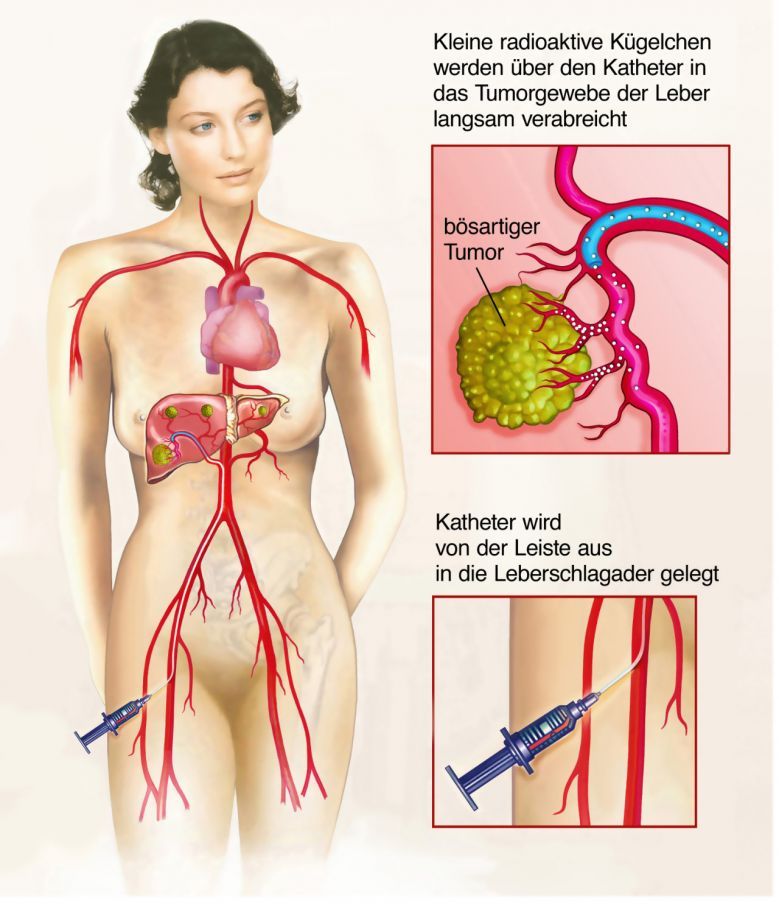
Healthy liver tissue is supplied with nutrients primarily via the portal vein (Vena portae). The hepatic artery (Arteria hepatica), on the other hand, plays only a minor role in supplying the liver. A cancerous tumor, however, receives its blood supply primarily via the hepatic artery.
SIRT takes advantage of this difference in blood supply. To do this, radioactively charged microspheres—made of glass or resin—are delivered into the hepatic artery via an inguinal catheter. The microspheres contain a radioactive isotope (an atom, in this case yttrium-90), which has a short radiation range of only two to eleven millimeters.
These microspheres travel directly to the tumor and are deposited in the smallest blood vessels within the tumor. In this way, they specifically expose the cancer cells there to a high dose of radiation, while healthy tissue is largely spared.
At the same time, the blood vessels that supply the tumor with nutrients are blocked, effectively “starving” the cancer cells. As a result of the treatment, the tumor usually shrinks; in a few cases, it is even completely destroyed.
The procedure usually takes about an hour and a half and is performed under local anesthesia.
Abdominal pain and nausea may occur during treatment. For this reason, the patient is given a preventive medication for nausea and a pain reliever intravenously. After SIRT treatment, patients undergo regular blood tests and imaging studies to monitor the success of the therapy.
Risks and Side Effects
Patients generally tolerate SIRT well. It is particularly gentle compared to chemotherapy or external radiation therapy. To date, no hair loss (alopecia) has been observed following SIRT treatment.
The radioactive particles used in the treatment have a range of only a few millimeters and do not penetrate other organs in the body. Consequently, there is no radiation exposure to the patient’s surroundings.
Nevertheless, as with any therapy, undesirable consequences may occur. Angiography may lead to side effects and complications such as
- bleeding,
- bruising, or
- infections
. In addition, a condition known as post-embolization syndrome may develop due to vascular occlusion and the rapid death of cancer cells (within one to five days). This syndrome is characterized by flu-like symptoms such as
- nausea and vomiting,
- fever,
- body aches,
- chills, and
- abdominal pain.
Accidental deposition of microspheres outside the liver can lead to
- gastritis or
- a stomach ulcer (ulcus)
.
In very rare cases, SIRT may result in
- pulmonary fibrosis (excessive formation of connective tissue in the lungs) or
- radiation-induced liver damage (radiogenically induced liver disease, or RILD for short)
may occur.
No allergic reactions to the microspheres have been observed to date.
Follow-up Care
On the first day after the procedure, the distribution of the microspheres is precisely documented using a scintigram. This allows physicians to detect deposits outside the liver at an early stage.
Patients typically stay in the hospital for one to two days. This allows the treating medical team to intervene immediately in the event of any complications.
Follow-up care also includes regular monitoring of liver function and the success of the treatment using imaging techniques such as
- computed tomography (CT) or
- magnetic resonance imaging (MRI).

About the medical author
Univ.-Prof. Dr. Dr. med. Thomas J. Vogl
Medical Author · Director · Frankfurt am Main
Sources
- https://radiologie-uni-frankfurt.de/fuer_patienten/interventionelle_radiologie/onkologische_interventionen/sirt/index_ger.html
- https://www.radiologie.de/
- S1-Leitlinie der Deutschen Gesellschaft für Nuklearmedizin e.V.: https://www.awmf.org/uploads/tx_szleitlinien/031-048l_S1_SIRT_maligner_Lebertumoren_2014-12.pdf