Colorectal cancer is a malignant tumor in the large intestine, small intestine or rectum. This tumor arises from the transformation of benign into malignant cells of the intestinal mucosa. In most cases, tumors develop in the large intestine or rectum. Tumors in the small intestine are extremely rare.
Doctors divide bowel cancer into the following technical terms:
- Colon carcinoma refers to a tumor of the large intestine. Colon surgery deals with this form of bowel cancer.
- Rectal cancer refers to cancer of the rectum. The tumor is located in the last part of the intestine.
- Colorectal carcinoma refers to tumors in the colon and rectum.
Colorectal cancer is the second most common cancer in Germany for both men and women (Robert Koch Institute 2006).
In Germany, around 73,000 people are diagnosed with bowel cancer every year and around 28,000 people die from it.
Statistically speaking, 6 out of every 100 people in Germany will develop bowel cancer in the course of their lives, i.e. one in seventeen of us.
As a rule, bowel cancer develops from benign bowel polyps. The degeneration from benign intestinal polyp (adenoma) to malignant cancer (carcinoma) takes many years (adenoma-carcinoma sequence).
The degeneration is based on many successive genetic changes (mutations) in the mucosal cells of the intestinal wall. These ultimately lead to the loss of growth control of the cells. This allows them to divide unhindered and spread malignantly.
The degeneration process takes place in several stages:
The mucosal cells begin to push over each other locally, gradually forming a small tumor known as an adenoma. The most common appearance of such an adenoma is the intestinal polyp. A polyp grows into the intestinal cavity as a visible bud. It can therefore be easily recognized and, if necessary, removed during a colonoscopy.

Removal of a polyp during a colonoscopy © Ortenau Klinikum, License: CC BY 3.0
If the polyp is not recognized, the genetic changes in the cells accumulate until they have finally become malignant cancer cells. If they begin to proliferate into the surrounding tissue at some point, this is known as "invasive" tumor growth.
The tumor cells slowly grow into the entire intestinal wall. In addition, individual cells can become detached and be carried by blood or lymphatic fluid to other parts of the body. There they can formmetastases.
This entire process, from polyp to cancer, is estimated to take 5-10 years. The risk of such degeneration increases with age; most bowel cancer patients are over 50 years old.
However, the genetic changes can also be inherited (approx. 20-30% of cases). As a result, the cancer can develop at a younger age. If there is a high incidence of bowel cancer in a family, particular caution is therefore required.
Other factors that can alter genes and thus promote the development of cancer include
- tobacco smoke
- increased alcohol consumption
- being overweight
- unhealthy lifestyle and diet (little fruit, vegetables and exercise)
- chronic inflammation(Crohn's disease, ulcerative colitis)
- various chemicals
- radioactive radiation
- UV radiation
There is also an increased risk of developing bowel cancer in the context of certain other cancers. These include breast or ovarian cancer, for example.
In the early stages of bowel cancer, there are usually no or very few symptoms. As the disease progresses, the following initial signs may appear:
- Changes in bowel habits (alternating between constipation and diarrhea, changes in stool consistency, color, odor or frequent urge to defecate)
- Blood in the stool
The detection of blood in the stool does not necessarily mean bowel cancer. Other diseases are much more common, such as
are the cause. Nevertheless, clarification should always be sought in these cases!
Other warning signs are
- sudden drop in performance
- weight loss
- night sweats
- fever
However, such symptoms are by no means proof of bowel cancer. They can also occur with other diseases. It is therefore advisable to undergo a thorough examination.
The earlier colorectal cancer is detected and treated, the better the chances of recovery. Regular screening examinations can almost completely prevent or cure bowel cancer. Everyone can and should take advantage of the bowel cancer screening program offered by the statutory health insurance companies from the age of 50. Up to the age of 55, you are entitled to a palpation examination of the rectum and a test for hidden blood in the stool every year.
After the age of 55, acolonoscopy is also offered as a preventive measure, even if there are no symptoms. If there is a family history of bowel cancer, the colonoscopy should be performed earlier in some circumstances. This is because colorectal cancer has a very good prognosis if detected early and can be completely cured.

If detected early, bowel cancer can be treated well. Prevention is therefore very important! © Wolfilser | AdobeStock
Examination methods for the prevention or detection of bowel cancer
First, the doctor performs a palpation examination of the rectum (digital rectal examination). He feels and assesses the rectum, the sphincter and the prostate. Any abnormal palpation findings must be clarified by means of a colonoscopy.
Further examinations may follow. The aim is to
- to determine whether bowel cancer is actually present (tumor detection),
- and if detected, how advanced it is (tumor staging).
In a detailed consultation, the doctor will ask about the current symptoms, concomitant diseases and risk factors.
Test for hidden blood in the stool (occult blood test, hemocult)
The rectum, sphincter and prostate can be palpated and assessed with a finger. Any abnormal palpation findings must be clarified by means of a colonoscopy.
Test for hidden blood in the stool (occult blood test, hemocult)
Three consecutive stool samples are examined in the laboratory for blood that cannot be seen with the naked eye. If blood is detected in the stool, a colonoscopy must be performed for further clarification.
Colonoscopy (colonoscopy)
Only a colonoscopy, in conjunction with the removal of a tissue sample, can reliably detect bowel cancer. The doctor can also detect intestinal polyps as possible precancerous lesions and remove them immediately.
Depending on the section of bowel examined and the endoscope (camera and light instrument) used, colonoscopy can be further subdivided into
- Colonoscopy (flexible endoscopy of the entire colon)
- Sigmoidoscopy (flexible endoscopy of the lower colon and rectum)
- Rectoscopy (rigid endoscopy of the rectum up to approx. 15-20 cm)

Colonoscopy with normal findings | License: CC-BY-SA-3.0
X-ray diagnostics, CT and MRI
These radiological procedures are not part of routine screening, but can be important for specific issues. Computed tomography (CT) or magnetic resonance imaging (MRI) involves taking tomographic images of the inside of the body. These are processed using special computer programs to create a three-dimensional view of the inside of the intestine.
In addition to the general assessment of the heart and lungs, X-rays of the lungs are also used to search for possible bowel cancer metastases in the lungs.
CT can be used to image not only the tumor itself, but also possible lymph node enlargements or metastases.
MRI does not use X-rays, but alternating magnetic fields. This technique enables the most precise depiction of the extent and anatomy of a tumor. This is important for surgical planning, e.g. for rectal cancer.
As with CT, enlarged lymph nodes or other organ metastases (especially in the liver) can also be easily detected.
X-ray examination (colon contrast enema)
In a colon contrast enema, the colon is filled with contrast medium via the anus and visualized on X-ray images. However, inflammation and smaller polyps are more difficult to assess than with a colonoscopy. In addition, there is the radiation exposure and the limited applicability of CT/MRI for patients with metal implants, pacemakers or claustrophobia.
Ultrasound examination of the abdomen
Ultrasound(sonography) is a simple and risk-free examination method for visualizing internal organs such as the liver, kidneys or spleen.
An ultrasound examination of the abdominal cavity (abdomen) is used to clarify whether bowel cancer metastases are present in other abdominal organs (e.g. liver).

Ultrasound is one of the imaging procedures used in cancer diagnostics © auremar | AdobeStock
Blood tests, including tumor markers (CEA)
Blood tests can be used to determine so-called tumor markers. Tumor markers are substances that are increasingly produced by tumor cells, but can also occur in healthy people.
A negative or normal tumor marker does not rule out cancer, nor does an elevated tumor marker prove it.
These values are therefore mainly used to monitor the course of the disease. An increase in the values after tumor surgery would indicate a recurrence of the disease.
The most important tumor marker for colorectal cancer is CEA (carcino-embryonic antigen).
Positron emission tomography (PET)
Cancer cells grow faster and therefore consume more energy and sugar than normal cells. Positron emission tomography makes this visible.
For this purpose, a labeled sugar is injected, which is absorbed more strongly by the active cancer cells than by normal cells. The different distribution of the labeled sugar is then visualized on a special examination image.
PET is not a routine examination for colorectal cancer, but is only used for specific issues. These include, for example
- response to chemotherapy or radiotherapy or
- recurrence diagnostics.
The results of the examination can also be used to assess the risk of surgery (operability). This also includes a thorough examination of the function of vital organs such as the heart and lungs.
Surgical removal of the tumor is the most important part of the treatment concept for colorectal cancer. Surgery is usually the only chance of a complete cure.
In general, however, a cure can only be achieved if the tumor cells have not yet spread to other organs. Only in certain constellations can a cure still be possible in these cases.
It is therefore very important that the exact extent of the disease (staging) is clarified before any operation.
Colon carcinoma (colon cancer)
In the case of colon carcinoma, surgery is performed as soon as possible after diagnosis and staging. The patient can only be cured if the tumor is completely removed.
To ensure this, the removed tumor is assessed and further examined by a pathologist after the operation. The pathologist checks both the cut edges of the specimen and the removed lymph nodes.
Depending on the results, additional chemotherapy may then be necessary.

© bilderzwerg / Fotolia
Rectal cancer (cancer of the rectum)
In the case of rectal cancer , the exact size and depth of penetration of the tumor must first be clarified after diagnosis.
In the case of smaller tumors, immediate surgical removal of the tumor is recommended. Large tumors are usually first treated with chemotherapy and radiotherapy (or radiotherapy alone) to reduce the size of the tumor.
This serves to simplify and reduce the risk of surgery and lowers the risk of tumor recurrence after surgery.
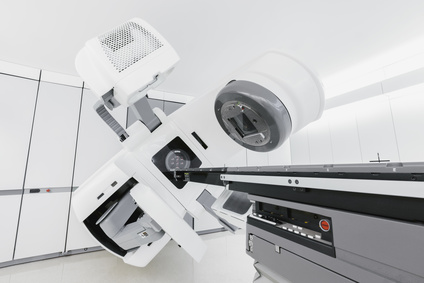
Medical linear accelerator for oncological radiotherapy © Thomas Hecker / Fotolia
Radiotherapy requires precise planning. It is carried out on an outpatient basis for a few minutes, 5 days a week, over a period of about 5 weeks. There are also side effects here, including
- diarrhea
- Skin irritation (dryness, redness)
- Occasional skin discoloration
- Hardening of the subcutaneous fatty tissue
Whether additional chemotherapy is necessary after the operation depends on various histopathological criteria.
However, radiotherapy (usually in combination with chemotherapy) is only used for rectal cancer. It is used before surgery (neoadjuvant) to shrink the tumor and after surgery (adjuvant) to prevent recurrences.
What information does the pathological examination of the surgical specimen provide?
The pathologist's work begins after the operation. They carry out a histological examination of surgical specimens. Both the specimen and all removed lymph nodes are sliced into very thin layers. This is followed by treatment with special stains that allow tumor tissue to be better identified.
Only these examinations can
- the exact size of the tumor,
- the depth of spread and
- the degree of differentiation
can be determined.
The pathologist also checks the edges of the incision to ensure that the tumor has been completely removed. He usually attends the operation. He can then instruct the surgeon to remove further tissue if necessary.

Tissue of the intestine at 400x magnification. Here the pathologist can directly recognize cancer cells © lukszczepanski | AdobeStock
Tumor classification
Important information can be obtained by processing the surgical specimen. This makes it possible to classify the tumor precisely.
This classification is known as the TNM classification. It reflects the individual extent of the tumor infestation for each individual patient. This is decisive for further therapy. In the TNM classification, 3 points are assessed as follows:
- T= Tumor: Used to assess the depth of penetration and extent of the tumor infestation on the individual intestinal sections (T1 to T4)
- N= Nodus (lymph node): Used to assess regional lymph node metastases (N0=none, N1=few to N2=numerous). Lymph node metastases are generally associated with a poorer prognosis for the patient, therefore subsequent chemotherapy is recommended from stage N1.
- M= Metastases: Used to assess distant metastases in other organs. The M stage is divided into M0 (=no metastases) or M1 (=metastases).
Tumor stage
Based on the tumor findings and the resulting TNM classification, the tumor is classified according to UICC criteria. A distinction is made between 4 stages I-IV:
- Stage I: T1 or T2 N0 M0
- Stage II: T3 or T4 N0 M0
- Stage III: any T N1 or N2 M0 (existing lymph node metastases)
- Stage IV: any T any N M1 (distant metastases present)
In addition, the "grading", which indicates the differentiation of the tumor, is also assessed. The degree of differentiation describes how similar the tumor tissue is to its original intestinal tissue. The more degenerated the tissue is, the more aggressive the cancer is:
- G1=well differentiated,
- G2=moderately differentiated and
- G3=poorly differentiated.
Another important factor for prognosis and therapy is the R classification. It describes whether residual tumor remains in the body (residual tumor status).
This involves assessing metastases remaining in the body and the cut edges of the surgical specimen. The greater the distance between the margins of the incision and the tumor, the better the prognosis for the patient:
- R0 = no tumor visible on the fine tissue
- R1 = Fine tissue evidence of residual tumor
- R2 = Residual tumor or metastases visible to the naked eye.
In tumor stage III - IV, additional recurrence or metastasis therapy is considered after colorectal cancer surgery. Tumor follow-up care is planned and organized with the involvement of all doctors involved. The prerequisites for this are
- a complete colonoscopy before or as soon as possible after the bowel operation and
- an assessment of the patient's general condition.
Whether and how often tumor follow-up care is indicated in individual cases can only be assessed once all findings are complete.
The aim of follow-up care is to detect as early as possible if
- the tumor starts to grow again (recurrence) or
- forms new metastases.
The risk of this is highest in the first two years after bowel cancer surgery. Regular check-ups are therefore essential. After that, the risk of recurrence decreases over time. The intervals between check-ups can therefore be extended further and further:
- for colon cancer, every 6 months for the first 3 years, and every year for the 4th and 5th year
- for rectal cancer, however, 3-monthly intervals are recommended in the first year.
After 5 years, colorectal cancer follow-up can usually be stopped.
What does tumor follow-up care for colorectal cancer involve?
Basic follow-up care includes
- a medical consultation
- a physical examination
- Checking laboratory parameters in the blood
- Determination of the tumor marker CEA
- an ultrasound examination of the abdominal cavity
- an X-ray of the lungs
Another fixed component of follow-up care is a colonoscopy.
In the case of colon cancer, it is recommended to have a colonoscopy once a year. Rectoscopy is recommended for rectal cancer, especially at the beginning of every follow-up examination.
Computed tomography (CT) is not routinely used in colorectal cancer aftercare. However, it can be helpful to record the initial status after the operation and to detect even the smallest changes quickly.
Nutrition after bowel cancer surgery
After bowel cancer surgery, the patient's diet can have a targeted influence on
- the consistency of the stool,
- flatulence and
- general well-being
general well-being. However, there are neither binding dietary recommendations nor blanket prohibitions.
Avoidance of certain foods
The basis of nutritional therapy is the "light whole food diet", i.e. the avoidance of foods and drinks that experience has shown often lead to intolerances. These include
- pulses
- mushrooms
- cabbage
- Raw onions
- garlic
- leeks
- fried foods
- wholemeal bread with whole grains
- freshly baked bread
- hard-boiled eggs
- Acidic foods
- Heavily fried foods
- smoked meats
- spicy food
- food and drinks that are too hot or too cold
- carbonated drinks
- unripe fruit
It is advisable to keep a food and symptoms diary to assess individual food tolerance.
Dietary recommendations
Sometimes an artificial bowel outlet is created in the small intestine (ileostomy or jejunostomy) during bowel cancer surgery. In this case, the function of the large intestine is lost, which leads to a reduced reabsorption of water and electrolytes. This results in thin, mushy stools and increased stool frequency.
It is important for the person concerned to know that every intake of food and drink leads to defecation. Eating and drinking slowly and chewing thoroughly can be very helpful here.
In the long term, it is advisable to change your eating habits to the Mediterranean diet. It not only prevents heart disease and obesity, but also certain forms of cancer. The Mediterranean diet includes
- daily and plentiful consumption of fruit, vegetables and salad
- a preference for vegetable fats such as rapeseed, olive or soybean oil
- reducing fat (dairy products in moderation, less red meat and sausages)
- Frequent consumption of fish (2-3 portions per week)
- Moderate consumption of alcohol
The Mediterranean diet also includes
- a healthy lifestyle,
- rest and time at mealtimes and
- regular physical exercise
with it.

The Mediterranean diet helps to support the body after bowel cancer treatment © aamulya | AdobeStock
Do I need chemotherapy after bowel cancer surgery?
If tumor-infested lymph nodes are detected, postoperative (adjuvant) chemotherapy is recommended.
In this case, the tumor itself has been completely removed. However, there is a risk (up to 50 %) that individual cancer cells have already spread to other parts of the body. These cannot be detected using current methods.
Large-scale studies have shown that prophylactic (adjuvant) chemotherapy significantly reduces the risk of recurrence.
Further indications for adjuvant chemotherapy would be, for example, in lower tumor stages if
- the tumor had ruptured during colorectal cancer surgery,
- the tumor has poor cell differentiation (G3) or
- fewer than 12 lymph nodes were removed during the operation.
In these cases, the experts must decide on an individual basis whether chemotherapy is indicated or not.
If the bowel cancer has only been discovered at a late stage, it has already metastasized. In this case, so-called palliative chemotherapy is indicated. A cure is then usually no longer possible. In this case, the aim of therapy is to improve the quality of life and prolong life.
In some cases, chemotherapy can reduce the size of bowel cancer metastases to such an extent that they can be surgically removed.
The target of chemotherapy are cells that - like cancer cells - divide rapidly. Unfortunately, this also affects healthy tissue with a high division rate, such as hair cells. The blood distributes the drugs, also known as cytostatics, throughout the body, making it a "systemic therapy".
Which drugs are used?
A large number of different drugs are now available. Oncologists select and combine them depending on the patient's individual case.
In most cases, several drugs are used according to a fixed, tried and tested regimen (e.g. FOLFOX regimen). Provided there are no health problems that prevent it, chemotherapy can be carried out on an outpatient basis. It may be necessary to implant a venous port catheter to administer the medication. This makes the treatment easier and more comfortable for the bowel cancer patient.
Adjuvant chemotherapy usually lasts six months. However, the duration of treatment can be extended in the metastatic stage depending on the response to therapy.
In these cases, so-called antibodies (e.g. cetuximab, bevacizumab) can be used in addition to the classic cytostatics. Antibodies bind to certain surface structures on cancer cells that are important for tumor growth and block them.
However, antibodies are not (yet) used in regular "adjuvant" therapy.
What side effects can occur with chemotherapy?
The possible side effects of chemotherapy are based on the fact that rapidly dividing healthy cells are also killed. However, they do not necessarily occur.
These include, for example, the bone marrow, in which the various blood cells are formed, so that in the course of the therapy
- anemia,
- bleeding and
- infections
can occur.
The white blood cells are most important for the defense against infection. They require regular monitoring (blood count) during chemotherapy. If their number falls too low, the therapy must be interrupted for a recovery period.
The digestive tract is also one of the rapidly dividing tissues. Various side effects can occur here during chemotherapy. These include
- nausea
- vomiting
- loss of appetite
- taste disturbances
- diarrhea
Fortunately, there are some very effective drugs (e.g. antiemetics) to combat these side effects.

In some cases, chemotherapy is used to treat bowel cancer © Elnur | AdobeStock
Another possible side effect of some cytostatic drugs is hair loss. However, complete hair loss is not to be expected with the drug combinations used to treat bowel cancer.
In any case, the hair will grow back after the end of treatment. The other side effects mentioned above also disappear after the end of treatment.
With the antibodies already mentioned, there are generally only a few side effects to worry about. As these are proteins, allergic reactions may occur in rare cases (e.g. acne-like skin rash with cetuximab).
In general, your oncologist should inform you about the side effects to be expected according to the treatment regimen. If necessary, he will treat them with additional medication.
Research has shown that there are no general factors that improve or worsen our quality of life. The term quality of life refers to the personal experience of cancer. This includes feelings and complaints in mental, physical and social life.
There are therefore major differences between different people in how they deal with the disease. Each patient copes differently with the stresses and strains that the disease or treatment can cause.
Many of these stresses are temporary, but they can also be persistent and require psychological treatment.
During treatment in hospital, the fear of the operation often takes center stage. It is important to ask as many questions as you want to know and understand during your discussions with the attending physician. Your own anxiety and worries should also be discussed. After being discharged from hospital, many demands often lead to excessive demands.
The main concern of many cancer patients is that the disease could recur or spread ("fear of progression"). This concern is normal and appropriate, but it can also get out of hand.
In this situation, it can be helpful to talk about it and approach the fear. The importance of coping with mental illness is increasingly recognized. As a result, there are now a variety of appropriate support options in psycho-oncology.
The overall prognosis for colorectal cancer depends primarily on how completely the tumor can be removed. The earlier the tumor is detected and removed, the better the chances of recovery.
Thanks to improved screening, more than 50% of cases can now be cured. However, if colorectal cancer metastases are already present in other organs or lymph nodes at the time of diagnosis, the prognosis is significantly worse.
Colorectal cancer specialists usually come from the visceral surgery and/or gastroenterology departments. These two specialist departments cooperate in the treatment of colorectal cancer, in particular with the following specialties
In addition to these medical specialties, pain and physiotherapy
- pain and physiotherapy,
- specialized oncological care and
- supportive therapy
also play a major role. Supportive therapy includes, among other things
The latter support affected bowel cancer patients and their relatives throughout the entire treatment process.