
Diagnosis is usually made through imaging using MRI or CT, along with a biopsy. Treatment includes surgery, radiation therapy, chemotherapy, and modern therapeutic options in neurosurgery and oncology.
The prognosis depends on the WHO grade, the extent to which the tumor has been removed, and the genetic characteristics of the tumor cells.
Gliomas – A Brief Definition
“Glioma” is the term used to describe a group of brain tumors that arise from glial cells within the central nervous system (CNS).
These are not the brain’s nerve cells themselves. They are supporting cells whose functions optimize neurophysiological processes and tasks—or, in fact, make them possible in the first place. In adults, gliomas tend to grow in the cerebrum, while in children they are more likely to develop in the brainstem and cerebellum. Gliomas account for about 30 to 50% of all intracranial brain tumors.
Men are affected slightly more often, with a ratio of 6:4. Most cases occur between the ages of 40 and 65. However, the actual cause of the disease remains largely unknown.
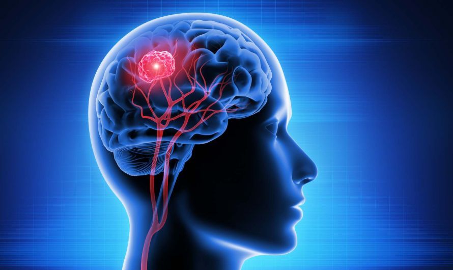
Tumors always develop when normal cell division goes awry. Cell division and metabolic activity carry out daily “renovation work,” including on the DNA’s genetic material.
In some cases, these DNA repairs cannot keep up with the damage, leading to mutations or defective DNA segments.
This can lead to the abnormal cells multiplying uncontrollably and forming diseased tissue. In medical terminology, this is referred to as a neoplasm. Among gliomas, there is a small subgroup whose genetic alterations have a genetic cause.
For example:
- neurofibromatosis or
- tuberous sclerosis
In addition, gliomas can occur as a long-term consequence of radiation therapy, meaning that a history of cancer is then a risk factor for new gliomas.
The WHO grading system determines the degree of malignancy of gliomas
According to the WHO grading system (World Health Organization classification), gliomas are classified into grades I through IV. This means that there are both benign and malignant forms. This WHO classification is used when doctors need to determine the degree of malignancy.
The degree of malignancy allows the following factors to be determined:
- Cellular origin
- Cell composition, and
- Growth patterns of gliomas
This makes it possible to assess how benign or malignant the tumor actually is.
The WHO classification distinguishes between four different grades of malignancy:
- WHO Grade I: benign (benign tumors)
- WHO Grade II: still benign
- WHO Grade III: already malignant
- WHO Grade IV: malignant (malignant tumors)
The WHO classification of gliomas into further subcategories
The subgroups of gliomas indicate exactly which cells they have developed from. In most cases, the cells of origin give the tumor class its name:
Astrocytomas: Astrocytomas arise from astrocytes. These cells are part of the central nervous system’s supporting structure and serve to separate the nervous tissue from the brain’s surface and the blood vessels. Astrocytomas can be further divided into subgroups based on their severity according to the WHO classification.
While pilocytic astrocytomas are classified as WHO Grade I, diffuse astrocytomas are classified as Grade II. Anaplastic astrocytomas are classified as Grade III.
Glioblastomas: These tumors also arise from astrocytes and represent a malignant, usually very aggressive form of astrocytoma. They are therefore classified as Grade IV.
Primary glioblastomas arise directly from healthy astrocytes. Secondary glioblastomas develop from pre-existing tumor tissue in this region of the brain.
This means that Grade II and III astrocytomas can transform into a glioblastoma.
Oligodendroglioma: When oligodendrocytes—which surround the individual nerve pathways in the brain—form a glioma, it is called an oligodendroglioma.
This brain tumor is classified as WHO grades II and III. It generally has a better prognosis than astrocytomas of the same grade.
Ependymomas: The human brain contains a kind of irrigation system. It consists of internal and external ventricles. Cerebrospinal fluid is produced there every day, which the body drains outward and reabsorbs. The internal ventricles are lined with ependymal cells, which are a subgroup of glial cells.
When these ependymal cells become malignant, doctors refer to them as ependymomas. They have a WHO grade between I and III. Ependymomas can develop both in the internal ventricles of the brain and in the central canal of the spinal cord.
Gangliogliomas: When ganglion or Schwann cells are affected by tumor growth, the result is a ganglioglioma. Since these cells are found throughout the central nervous system, gangliogliomas can occur in many locations.
They most commonly grow in the cerebellum, hypothalamus, or temporal lobes. They are mostly Grade I, but can also occur as Grade II or III. In addition, there are other diffuse gliomas as well as mixed forms of the above classifications.
Signs and Symptoms of Gliomas
Gliomas can develop gradually or appear suddenly. The symptoms are often varied. This is primarily because gliomas develop in different parts of the central nervous system. They can therefore affect various functions.
Often, gliomas have been growing for some time before symptoms become visible or noticeable. Unlike with some other cancers, early detection is nearly impossible. There are no specific tumor markers for gliomas; diagnosis relies solely on imaging techniques. The initial signs and symptoms of gliomas can vary widely. They are often very nonspecific and depend on the tumor’s location.
The following symptoms may occur:
- Headaches—especially in the morning and at night
- Nausea and vomiting
- Fatigue and insomnia
- Decreased performance
- Seizures
- Personality changes
- Speech difficulties
- Symptoms of paralysis
- Sensory disturbances
- Coordination problems and unsteady gait
- Visual disturbances
How Gliomas Are Diagnosed
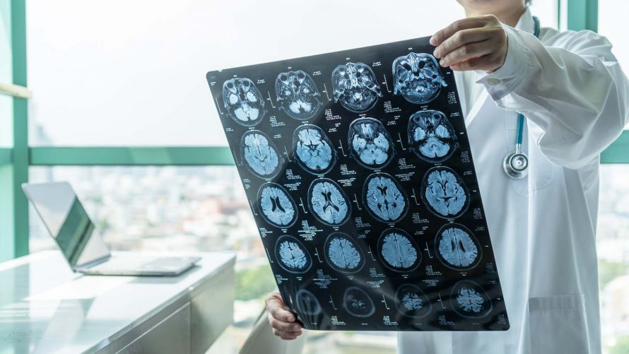
If symptoms raise suspicion of a brain tumor or glioma, prompt diagnosis is necessary. This is done using MRI.
Often, doctors initially use a CT scan to rule out an acute stroke. However, only MRI provides a detailed image of the brain’s fine tissue structures.
Computed tomography
Computed tomography (CT) scans are a readily available imaging modality. They allow doctors to get an overview of the situation and rule out strokes.
MRI with contrast agent: This involves imaging in a magnetic resonance imaging (MRI) scanner. In WHO Grade III and IV gliomas, the tumor cells typically take up the contrast agent.
This makes the mass even more clearly visible. For surgical planning, MRI sequences are performed in 3D and occasionally in 4D.
In addition to the tumor, this allows functional centers and nerve tracts to be identified and visualized in relation to the tumor.
This information is crucial for performing a precise surgery. For diffuse tumors, metabolic imaging (PET, positron emission tomography) is helpful. In this procedure, particularly active areas are visualized using radioactive amino acids.
Biopsy
Cells are collected during a minor surgical procedure. Doctors can then have the cells examined and analyzed in the laboratory.
This provides information about the origin and the degree of malignancy. The definitive diagnosis—which is essential for optimal treatment—is only possible through this histopathological examination. Therefore, surgery or a biopsy to obtain tissue is necessary in every case.
Treating and Combating Gliomas
Since some forms of gliomas are highly malignant and grow rapidly, early treatment is necessary.
Treatment is individualized for each patient and tailored to the patient’s specific condition.
The following factors play an important role in this:
- Age
- General health and
- degree of malignancy, as well as
- size of the tumor
Like most other brain tumors, gliomas can be treated with surgery, radiation therapy, and/or chemotherapy:
Surgery
The goal of surgery to treat gliomas is to resect the tumor as completely as possible and to obtain tissue samples for histopathological examination.
In many cases of WHO Grade I gliomas, complete tumor removal is possible. The location of the glioma is often problematic, as it prevents macroscopically complete removal without risking subsequent damage. The guiding principle here is “function over resection.” In gliomas, therefore, the extent of resection often determines the prognosis.
For this reason, surgeons employ a wide variety of techniques during surgery to optimize the resection outcome while preserving vital brain tissue. Neurosurgery utilizes intraoperative imaging techniques such as MRI and ultrasound combined with intraoperative 3D navigation.
In addition, surgeons can monitor various brain functions—such as motor function, various cranial nerves, or sensory perception—using intraoperative electrophysiology. The distance to nerve pathways is tracked in real time during surgery, much like an electrical radar. This allows the surgeon to perform the surgery as thoroughly as possible while preserving function. In some cases, awake surgery is also used to monitor speech function.
Follow-up Chemotherapy and/or Radiation Therapy
Once the final diagnosis has been confirmed by histopathology, an interdisciplinary team convenes to discuss the case.
The team usually consists of:
- Neurosurgeons
- Neuroradiologists
- Oncologists
- Radiation oncologists, and
- Neurologists
The medical professionals discuss each patient’s case individually as a team. Depending on the diagnosis, they develop the best possible treatment plan. Depending on the WHO class and WHO grade, subsequent radiation therapy or chemotherapy may be appropriate. If necessary, these treatments are administered in combination.
Tumor Treating Fields
In the case of glioblastomas, doctors can also utilize what are known as tumor-treating fields (TTF).
A cap generates magnetic fields that can inhibit tumor cell growth. Studies have demonstrated the effectiveness of this method. Therefore, doctors should always consider it as an option in cases of glioblastoma.
Risks and Prognosis
The prognosis for a glioma patient depends on the following factors:
- Location
- Size
- Biological characteristics of the glioma
If left untreated, malignant gliomas often grow rapidly and are destructive. It is therefore particularly important to make an early diagnosis and begin treatment.
Optimal microsurgical tumor resection, utilizing all available technical resources and methods, is the key to improving the long-term prognosis.
FAQ
What is a glioma?
A glioma is a tumor of the central nervous system that originates in the brain. The tumor develops from a glial cell and is one of the most common types of brain tumors. Gliomas can be benign or malignant and may grow with varying degrees of diffuseness.
What symptoms do gliomas cause?
The symptoms of gliomas depend on the tumor’s location in the brain. Common symptoms include headaches, neurological deficits, nausea and vomiting, or epileptic seizures. Changes in quality of life and difficulty concentrating are also possible.
How are gliomas diagnosed?
Diagnosis and treatment involve MRI, CT, and other imaging procedures. A biopsy is often performed as a supplementary measure to examine the tissue and tumor cells more closely. The WHO classification system helps categorize gliomas according to WHO grade.
What treatment options are available for gliomas?
Treatment for gliomas includes surgery, radiation therapy, and chemotherapy. Specialists in neurosurgery, neurology, and oncology work together to determine the appropriate treatment options. The goal is, if possible, to completely remove the tumor.
What is the prognosis for a glioma?
The prognosis depends on the WHO grade, the location, and the type of tumor. Low-grade gliomas, such as Grade 1 or Grade 2 gliomas, generally have a better prognosis than malignant tumors such as glioblastoma or Grade 4 tumors. Patients between the ages of 40 and 65 are particularly prone to developing certain types of gliomas.
Share this article
Sources
- S2k-Leitlinie „Gliome“ (AWMF-Register-Nr. 030-099), Stand 27.07.2021: register.awmf.org/de/leitlinien/detail/030-099
- https://www.aerzteblatt.de/archiv/die-gliome-des-erwachsenen-0e9d6ae1-6d7a-4158-9c27-0692e081a2ca
- https://www.onkopedia.com/de/onkopedia/guidelines/gliome-im-erwachsenenalter/@@guideline/html/index.html
- https://flexikon.doccheck.com/de/Gliom
- https://www.onko-portal.de/basis-informationen-krebs/krebsarten/hirntumor/ependymom.html
- https://www.hirntumorhilfe.de/hirntumor/tumorarten/
- https://www.aerzteblatt.de/archiv/behandlung-von-gliomen-im-erwachsenenalter-5d14e5ef-8f3e-43d2-b027-06b151e6deaa