An oligodendroglioma is a brain tumor that occurs primarily in adults. These gliomas are associated with various symptoms and can be classified into four WHO grades. Early diagnosis and prompt treatment significantly improve the prognosis for this disease.
Here you will find further information as well as a list of selected specialists and centers for the treatment of oligodendrogliomas.
What are oligodendrogliomas?
Oligodendrogliomas are glial tumors—that is, brain tumors—that grow diffusely into the surrounding brain tissue. Astrocytomas and oligodendrogliomas are among the most common diffuse gliomas.
Under a microscope, their tumor cells resemble oligodendrocytes, which are also found in the brain. Oligodendrocytes are support cells that form myelin sheaths. Myelin sheaths are lipid-rich layers that form around the axons of certain nerve cells. Oligodendrogliomas, however, do not form these myelin sheaths.
Oligodendrogliomas most commonly affect adults between the ages of 35 and 50. However, younger patients can also develop this condition. This brain tumor occurs slightly more frequently in men than in women.
Oligodendrogliomas usually, but not always, develop in the frontal lobe of the cerebrum. Less commonly, these tumors are located in the brainstem or the cerebellum.
What does the WHO grade classification indicate?
The WHO (World Health Organization) classifies oligodendrogliomas into different grades. This classification provides information about the prognosis and the recommended treatment options.
A common criterion for classifying tumors is the degree of malignancy. This refers to whether the individual tumor is benign or malignant. Malignancy encompasses factors such as the tumor’s growth rate and its tendency to spread into surrounding tissue.
- WHO Grade I: Benign oligodendrogliomas that grow only slowly and are therefore associated with a very good prognosis.
- WHO Grade II: Grade II oligodendrogliomas grow slowly, tend to recur, and can develop into higher-grade tumors.
- WHO Grade III: Malignant oligodendrogliomas; following surgical removal, chemotherapy or radiation therapy is recommended to prevent recurrence (the reappearance of the tumor).
- WHO Grade IV: Highly malignant oligodendrogliomas that grow rapidly and also require subsequent radiation therapy or chemotherapy.
Molecularly, oligodendrogliomas are characterized by an IDH mutation as well as alterations in chromosomes 1p and 19q, which are considered important diagnostic features. As the WHO grade increases, the tumor’s cell density increases. Starting at WHO Grade 3, microvascular proliferations may also occur in the tumor tissue, indicating more aggressive growth.
The final WHO classification is determined after evaluation of the biopsy and molecular pathological examinations.
What symptoms does an oligodendroglioma commonly cause?
An oligodendroglioma often does not cause noticeable symptoms until later in the course of the disease. These primarily include:
- headaches, nausea, and vomiting (indicating increased intracranial pressure)
- neurological deficits and epileptic seizures
- Symptoms of a stroke if the glioma hemorrhages (unilateral paralysis, speech difficulties, etc.)
There is a difference between ordinary headaches and headaches caused by a brain tumor.
Headaches caused by a glioma occur very suddenly. They become increasingly severe within days or weeks. Pain relievers usually do not help or only provide short-term relief. Often, the pain worsens when lying down and subsides spontaneously when sitting or standing upright.
If the brain tumor disrupts the flow of cerebrospinal fluid, symptoms of hydrocephalus (“water on the brain”) may also occur:
- Memory problems
- Fatigue
- Dizziness
- Nausea
If you notice these symptoms in yourself or a loved one, it is advisable to see a doctor as soon as possible.
How are oligodendrogliomas diagnosed?
The doctor first conducts a medical history interview with the patient. During this interview, the doctor asks the patient about their symptoms and medical history. This often allows the doctor to make a preliminary diagnosis and provides clues as to the causes of the condition.
If an oligodendroglioma is suspected, the next step is a neurological examination. This usually reveals the first clear signs of a brain tumor.
This suspicion can be confirmed with a CT (computed tomography) scan. In this procedure, X-rays pass through the brain and produce many individual cross-sectional images. The doctor examines the images for unusual accumulations of tissue.
However, an MRI (magnetic resonance imaging) provides the most detailed overview. This allows the doctor to best identify any possible irregularities in the brain’s structures.
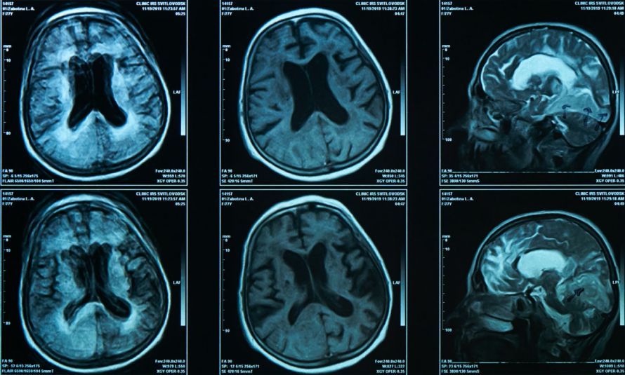
Often, the examining physician confirms the diagnosis by ordering a biopsy. To do this, they remove a tissue sample and have it examined in a laboratory.
Other diagnostic methods that are useful when a brain tumor is suspected include:
- an EEG (electroencephalogram) of the brain, which pinpoints the exact location of the tumor,
- a cerebrospinal fluid (CSF) analysis to rule out other (inflammatory) diseases of the nervous system.
The doctor decides which of the aforementioned tests are appropriate on a case-by-case basis.
Are there ways to detect it early?
Currently, no specific risk factors are known to cause an oligodendroglioma. Neither diet, genetic factors (except for a few isolated cases), nor a specific mutation has yet been conclusively proven to be a concrete cause. Therefore, there are no early detection programs or preventive measures that can detect or prevent an oligodendroglioma at an early stage.
To make matters worse, the symptoms of a brain tumor often do not appear until the disease is advanced. This makes it all the more important to see a doctor immediately if you experience these symptoms. A diagnosis can only be made and appropriate treatment initiated with the help of imaging techniques.
So if you notice any of the symptoms described above, it’s best to have them evaluated by a doctor. In most cases, the earlier treatment begins, the better the prognosis.
What are the treatment options for oligodendrogliomas?
Treatment depends on the WHO grade of the oligodendroglioma.
For Grade 2 brain tumors, doctors recommend resection of as much of the tumor tissue as possible. If the tumor grows back after surgery or if there is a high risk of recurrence, chemotherapy and radiation therapy are recommended afterward. The surgical approach always depends on the tumor’s location in the brain. These treatments are intended to prevent the tumor from growing back.
Doctors also treat Grade 3 WHO oligodendrogliomas with surgery followed by chemotherapy. The PCV regimen includes the active ingredients procarbazine, lomustine, and vincristine; temozolomide is used as an alternative.
In cases of recurrence or complicated disease progression, clinical trials may represent another treatment option. The treating physician also informs patients about possible side effects of the respective therapy.
Rehabilitation follows the removal of the tumorous tissue. Especially if the disease has already led to neurological changes, rehabilitation can improve the patient’s quality of life. Options may include, for example,
- speech therapy for speech impairments,
- occupational therapy and physical therapy for physical symptoms,
- neurocognitive training, such as using special computer programs.
The treating physician advises patients on which therapies are likely to yield the desired results.
Which medical specialties treat oligodendrogliomas?
An oligodendroglioma is often accompanied by numerous symptoms. The diagnosis and treatment are also complicated and multifaceted.
When it comes to brain tumors, specialists from various fields work together in an interdisciplinary manner.
The first point of contact for neurological symptoms is a neurologist. The neurologist conducts the initial examination and neurological tests to narrow down the possible causes of the condition.
Radiation oncologists are specialists in the field of diagnostic imaging for cancer.
Further diagnostic tests and subsequent treatment are carried out by physicians
- in oncology (cancer specialists),
- neurosurgery, and
- neuro-oncology
. Ideally, an entire team of specialists comprising these doctors works together to achieve the best possible treatment outcome.
Conclusion
Oligodendrogliomas are brain tumors that require prompt treatment. If you suddenly experience severe headaches or neurological symptoms, see a doctor as soon as possible.
The doctor will conduct an initial examination and, if necessary, order further diagnostic tests. Prompt treatment is usually associated with a better prognosis.
FAQ
What is the difference between a grade 2 and a grade 3 oligodendroglioma?
A grade 2 oligodendroglioma grows slowly and tends to recur, whereas a grade 3 oligodendroglioma (anaplastic oligodendroglioma) is already malignant and requires radiation therapy and chemotherapy following surgery.
What drugs are used in chemotherapy?
Chemotherapy often involves drugs from the PCV regimen, such as procarbazine, lomustine, and vincristine, or alternatively, temozolomide.
How is an oligodendroglioma reliably diagnosed?
In addition to a clinical neurological examination, MRI imaging provides the most detailed overview; a biopsy definitively confirms the diagnosis.
What side effects can occur after treatment?
Depending on the therapy, side effects such as fatigue, neurological deficits, or speech impairments may occur—which is why speech therapy, occupational therapy, or physical therapy are often recommended afterward.
Share this article

Sources
- https://www.krebsgesellschaft.de
- https://www.hirntumorhilfe.de/
- https://flexikon.doccheck.com/de/Oligodendrogliom
- https://flexikon.doccheck.com/de/Neoplasie
- https://www.krebsgesellschaft.de
- https://www.krebsgesellschaft.de
- http://www.neurochirurgie.insel.ch
- S2k-Leitlinie „Gliome“ (AWMF-Register-Nr. 030-099), Stand 27.07.2021: register.awmf.org/de/leitlinien/detail/030-099
- https://neurochirurgie.uk-koeln.de