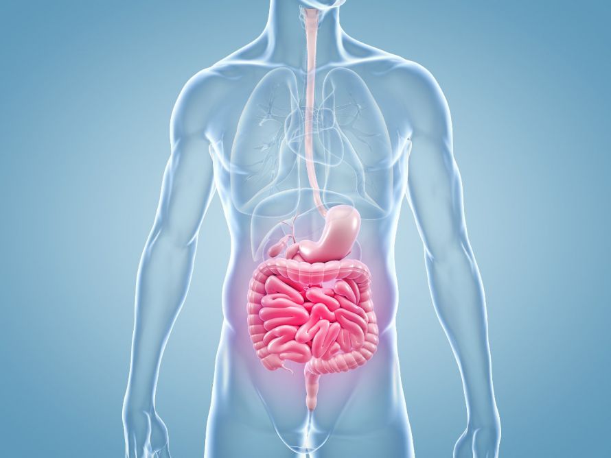
A bowel perforation is a hole in the intestinal wall—sometimes more than one. When a bowel perforation occurs, air and intestinal contents (feces, chyme, digestive juices, pus) leak into the abdominal cavity. This often leads to peritonitis and can be life-threatening. In short: A bowel perforation is a medical emergency.
Here you will find concise information on causes, symptoms, diagnosis, and treatment, as well as a list of selected specialists in visceral surgery.
What are the possible causes of a bowel perforation?
Common causes/mechanisms of perforation in the gastrointestinal tract:
- Intestinal distension / intestinal obstruction (ileus): Overstretching → tear in the intestinal wall; Ogilvie syndrome (acute colic pseudo-obstruction) can also contribute to intestinal perforations.
- Acute inflammatory: suppurative appendicitis with rupture, gastric ulcer (chemical irritation from acid) leading to perforation.
- Chronic inflammatory: inflammatory bowel diseases such as Crohn’s disease or ulcerative colitis/diverticulitis (especially the colon/sigma) – inflamed tissue tears more easily.
- Circulatory disorders: Circulatory disorders (intestinal infarction) → tissue necrosis → perforation.
- Trauma/foreign bodies: blunt abdominal trauma; swallowed or inserted foreign bodies (e.g., toothpicks) injure the intestinal mucosa or intestinal lumen.
- Tumors: Decaying carcinoma (stomach/intestine) leading to perforation due to mechanical forces or reduced blood flow.
- Medical procedures: Injury during endoscopy (colonoscopy, gastroscopy) or surgery (rare, but possible).
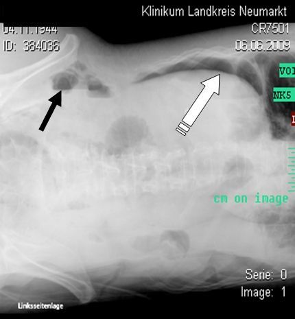
Symptoms and consequences of intestinal perforation
Symptoms of intestinal perforation vary depending on the location and cause, but typically result in severe pain. Example: appendicitis—initial pain in the right lower abdomen, followed by brief relief upon perforation, then rapid onset of signs of peritonitis.
Additional signs/course:
- Sudden onset of pain, abdominal wall rigidity, possibly nausea and vomiting
- Chemical irritation (stomach acid) → severe inflammatory reaction; intestinal contents leak out → bacterial infection
- Fever, progressive intestinal paralysis, lab findings: ↑ white blood cells, CRP, procalcitonin
- If left untreated, there is a risk of sepsis, circulatory, respiratory, and renal failure
Other segments: Esophageal perforation can spread to the mediastinum; small and large bowel perforations vary in severity depending on the intestinal segment. Fistula formation is possible with prolonged disease progression.
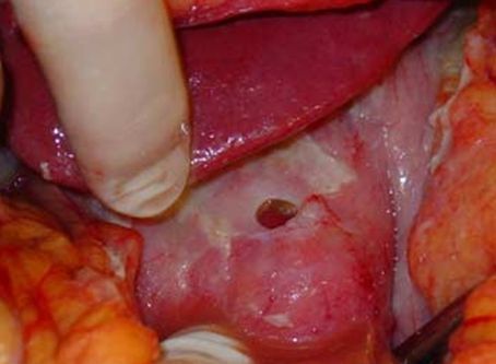
Even before perforation occurs, the underlying conditions can cause pain. If a bowel perforation occurs, allowing intestinal contents (feces, chyme, digestive juices) to enter the abdominal cavity and air to escape, severe pain sets in suddenly.
This is caused by chemical irritation of the peritoneum—including from stomach acid—whose pain-sensitive nerves trigger the most intense pain response (often accompanied by defensive muscle tension). Temporary relief, such as that described in cases of perforated appendicitis, quickly gives way to peritonitis.
In a second phase, the ongoing contamination intensifies the inflammatory response. If stool from the large intestine enters the abdominal cavity, this additionally leads to a massive bacterial infection. Typical accompanying symptoms include fever, nausea/vomiting, and progressive intestinal paralysis (paralytic ileus).
Laboratory tests reveal elevated inflammatory markers (e.g., white blood cell count, CRP, procalcitonin). If treatment is not provided or is delayed, there is a risk of sepsis (blood poisoning) with systemic deterioration: circulatory and respiratory failure, decreased urine output, and even multiple organ failure.
Older patients, as well as people with cancer or other severe intestinal diseases/immunosuppression, are particularly at risk—in these cases, the condition can quickly become life-threatening.
Diagnosis of Intestinal Perforation
Peritonitis is primarily a clinical diagnosis: medical history, physical examination (tenderness, guarding), and vital signs are crucial. This is followed by targeted imaging and laboratory diagnostics:
- Ultrasound examination: fast, repeatable; free fluid, abscesses.
- Conventional X-ray: detection/exclusion of free air (subdiaphragmatic).
- CT / Computed Tomography (with oral/IV contrast): most often the most informative (CT shows air, bowel loops, leakage, location, and cause of the perforation).
- Endoscopy (colonoscopy, upper GI endoscopy) should be performed only selectively during the acute phase; consider the risks.
- Laboratory: Inflammatory markers, organ function; urinalysis; pregnancy test if indicated.
Goal: To quickly determine whether immediate surgery is necessary and which treatment (including antibiotics) is appropriate.
When is immediate surgery not necessary in cases of bowel perforation?
Not every perforation requires surgery. In rare, exceptional cases, doctors may adopt a wait-and-see approach. If “free air” is incidentally detected on an X-ray—which is a clear indication of a possible perforation—doctors may choose to wait and see. However, this is only appropriate if the patient has no symptoms and is in good health.
Appendicitis can lead to the formation of a localized abscess. In such cases, it is also possible to stabilize the condition by removing the appendix in about 6 weeks.
However, this is only possible if:
- the patient is in good condition
- there is evidence of a regional abscess
- antibiotic prophylaxis is in place
As a supportive measure, interventional drainage of the abscess under ultrasound or CT guidance may be necessary.
A similar approach can be taken in cases of a covered perforation, such as sigmoid diverticulitis.
In this case, although perforation has occurred, it is covered by adjacent structures—the diverticula. In the absence of peritonitis, physicians may also adopt a wait-and-see approach here.
Rare exceptions (provided close monitoring is in place):
- Incidental finding of “free air” without symptoms, stable condition.
- Covered perforation (e.g., sigmoid diverticulitis with limited leakage of intestinal contents), no diffuse peritonitis.
- Appendicitis with a localized abscess: drainage (interventional under ultrasound/CT guidance) + antibiotics, followed by appendectomy.
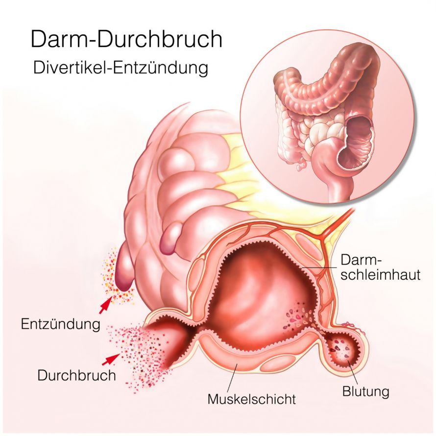
Surgical Management of a Perforation
A bowel perforation is an acute emergency. The goal of surgery is always rapid debridement of the site: closing the hole in the bowel wall or removing the affected segment of the bowel, clearing contamination caused by bowel contents entering the abdominal cavity, and controlling peritonitis.
Intraoperative Assessment—What Determines the Strategy?
Visceral surgeons systematically evaluate:
- Location (small intestine vs. large intestine/sigmoid colon, stomach, rarely the esophagus), size of the defect, condition of the intestinal mucosa and the intestinal lumen
- Extent of contamination in the abdominal cavity (feces, bile, chyme, pus, free air)
- Blood supply and vitality of the bowel loops (ischemia/infarction?)
- Concomitant conditions/sepsis, immunosuppression, and the patient’s circulatory stability
- Cause of perforation (e.g., diverticulitis, Crohn’s disease, ulcer, foreign body/foreign bodies introduced during endoscopy/colonoscopy, tumor)
Standard steps (source control)
-
Exploration of the abdominal cavity, visualization of all affected bowel loops
-
Aspiration of contaminated fluid (if intestinal contents have entered the abdominal cavity) and generous irrigation of the abdominal cavity
-
Incision and drainage of abscesses/foci between the bowel loops; placement of drains (early warning signs of suture leakage)
-
Reconstructive procedure depending on findings: suture closure, resection + anastomosis, or stoma creation (temporary artificial bowel outlet)
Closure with sutures
The principle of any surgery for a perforation is to close the hole. This can only be achieved in exceptional cases by simple closure with sutures or with the aid of so-called stapled sutures (steel).
Such closure is appropriate and possible in cases of:
- uncomplicated gastric perforations at the pylorus
- a hole in the small intestine caused by a foreign body, or
- fresh perforations or ruptures of the esophageal wall
Perforations occurring during diagnostic gastroscopy or colonoscopy can also often be sutured closed. In these cases, the bowel is clear due to prior bowel preparation. Doctors often detect this early and can therefore perform surgery promptly.
Two-stage procedure
The likelihood of successful suturing decreases the more time that passes after the intestinal perforation. It also decreases as the inflammation progresses.
The tissue loses its structural integrity due to the inflammation. As a result, doctors can no longer suture the tissue.
It is necessary to debride the tissue—that is, to excise portions of it—to achieve stable conditions suitable for suturing.
This has its anatomical limits and often necessitates the removal of entire sections of the organ. The resulting surgeries are correspondingly complex.
In the simplest case, doctors remove only the inflamed appendix. The challenge here is ensuring a secure closure of the resection site at the base (where the appendix joins the large intestine). However, if the inflammation has not significantly affected the beginning of the large intestine, this should be quite feasible.
In some cases, the temporary creation of a colostomy may be necessary. In this way, doctors divert stool around the problematic section, allowing it to stabilize.
Restoration of bowel function can take place weeks later during a second surgery once conditions have stabilized.
Extremely poor circulatory conditions or severe infections may also necessitate such a two-stage procedure.
Removal of Organs
In cases of primary colitis or inflammation of diverticula in the colon, doctors must remove the inflamed section of the colon.
Here, too, the nature of the underlying disease and the inflammation plays a decisive role. In Europe, diverticula and their complications most commonly occur in the left side of the colon, particularly in the sigmoid colon. It is therefore advisable, in the event of a perforation, to remove the affected portion—usually the sigmoid colon—along with the perforation.
Ulcerative colitis tends to cause inflammatory changes throughout the entire large intestine and can also become malignant over the years. In extreme cases, it may be necessary to remove the entire large intestine.
In the case of the other specific inflammatory bowel disease, Crohn’s disease, however, only minimal resection of the bowel is necessary.
In the case of a perforation within a tumor—especially a malignant tumor—suturing is not an option. The goal must be to remove the tumor, if possible.
This applies to both the stomach and the intestine. However, the spread of tumor cells through the perforation has an extremely negative impact on the further course of the disease.
Stomach ulcers that perforate in the region where the stomach meets the duodenum are usually inflammatory in nature. Very often, they are caused by infection with the bacterium Helicobacter pylori.
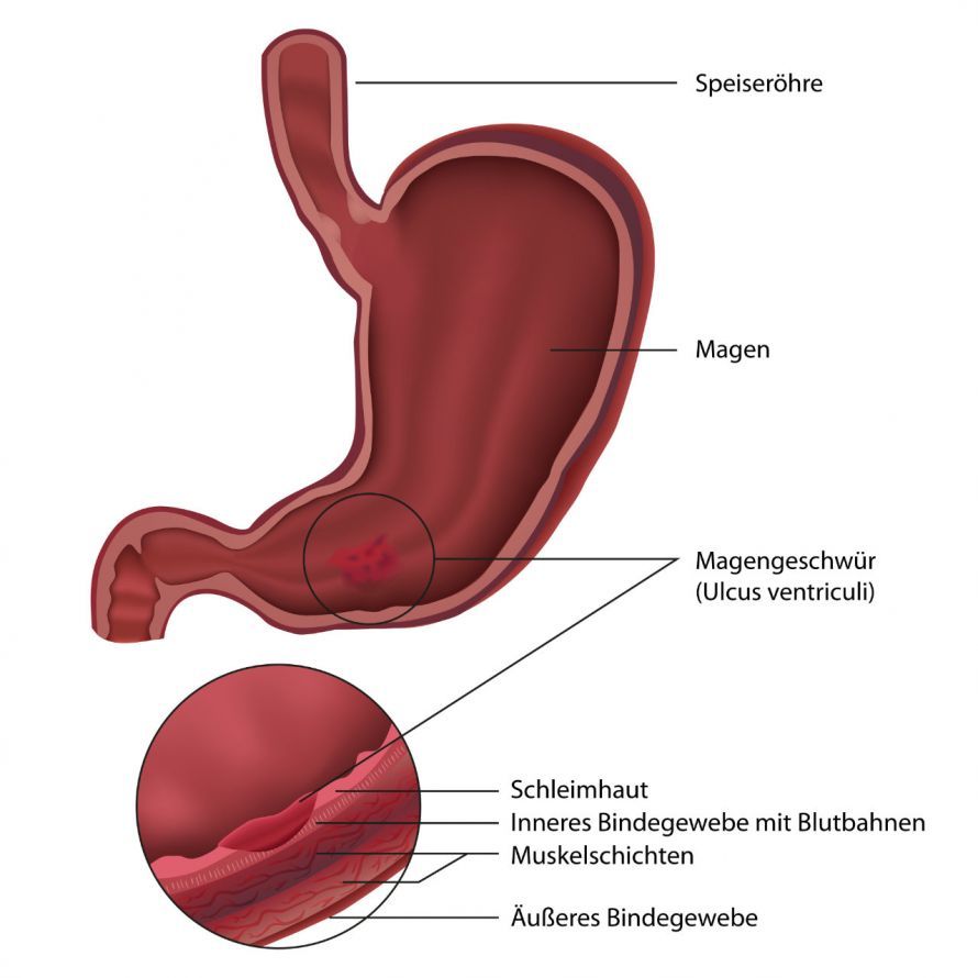
In the upper portions of the stomach, however, it is not uncommon for the lesion to be a previously undiagnosed, perforated carcinoma. In these cases, excision of the lesion followed by closure should be performed. Simple suturing is not recommended here.
If a tumor is clearly confirmed, partial removal of the stomach (B-I or B-II resection) may be necessary. In severe cases, complete removal of the stomach (gastrectomy) may even be indicated.
Traumatic injuries, such as those resulting from accidents, can cause very complex injuries. Organ ruptures, such as those involving the spleen or liver, lead to extreme bleeding. The immediate priority is then to address the shock caused by blood loss.
VAC Dressings for Intestinal Perforation
In cases of severe inflammation, multiple surgeries involving debridement of the abdominal cavity may be necessary. It is also essential to check the sutures for leaks.
In these cases, temporary abdominal wall closure using films and sponges has proven effective. These so-called VAC dressings (vacuum-assisted closure) drain inflammatory fluid from the abdominal cavity using negative pressure.
The dressing can be changed under anesthesia in the intensive care unit or in the operating room. This is done every 3–4 days.
Once the patient’s condition has stabilized, doctors can then close the abdomen. If necessary, they may also need to apply a VAC dressing to the abdominal wall. They often change this dressing without anesthesia.
Antibiotic Therapy (Antibiotic Treatment)
In addition to surgical debridement (removal of the cause of the perforation and irrigation of the abdominal cavity), antimicrobial therapy is almost always required to control the infection and peritonitis and to prevent or treat sepsis.
- Initial treatment strategy: Immediately after diagnosis, broad-spectrum therapy is initiated to cover typical gut pathogens (aerobic/anaerobic). The choice of antibiotic and dosage depend on the type and extent of the perforation, the degree of contamination in the abdominal cavity, and any comorbidities (e.g., immunosuppression).
- Microbiology & De-escalation: Samples are collected during surgery. Results are typically available after 2–3 days—at which point therapy is switched to a targeted, appropriate antibiotic (de-escalation).
- Duration of Therapy: The duration of treatment depends on the clinical course. After successful debridement of the infection site and stabilization of the patient’s condition, treatment often lasts only a few days; in complicated cases (abscesses, pneumonia, recurrent fever episodes), longer-term administration is necessary.
- Special considerations: Additional foci of infection (e.g., abscesses) should—if possible—be drained under ultrasound or CT (computed tomography) guidance; this can shorten the course of antibiotic therapy and improve control of the infection.
Open abdominal surgery or laparoscopy?
Which surgical approach is appropriate depends less on the type of incision than on whether the underlying cause can be resolved. Both approaches share the same goal: safe debridement of the infection site, repair or closure of the defect, irrigation of the abdominal cavity, and placement of drains if necessary.
- Laparoscopy (keyhole surgery):
Suitable when the situation is straightforward (recent perforation, limited contamination). A gastric or small bowel perforation can often be sutured or resected laparoscopically; lavage of the abdominal cavity is also reliably possible. - Open abdominal incision:
Required in cases of long-standing peritonitis, diffuse fecal peritonitis, the need for major bowel or gastric resection, or when the visual field or tissue quality is insufficient for laparoscopic surgery. Conversion (switching from laparoscopic to open surgery) is possible at any time—safety takes precedence over minimally invasive techniques.
Treatment options—depending on the findings:
- No surgery, but antibiotic therapy (+ close monitoring) in very select cases (e.g., covered perforation without diffuse peritonitis).
- No surgery, but interventional drainage (abscess) guided by CT or ultrasound.
- Laparoscopic surgery (suturing/resection, irrigation, drains).
- Conventional (open) surgery with suturing or resection + anastomosis.
- Open surgery followed by a second procedure, e.g., repositioning of a colostomy.
- Open surgery with temporary abdominal wall closure (e.g., VAC dressing) and multiple revisions for irrigation—followed by secondary closure.
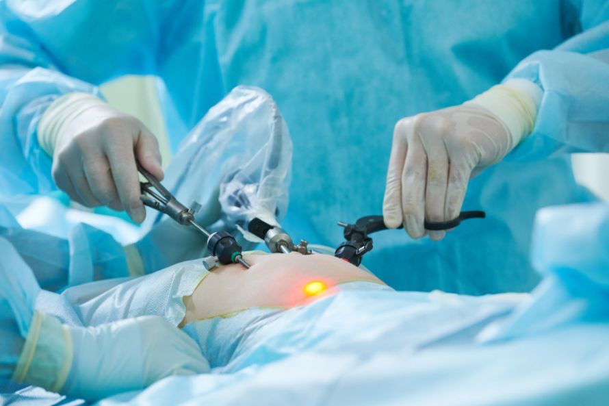
In laparoscopic surgery, several tiny incisions are sufficient instead of a single large incision © Kadmy | AdobeStock
Risks and Possible Complications of Intestinal Perforation Surgery
Risk factors (pre- and perioperative) include known gastric ulcers, intestinal ulcers, sigmoid diverticula/diverticulitis, and inflammatory bowel diseases requiring long-term corticosteroid therapy. In the large intestine, diverticulitis is considered the most common cause of bowel perforations; in the upper GI tract, ulcers and iatrogenic injuries are the primary causes. The resulting peritonitis is a potentially life-threatening condition and requires rapid debridement of the affected area.
The extent of treatment and the course of the disease can vary greatly: from a few days of hospitalization to complex intensive care management involving mechanical ventilation, renal replacement therapy, and multiple reoperations. An interdisciplinary approach involving visceral surgery, internal medicine, and critical care medicine is often required—a point that is also emphasized in standard textbooks (e.g., Thieme, De Gruyter) and patient-oriented health resources.
Typical Early and Late Complications
Early complications (in the operating room or shortly thereafter):
- Bleeding, wound infection, intra-abdominal abscess (in the abdomen).
- Suture/anastomotic insufficiency: Leakage with escape of intestinal contents into the abdominal cavity → risk of peritonitis and sepsis.
- Fistula formation (e.g., enterocutaneous fistula) in cases of prolonged leakage.
- Injury to or narrowing of the intestinal lumen (e.g., due to swelling or stapled sutures) with a risk of obstruction.
Late complications:
- Adhesions (bridges) with a risk of (sub)ileus or bowel obstruction even years later.
- Stenoses in the area of the suture (narrowing of the lumen), chronic abdominal pain.
- Incisional hernia, recurrent abscesses, persistent/recurrent fistulas.
Checking the integrity of the sutures
The integrity of the suture/anastomosis can be checked using a water-soluble contrast agent: orally, via a tube “from above,” or as an enema “from below.” Depending on the situation, a CT scan may also be performed. If a leak is detected, repeat surgery or at least drainage/intervention is often necessary.
Can a perforation in the gastrointestinal tract be prevented?
Intestinal perforation cannot always be prevented, but the risk can be significantly reduced—primarily through consistent treatment of underlying conditions and by avoiding typical triggers.
Consistently treat underlying conditions
- Stomach ulcers: Treat early (including Helicobacter pylori eradication) and monitor the healing process via endoscopy.
- Use NSAIDs (anti-inflammatory/pain medications) for as short a time as possible and at low doses; consider acid suppression (PPIs) if necessary.
- Manage inflammatory bowel diseases (ulcerative colitis, Crohn’s disease) according to guidelines to prevent flare-ups and complications (including fistula formation and perforation).
- Diverticulitis (in the large intestine/sigma—the most common cause of bowel perforations): Monitor the course closely. Following imaging (interval colonoscopy, CT), consult with the treatment team to determine whether elective resection is appropriate before a new flare-up or perforation occurs.
Standard textbooks on internal medicine and visceral surgery (e.g., Thieme, De Gruyter) as well as patient-oriented health resources emphasize the importance of low-inflammation disease management for prevention.
Reduce mechanical risks
- Avoid foreign bodies: Do not swallow hard or sharp fish bones, toothpicks, or similar objects; exercise particular caution in cases of intestinal narrowing (strictures).
- Actively manage constipation (drink plenty of fluids, eat a high-fiber diet, exercise) to avoid pressure spikes in the bowel.
- Prevent abdominal injuries: Fasten your seatbelt correctly; follow occupational safety guidelines (trauma prevention in the abdomen).
Take early warning signs seriously—“acute abdomen”
- Sudden severe pain, abdominal rigidity, fever, persistent nausea/vomiting, or symptoms of shock are warning signs.
- If you experience these signs of an acute abdomen, seek immediate medical evaluation (emergency). Early diagnosis prevents intestinal contents from entering the abdominal cavity and causing peritonitis or sepsis.
Who will care for me?
- Prevention and follow-up care are provided through an interdisciplinary approach involving primary care physicians, internal medicine/gastroenterology (including medication and endoscopies), and visceral surgery (surgical strategies).
- Regular follow-up visits after severe inflammation or surgery help detect long-term complications (e.g., strictures, fistulas) early on.
It’s better to go to the doctor’s office or emergency room once too soon than once too late—because a bowel perforation is a medical emergency. Other causes with similar symptoms (e.g., heart attack, ruptured ectopic pregnancy, ileus without perforation) must be ruled out through differential diagnosis; repeated examinations and monitoring are standard practice in unclear cases.
Share this article