News
Shunt – an artificial connection used in medicine

Sabine Schneider ·
A shunt is an artificially created connection between two parts of the body, usually between blood vessels. It is used to specifically reroute blood flow or to provide access for medical procedures—such as dialysis or in cases of portosystemic shunts in the liver. The term derives from the English verb “to shunt,” meaning “to divert.”
Medical Significance of Shunts
Shunts are used in various medical specialties. They can either occur naturally or be surgically created. Typical applications include:
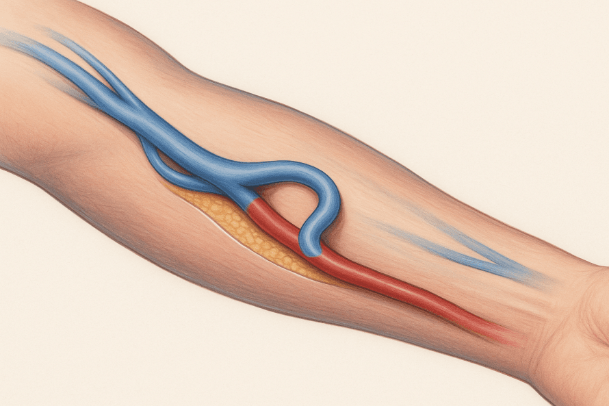
- Dialysis shunt (AV fistula): A connection between an artery and a vein in the forearm to create permanent access for hemodialysis treatments.
- Portosystemic shunt: Used in liver diseases such as portal hypertension to reduce pressure in the portal venous system.
- Ventriculoperitoneal shunt: Used in cases of hydrocephalus (water on the brain) to drain excess cerebrospinal fluid into the abdominal cavity.
- Cardiovascular shunts: Used to bypass blocked blood vessels or in cases of congenital heart defects.
Shunt Placement
Shunt placement is performed under sterile conditions, usually in a hospital by specialized surgeons, nephrologists, or radiologists. Depending on the type of shunt, the procedure may be performed under local or general anesthesia.
Dialysis Shunt (AV Fistula)
In this procedure, an artery is connected directly to a vein, often in the forearm. The arterial pressure increases blood flow to the vein, causing it to dilate; after a few weeks, it is suitable for regular dialysis access. The procedure usually takes about an hour.
Portosystemic Shunt
If there is increased pressure in the portal venous system (e.g., due to liver cirrhosis), a shunt is created between the portal vein and the inferior vena cava. This can be performed surgically or radiologically—in the latter case, it is referred to as a TIPS (transjugular intrahepatic portosystemic shunt). The goal is to relieve pressure on the portal circulation.
Shunt for Hydrocephalus
A ventriculoperitoneal shunt is implanted by neurosurgeons. Excess cerebrospinal fluid is drained from the cerebral ventricles into the abdominal cavity via a tube system, where it is reabsorbed.
Follow-up Care and Monitoring
Regular follow-up is necessary after placement to ensure the shunt is functioning properly and remains patent.
- For dialysis shunts, blood flow is regularly monitored to detect narrowing or thrombosis at an early stage.
- For portosystemic shunts, follow-up examinations are performed using ultrasound or CT scans to assess patency and the direction of flow.
- For ventricular shunts, imaging studies are necessary to ensure proper drainage of cerebrospinal fluid.
Patients should watch for swelling, redness, pain, or functional abnormalities in the area of the shunt and have these evaluated by a doctor immediately.
Risks and Possible Complications
As with all procedures, there are risks:
- Bleeding or infection at the insertion site
- Thrombosis or shunt occlusion
- Excessive blood flow (in AV fistulas)
- Leaks or malfunctions in shunt systems
Careful hygiene and regular medical checkups significantly reduce the risk of complications.
Prognosis
A properly implanted and well-maintained shunt can function reliably for many years. Especially for dialysis patients, it forms the basis for successful long-term therapy. The outcomes are also generally very good for portosystemic or ventricular shunts, provided that regular checkups are performed and potential complications are detected early.
Share this article
Verified expertise
Recommended specialists
12 specialists
12 results




Dr. med. Konstantinos Verginis
Duisburg



Dr. med. (syr) Amer Jomha
Bad Hersfeld

Dr Thomas Riester
Karlsbad







1 / 2
News
Read next
- News
Scabies: A contagious skin disease that is easily treatable
Oct 26, 2026
Severe itching and minor skin changes should be taken seriously
Read more - News
Heat Rash: What's Behind the Itchy Blisters
Oct 23, 2026
Heat Rash: Heat and Sweat Can Disrupt the Skin's Balance
Read more - News
Enlarged Prostate – When Medication Is Sufficient and When Surgery Is Recommended
Oct 21, 2026
Why Many Men Ignore Urinary Problems for Too Long
Read more