In shoulder joint-preserving surgery, so-called minimally invasive or arthroscopic surgical methods are used. These are surgical treatment options employed to restore the shoulder joint’s original function. They have now largely replaced most procedures that were previously performed using open surgery. Very few reconstructive procedures on the shoulder joint are still performed as open surgeries today. However, there are still important open procedures that cannot yet be performed arthroscopically.
Here you will find further information as well as a selection of specialists and centers for shoulder joint-preserving surgery.
The Anatomy of the Shoulder Joint
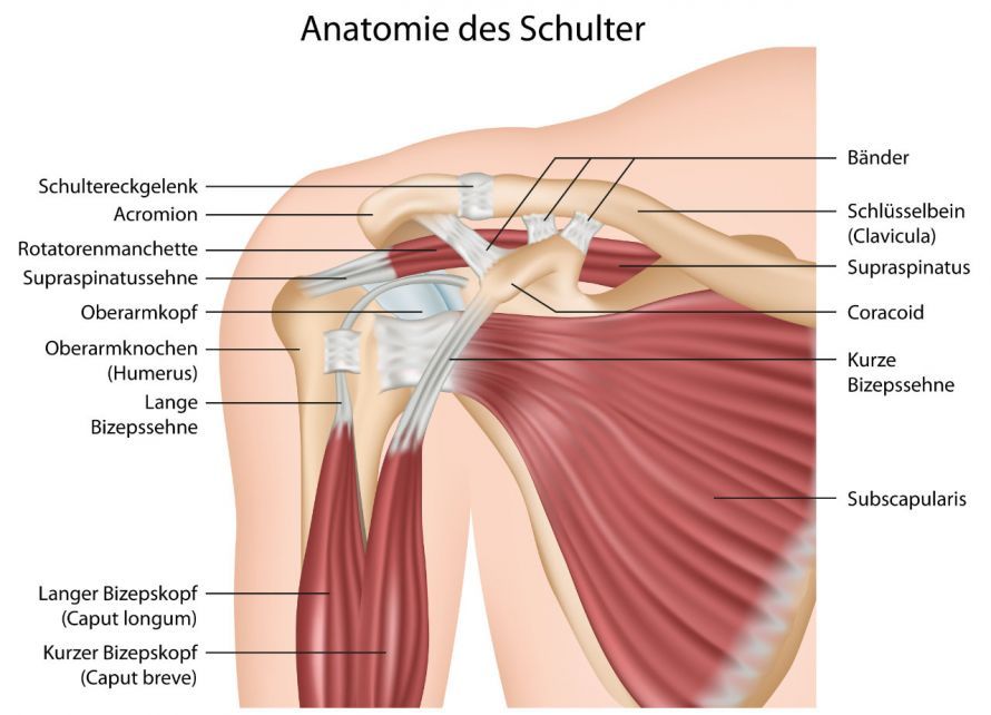
The shoulder joint connects the upper arm to the shoulder blade. The joint between these two structures consists of the head of the humerus and the glenoid cavity of the shoulder blade. The shoulder joint’s high range of motion requires particularly strong support from
- muscles,
- ligaments, and
- tendons
: These ensure that the head of the humerus remains stable in the glenoid cavity even during wide ranges of motion.
Due to the high stress they endure, the protective and stabilizing structures of the shoulder joint are prone to signs of wear and tear. This results in what is known as joint degeneration.
In addition, falls onto the shoulder often lead to dislocations of the joint, despite the good support provided by muscles and ligaments. The shoulder joint is particularly prone to dislocations. Unlike the knee joint, stabilization is provided mainly by muscles and ligaments, rather than by bony guidance.
When is minimally invasive shoulder surgery used?
Accidents or signs of wear and tear are the most common causes of shoulder problems. The cause of the symptoms can usually be determined through a thorough clinical examination and appropriate imaging diagnostics.
Various imaging techniques are used as part of the diagnostic process. A series of X-rays is essential. Depending on the specific changes present, cross-sectional images such as magnetic resonance imaging (MRI) and/or computed tomography (CT) can also provide important insights.
The physician’s experience in this field is also of great importance. Only then can an optimal diagnosis be made.
As a rule, conservative physical therapy is the first treatment option. If this is unsuccessful or cannot be used, shoulder arthroscopy is often performed. This allows the doctor to confirm the diagnosis and treat the underlying cause immediately.
This usually requires general anesthesia. Through small incisions in the shoulder area, the surgeon inserts a small camera and tiny instruments. These allow the surgeon to assess the condition of the joint and perform various procedures.
Common indications for arthroscopic surgery on the shoulder joint include, for example:
- Instability of the shoulder joint due to a sports injury or wear and tear of stabilizing structures such as the shoulder capsule, ligaments, and muscles (for example: shoulder dislocation, dislocated shoulder joint)
- Tears in key muscles of the shoulder joint that are essential for stabilization and function (rotator cuff tear)
- Calcifications in the shoulder joint (calcific tendinitis) as well as various painful conditions in the shoulder region: Frozen shoulder, entrapment of tendons and/or muscles in the shoulder joint (impingement syndrome), degenerative joint disease of the acromioclavicular (AC) joint (AC joint osteoarthritis)
- Avascular necrosis of the humeral head, in which bone material breaks down due to a slowed conversion of cartilage into bone tissue. As a result, bone particles break off and alter the structure of the joint surfaces
- Omarthrosis (shoulder osteoarthritis): degenerative disease of the shoulder joint
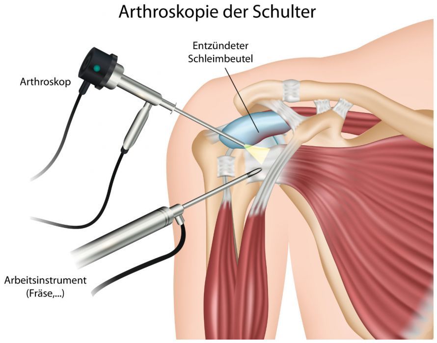
Schematic illustration of minimally invasive shoulder arthroscopy © bilderzwerg | AdobeStock
Surgical Procedures in Minimally Invasive Shoulder Surgery
If shoulder surgery is unavoidable, there are various options for surgical intervention:
Especially following traumatic dislocations, surgery to repair the damaged, torn, and displaced joint lip (labrum) and surgical tightening of the joint capsule (capsular shift) is often unavoidable.
Tendon tears can be surgically repaired using secure anchoring by suturing the tendon to the bone (tendon repair).
In cases of tendon calcifications, the surgeon opens the calcific pocket and removes the calcified material. Adhesions in the joint capsule can be treated in a similar manner.
In decompression surgery, the surgeon creates space by
- removing the inflamed bursa and
- shaving down the acromion
to relieve pressure on the tendons. In recent years, this procedure has been performed too frequently and without due consideration for shoulder pain of any kind. Due to improper application, it subsequently fell into disrepute in the tabloid press.
A true impingement syndrome caused by bony changes (bony impingement) is the exception. In principle, it should first be addressed through conservative measures such as physical and manual therapy. These include
- stretching the joint capsule, as well as
- strengthening and balancing the muscles of the rotator cuff, the scapular stabilizers, and the deltoid muscle.
Thus, subacromial decompression alone is now a rare exception. It is most often performed in combination with other procedures (e.g., rotator cuff repair).
Cartilage smoothing (chondroplasty) is a procedure for treating cartilage damage that is performed during arthroscopy. Depending on the extent of the damage, either the superficial cartilage tissue can be smoothed or a more in-depth procedure can be performed. In the latter case, the bone tissue is stimulated to regenerate cartilage (microfracturing).
Arthroscopic treatment of early-stage shoulder osteoarthritis, as part of a so-called Comprehensive Arthroscopic Management (CAM) procedure, also includes
- the removal of the inflamed synovial membrane and the bursa,
- repair of the long biceps tendon, and
- if necessary, resection (removal) of the acromioclavicular joint.
In nearly all types of rotator cuff (RC) tears, the long biceps tendon (LBT) causes significant pain due to its destabilization. Therefore, repositioning or removal of the long biceps tendon is almost always necessary in such cases.
Specialists from the fields of
decide on a case-by-case basis which surgical method appears most appropriate.
After Surgery: Possible Complications and Follow-Up Care
Minimally invasive shoulder surgery is not associated with any particular risks. It usually proceeds without complications. As with all surgical procedures, however, there is a small risk of
- bone or nerve injuries, as well as
- postoperative bleeding and bruising in the treated area.
Swelling around the incision site is normal after surgery and will gradually subside. Pain and limited range of motion are also among the possible consequences of shoulder surgery. Your doctor will therefore prescribe pain medication, which you should take as directed.
A particular concern is postoperative joint stiffness. Excessive inflammation and scarring can cause patients to experience increased pain and long-lasting limited range of motion. This is effectively managed with a 3–4-week course of corticosteroid therapy.
After shoulder joint surgery, it is usually necessary to immobilize the joint for three to six weeks. During this time, you should take it easy and avoid any movements that put strain on the joint. You should therefore avoid leaning on the affected side, lifting heavy objects, and similar activities.
Immediate rehabilitation, on the other hand, is beneficial shortly after surgery and helps relax the muscles. If healing progresses well and as soon as you are physically able, you should begin a targeted rehabilitation program. This program will be tailored for you according to physical therapy guidelines.
The duration of immobilization varies depending on the condition and surgical technique, and mobilization is adjusted accordingly. You should always ask your surgeon for a detailed and personalized postoperative care plan.
Conclusion
A timely diagnosis facilitates a gentle surgical approach—usually minimally invasive—to restore the natural joint. Joint-preserving shoulder surgery is performed by specialized physicians who take the time to develop an individualized treatment plan and are available to answer any questions you may have on the subject.

About the medical author
Asst Lect. Dr Mike H. Baums
Medical Author · Senior Consultant · Dorsten
Sources
- Deutsche Gesellschaft für Unfallchirurgie: Schultergelenk Erstluxation. https://www.awmf.org/uploads/tx_szleitlinien/012-012l_S1Schultergelenk-Erstluxation_2018-07.pdf
- Deutsche Gesellschaft für Orthopädie und Orthopädische Chirurgie (DGOOC): S2e-Leitlinie „Rotatorenmanschette. https://www.awmf.org/uploads/tx_szleitlinien/033-041l_S2e_Rotatorenmanschette_2017-04_02.pdf