Dunbar syndrome is a rare compression syndrome in which the celiac trunk is constricted by the medial arcuate ligament, which typically runs deep. This anatomical feature can impair blood flow in the superior epigastric artery and often leads to postprandial upper abdominal pain, nausea, and weight loss. Because the symptoms are nonspecific, Dunbar syndrome is often diagnosed late and initially mistaken for gastroenterological or functional disorders. A characteristic feature is the combination of compression, symptoms, and typical diagnostic findings, such as the evaluation of the celiac trunk using duplex ultrasound or color-coded Doppler ultrasound. Treatment usually involves surgical division of the constricting ligament—often using minimally invasive techniques—to achieve a lasting improvement in blood flow.
What is Dunbar syndrome?
In Dunbar syndrome, a fibrous band (ligament) originating from the diaphragm presses on the celiac trunk, a major abdominal artery. This band is actually part of normal anatomy—but in some people, it runs so deep that it compresses the artery.
The result: blood flow is impeded. Those affected notice this especially after eating, when the gastrointestinal tract requires increased blood flow. In many cases, Dunbar syndrome causes severe abdominal discomfort, often accompanied by weight loss because patients avoid eating out of fear of pain.
Since the syndrome is very rare, it is often not diagnosed until late in the course of the disease. Many patients are initially given other diagnoses, such as irritable bowel syndrome or psychosomatic symptoms.
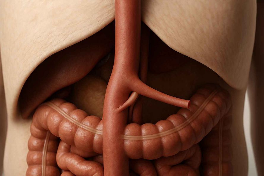
3D illustration of the celiac trunk in the upper abdomen with compression by the arcuate ligament—a typical anatomical finding in Dunbar syndrome.
How does Dunbar syndrome develop? (Pathophysiology)
Dunbar syndrome results from an anatomical abnormality in which the medial arcuate ligament—which typically lies too low—compresses the celiac trunk through the ligament. This compression of the celiac trunk and the surrounding celiac plexus leads to restricted blood flow in the superior epigastric artery. A characteristic feature is that the constriction often worsens postprandially, that is, after eating. This results in epigastric pain, nausea, a feeling of fullness, or nausea and vomiting. Dunbar syndrome is caused by compression of the celiac ganglion by the fibrous ligament. This combination of reduced perfusion and neurogenic irritation explains the typical symptoms. Harjola-Marable syndrome (also known as ligamentum arcuatum syndrome) occurs more frequently in slim, athletic patients. Important: Anatomical compression alone is not sufficient—Dunbar syndrome arises only from the combination of an anatomical constriction and corresponding symptoms.
Typical Symptoms of Dunbar Syndrome
The symptoms of Dunbar syndrome often occur after eating and can vary in intensity. The most common symptoms include:
- Abdominal pain in the upper abdomen, often described as stabbing or burning
- Nausea and a feeling of fullness
- Loss of appetite
- Weight loss, often unintentional
- Less commonly: back pain or diarrhea
Many of these symptoms can also occur with other gastrointestinal disorders—which makes diagnosis difficult. However, the connection between the symptoms and food intake is particularly characteristic of Dunbar syndrome.
Differential Diagnosis from Other Causes of Upper Abdominal Pain
Since Dunbar syndrome causes symptoms that resemble those of many other conditions, clear differentiation is essential. It is often initially confused with gastritis, ulcers, gastroparesis, irritable bowel syndrome, or psychosomatic symptoms. Biliary tract disorders can also cause nausea, diarrhea, or pain after eating. Only by correlating the symptoms with compression of the celiac trunk can a clinically relevant compression syndrome be confirmed. Confirmation is possible only if both the anatomical narrowing and the typical postprandial symptoms are present.
Causes and Risk Factors
Dunbar syndrome is caused by the arcuate ligament lying too low. Why this occurs in some people has not yet been fully clarified. Genetic factors or a certain posture may play a role.
Possible risk factors include:
- Slender or athletic individuals, as they have less protective fatty tissue surrounding the vessels
- Anatomical peculiarities in the area of the diaphragm
- According to studies, young women are affected more frequently than men
However, this is not a condition caused by external factors such as diet or lifestyle.
When is treatment necessary? (Indications for surgery)
Treatment is necessary when the compression caused by compression of the celiac trunk becomes clinically significant. The severity of the symptoms and their relationship to food intake are decisive factors. Typical indications for surgery include epigastric pain, significant weight loss, nausea, or impaired quality of life. The diagnosis is confirmed by identifying the stricture and its hemodynamic significance using imaging techniques. Only when a clear clinical significance is established through the correlation of symptoms and duplex findings do professional societies recommend surgical therapy—often minimally invasive (laparoscopic)—which is considered first-line treatment.
Examination and Diagnosis
Since the symptoms of Dunbar syndrome are nonspecific, targeted diagnostic testing is required to definitively diagnose the disease.
The examination usually includes:
- Doppler ultrasound: Here, the physician measures blood flow in the affected artery
- Computed tomography (CT) or magnetic resonance imaging (MRI) with contrast dye in both inspiratory and expiratory positions: The vessels are visualized in a lateral view using contrast medium, and the typical hockey-stick-shaped configuration of the celiac trunk is depicted during exhalation.
It is important to consider the narrowing in relation to the symptoms—anatomical compression alone is not sufficient for a diagnosis. Only when symptoms are present is the condition referred to as clinically relevant Dunbar syndrome.
Diagnostics in Detail—How Is Dunbar Syndrome Detected?
Diagnosing Dunbar syndrome requires a combination of clinical evaluation and imaging techniques. Examination of the celiac trunk using duplex sonography is particularly important, as it visualizes flow velocities and pressure differences. In addition, CT or MRI angiography, color-coded Doppler examinations (Doppler sonography), and functional imaging during inhalation and exhalation are used. A characteristic finding is the “hockey stick” bend in the region of the celiac trunk. A purely anatomical narrowing is not sufficient—it becomes diagnostically relevant only if it explains the symptoms. Modern diagnostic techniques also support surgical planning by assessing the extent, location, and possible involvement of the plexus and nerve network.
Treatment Options and Specialists
Treatment for Dunbar syndrome depends on the severity of the symptoms. In severe cases, surgery is usually the only effective option.
The goal of treatment is to relieve the compression. To do this, the surgeon surgically cuts through the constricting ligament. This can be done in two ways:
- Open surgery via an abdominal incision
- Minimally invasive surgery (keyhole technique), often via laparoscopy
In some cases, a vascular support system (stent) is also inserted to keep the celiac trunk open.
These procedures are primarily performed by specialists in vascular surgery or visceral surgery (abdominal surgery). The prognosis after successful surgery is generally very good—many patients report a significant improvement in their symptoms.
Minimally invasive or open—which procedure is appropriate?
Treatment involves surgically dividing the constricting ligament, which improves blood flow and reduces symptoms. A minimally invasive approach—usually laparoscopic—is now recommended as first-line therapy. This procedure allows for precise decompression and protects the celiac plexus. Open surgery via laparotomy is particularly indicated when there are severe adhesions or when additional vascular support (a stent) is necessary. In severe cases, a stent is additionally inserted to permanently keep the celiac trunk and the truncus open. The choice of procedure depends on symptoms, anatomy, and prior diagnoses.
Follow-up Care – What Happens After Surgery?
After surgical treatment, many patients require only a short recovery period. Early follow-up visits are important to ensure the success of the decompression. A duplex ultrasound is used to verify that the narrowing has been resolved and that no new scar tissue is forming. Symptoms usually improve within a few weeks—especially postprandial pain. Complications are rare but may include bleeding, recurrent narrowing, or irritation of the celiac plexus. Structured follow-up care improves long-term outcomes, particularly in cases of previously severe abdominal symptoms.
Course and Prognosis
If left untreated, Dunbar syndrome can lead to a permanent impairment of quality of life. The pain and fear of eating severely limit many affected individuals.
With successful treatment—particularly surgery—symptoms improve significantly in most cases. Studies show that 60 to 90 percent of patients are symptom-free or show significant improvement after the procedure.
A relapse is rare but possible—for example, if scar tissue forms again or the celiac trunk remains unstable.
Conclusion: Not common, but should be taken seriously
Dunbar syndrome is a rare but treatable condition. If you frequently experience abdominal pain, nausea, or weight loss after eating, you should have a doctor investigate the cause—especially if other diagnoses do not provide a solution.
Specialists in vascular surgery or abdominal surgery are responsible for treating this condition. The diagnosis is usually made using imaging techniques that visualize blood flow in the abdominal aorta. Surgery can specifically address the cause of the symptoms and help many patients regain a better quality of life.
Glossary
- Celiac trunk: The main artery in the abdomen that supplies the stomach, liver, and spleen
- Compression: Narrowing or squeezing of an organ or blood vessel
- Ligamentum arcuatum: A band of connective tissue connecting the diaphragm to the spine
- Vascular surgery: A medical specialty focused on the surgical treatment of blood vessels
- Compression syndromes: Conditions in which tissues or blood vessels are impaired by external pressure
FAQ: Frequently Asked Questions About Dunbar Syndrome
What causes Dunbar syndrome?
Dunbar syndrome is caused by compression of the celiac trunk by the medial arcuate ligament. This anatomical feature simultaneously compresses the celiac plexus, which can impair blood flow and cause epigastric pain. Dunbar syndrome typically results from a combination of mechanical constriction and dysfunction of the celiac ganglion caused by the fibrous ligament.
What are the symptoms of Dunbar syndrome?
Dunbar syndrome causes symptoms such as postprandial upper abdominal pain, a feeling of fullness, nausea, loss of appetite, and often subsequent weight loss. Some patients also report back pain, nausea, diarrhea, or discomfort throughout the entire abdomen. Crucially, these symptoms correlate with compression of the celiac trunk.
How is Dunbar syndrome diagnosed?
Diagnosis involves examining the celiac trunk using duplex sonography, color-coded ultrasound, and Doppler sonography to assess blood flow and the narrowing. CT or MRI angiography during inspiration and expiration helps confirm the typical narrowing. A clinically relevant diagnosis can only be made by correlating symptoms with the narrowing. The diagnostic workup also serves to plan surgery, for example, to visualize the celiac trunk using duplex sonography and to assess involvement of the nerve plexus.
What complications can occur?
Without treatment, clinically significant celiac trunk compression syndrome can lead to chronic pain, malnutrition, or functional disorders such as gastroparesis. Vascular complications, such as a marked reduction in blood flow or irritation of the celiac plexus, occur rarely. Postoperative complications are rare and include scarring, recurrent narrowing, or disorders in the abdominal region.
What treatment is considered effective for Dunbar syndrome?
Treatment consists of surgical division of the constricting ligament, usually performed laparoscopically, and is considered the first-line therapy. The goal is permanent decompression and improvement of blood flow to the celiac trunk and surrounding tissues. In select cases, a stent may also be necessary if significant vascular narrowing persists despite the resolution of the compression. The procedure can be performed via laparotomy or using a minimally invasive approach, depending on the patient’s anatomy and prior findings.
What are the chances of success after surgery?
The prognosis is very good: In 60 to 90 percent of cases, patients report significant or complete relief from symptoms. Follow-up duplex ultrasound examinations are important to detect any recurrence of narrowing at an early stage. Recurrence is rare but may occasionally occur in cases of excessive scar tissue or neurogenic irritation of the plexus. Overall, the procedure is considered a safe and effective treatment for celiac artery compression syndrome.
What is Harjola-Marable syndrome?
Harjola-Marable syndrome is an alternative term for Dunbar syndrome and describes the same clinical picture. The term is used particularly in the international literature and emphasizes the connection between compression caused by ligamentum arcuatum syndrome and typical epigastric symptoms.
Share this article

About the medical author
assoc. Prof. PD Dr. Markus Klinger MBA, FEBVS
Vienna
View full expert profile →