An aortic aneurysm is a pathological, sac-like dilation of the aorta, the largest artery in the human body. The bulge develops due to a weakening of the vessel wall and may increase in diameter over time. Many patients do not notice any symptoms at first, but if the aneurysm ruptures (aortic rupture), it can be acutely life-threatening. Abdominal aortic aneurysms are particularly common, while thoracic aortic aneurysms in the chest cavity are less common. Atherosclerosis and high blood pressure are considered major risk factors, as are genetic disorders such as Marfan syndrome. The diagnosis is usually made using ultrasound or a CT scan. Depending on the size of the aneurysm and the patient’s symptoms, treatment ranges from regular monitoring to surgery involving the placement of a vascular prosthesis or a stent.
What is an aortic aneurysm?
An aortic aneurysm is a pathological bulge in the aorta. In this condition, a section of the vessel wall permanently dilates and loses stability. Doctors diagnose an aneurysm when the diameter of the aorta at a specific point is more than 50 percent larger than normal. The cause is usually damage to or weakening of the vessel wall, such as from atherosclerosis, high blood pressure, or connective tissue disorders like Marfan syndrome.
Depending on the location, a distinction is made between a thoracic aortic aneurysm in the chest area and an abdominal aortic aneurysm in the abdomen. Both forms can go unnoticed for years until they become apparent due to their size or complications. If the vessel wall ruptures (aortic rupture), it results in a medical emergency that is life-threatening without immediate surgery.
Causes and Development of an Aortic Aneurysm
Men are significantly more likely to develop an aortic aneurysm than women. In cases where the condition runs in families, genetic factors are considered a possible cause.
A major cause of aortic aneurysm formation is atherosclerosis, the “hardening” of the arteries. Similarly, high blood pressure over time can weaken the arterial wall. 90–95% of all aortic aneurysms are caused by these cardiovascular diseases.
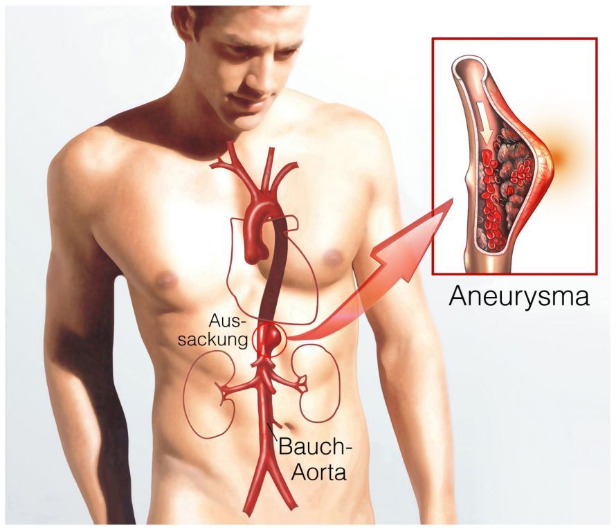
In patients, so-called plaques (calcium deposits) are found in the vessel wall. These damage the wall structure and cause the vessel to harden (sclerosis). This reduces the aorta’s elasticity, leading to the formation of a bulge.
If elevated blood pressure is added to the wall damage, the blood vessel may eventually rupture. If the vessel wall only tears without rupturing completely, doctors refer to this as an aortic dissection. In this case, blood flows between the layers of tissue in the aorta.
Aneurysm rupture is a serious complication. Large amounts of blood immediately flow into the chest or abdominal cavity. The resulting loss of blood volume in the circulatory system quickly leads to shock.
Shock is a life-threatening condition. Fewer than 10% of patients with an aortic rupture survive this emergency. Another 10–15 percent reach the hospital alive, but only about half of them are saved by emergency surgery. The risk of rupture increases as the diameter of the aortic aneurysm increases. The risk is particularly high when the diameter exceeds 5 cm.
In addition, there are also cases of patients who suffer from a congenital connective tissue disorder. Such connective tissue disorders may include:
- Marfan syndrome
- Ehlers-Danlos syndrome
- Cystic median necrosis of Erdheim-Gsell
What they all have in common is a structural defect in the connective tissue of the blood vessel wall. This causes the vessel wall to become less stable and to give way.
Inflammatory diseases can also weaken the blood vessels and lead to an aortic aneurysm.
There is an increased risk in cases of:
- Late-stage syphilis
- Takayasu arteritis
Less common causes of an aortic aneurysm include:
- congenital malformations
- hormonal changes, e.g., during pregnancy
- Injuries to the chest area
- Exposure to harmful substances (e.g., cocaine)
Symptoms of an aortic aneurysm
An aortic aneurysm is often asymptomatic. However, the following symptoms may also occur:
- Hoarseness
- Shortness of breath
- Shoulder pain
- Back problems
- Coughing up blood
Most often, however, a doctor discovers an aortic aneurysm incidentally during an X-ray examination.
Thoracic aortic aneurysm
An aortic aneurysm in the chest area is usually caused by existing atherosclerosis, high blood pressure, or inflammatory processes in the arterial wall. Similarly, injuries to the chest—whether sudden (acute) or occurring over several years (chronic)—can lead to an aortic aneurysm of the thoracic aorta.

A specific form is aortic dissection. In this condition, a tear occurs in the wall of the aorta. As a result, blood flows between the various layers of the aortic wall. In the early stages, there are often no symptoms. However, as the aortic aneurysm continues to grow, the following symptoms may occur:
- Chest pain
- Difficulty swallowing
- Hoarseness
- Paralysis of the legs
- Kidney problems
Abdominal Aortic Aneurysm
An abdominal aortic aneurysm usually presents with abdominal pain. Diffuse back pain resembling “lumbago” or renal colic is equally likely. Very large aneurysms may protrude from the abdomen as a pulsating mass.

Abdominal pain can be a sign of an abdominal aortic aneurysm
If the abdominal aneurysm ruptures, a life-threatening state of shock sets in rapidly. Patients often describe severe, “excruciating” pain and abdominal pain that radiates into the back. Nausea and vomiting also occur.
Due to the massive blood loss following a rupture, the person bleeds to death internally within a very short time. Life can only be saved through immediate surgery.
Diagnosis of an aortic aneurysm
The diagnosis is made through a clinical examination involving palpation of the abdomen and an ultrasound. Aortic aneurysms are usually discovered incidentally during routine abdominal ultrasound examinations. Larger aneurysms must be evaluated using computed tomography (CT) and contrast dye.
To better plan treatment, vascular surgeons must review and evaluate the CT images. A report from the radiologist alone is not sufficient.
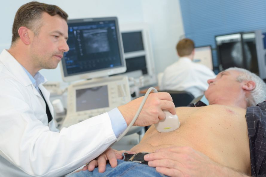
The doctor can diagnose an aortic aneurysm using ultrasound
An aortic aneurysm typically grows larger over the years. The risk of rupture must therefore always be considered. For this reason, even small aneurysms are regularly monitored with ultrasound. Although a small aortic aneurysm does not rupture frequently, it can cause pain.
Before surgery for an aortic aneurysm, blood tests are performed. Thyroid hormone levels and kidney function are of particular interest here. These values are checked again shortly before a CT scan or treatment.
The patient must be physically fit to successfully undergo the surgery. This can be determined using an ECG and a pulmonary function test. If the patient also has heart problems, a cardiologist will assess the patient’s suitability for the procedure.
What is the prognosis for an aortic aneurysm?
Depending on its severity and location, an aortic aneurysm can be a life-threatening condition. It is not always operable, and as the aneurysm continues to grow, the risk of rupture also increases.
Patients are usually afraid of bleeding. This significantly impairs their quality of life. They often avoid exercise and sudden movements, which contributes to risk factors such as obesity and atherosclerosis.
In many cases, patients are treated with medication. This reduces the risk of the aortic aneurysm progressing and slows the progression of the disease.
Severe cases of aortic aneurysms are usually treated surgically. Complications can occur during surgery. These are not always predictable in advance, so a careful risk-benefit assessment is always performed.
In any case, the doctor should inform the patient that an aortic aneurysm can reduce life expectancy as the patient ages.
Can an aortic aneurysm resolve on its own?
The condition progresses slowly over the course of years and is not reversible.
If the aortic aneurysm is smaller than 4 cm, a “watch-and-wait” approach is usually taken. The patient must then undergo an annual ultrasound examination. For aneurysms larger than this, follow-up examinations are conducted every six months.
There are a number of risk factors that contribute to the further growth of an aortic aneurysm:
- Diabetes mellitus
- High blood pressure (hypertension)
- High cholesterol and triglyceride levels
- Obesity
- Tobacco and alcohol
Those affected should eat a healthy and varied diet to avoid putting additional strain on their metabolism and blood vessels. This is because the combination of poor diet and atherosclerosis is a major cause of the formation and further growth of an aortic aneurysm.
Does an aortic aneurysm require surgery?
The larger the aortic aneurysm becomes over time, the greater the risk of a rupture in the aorta. Surgery is recommended if the diameter exceeds 5.5 cm in the thoracic aorta or 5 cm in the abdominal aorta.
For smaller diameters, the treating physician will make a decision based on the symptoms and the type of aortic aneurysm. Any blood clots that may have formed are also important factors in the assessment of the aortic aneurysm. Blood stasis can not only lead to reduced blood flow to tissues and organs but can also cause the aneurysm to swell further.
Aortic Aneurysm Surgery Procedures
For many years, the technique of minimally invasive treatment has continued to evolve. Under certain conditions, the patient can avoid an incision in the chest or abdomen to expose the aorta. Instead, the doctor makes a small puncture in the femoral arteries and advances a catheter into the aortic aneurysm.
There, the doctor then implants the aortic stent. The stent replaces and relieves pressure on the blood vessel. This minimally invasive surgery is gentler on the patient. However, stents do not last indefinitely. Several follow-up surgeries may therefore be necessary. Minimally invasive treatment is particularly beneficial for older patients, who often suffer from comorbidities. These factors increase the risk associated with open surgery.
For certain types of aortic aneurysms, the minimally invasive method cannot be used. This is the case when there are problems with the stent graft or in an acute emergency involving a ruptured aortic aneurysm. In these cases, open surgery is often necessary.
The surgeon exposes the abdominal aorta through an incision in the abdomen or flank. The surgeon then replaces the section containing the aortic aneurysm with a plastic tube, known as an aortic prosthesis.
Open surgery is always an option and offers long-term durability. The decision regarding which of the two treatment methods to use is made in consultation with the patient. In making this decision, the doctor considers:
- comorbidities,
- lifestyle factors, and
- the patient’s personal preferences
of the patient.
Procedure for Aortic Aneurysm Surgery
How is the surgery to implant a stent for an aortic aneurysm performed? The following sections explain the procedure in detail:
Before the Surgery
Before the surgery, several tests are performed to help plan the procedure more effectively. The doctor then decides which stent is most suitable. He or she subsequently discusses the plan with the patient.
Surgery for a Thoracic Aortic Aneurysm
Before the surgery begins, the patient’s groin area is cleaned and disinfected. The area is then numbed with a local anesthetic, or the patient is placed under general anesthesia. The doctor then makes a small incision in the groin area. He inserts a catheter and the stent into the artery in the groin.
The success of this procedure is verified using X-ray imaging (angiography). On the X-ray image, the surgeon can clearly see the location of the aortic aneurysm and advance the catheter to that point. The stent is then placed inside the aneurysm and adjusted to fit the diameter of the blood vessel.
Finally, the catheter is removed from the artery; the stent remains in place. If the aortic aneurysm is very large, the doctor may place multiple stents.
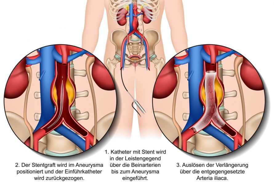
Minimally invasive stent surgery for an aortic aneurysm © bilderzwerg | AdobeStock
Surgery for an Abdominal Aortic Aneurysm
As with the thoracic aorta, various procedures are available for treating an aortic aneurysm in the abdomen. Open surgery is performed on the exposed abdominal aorta after the abdominal cavity has been opened.
The artery is clamped above and below the aortic aneurysm to temporarily stop blood flow. The aneurysm sac is then opened, and a vascular graft is inserted as a prosthesis. Depending on the size of the aortic aneurysm, either a tubular or a bifurcated prosthesis is implanted.
Open surgery is an option for all patients without restriction.
The stent procedure, on the other hand, is not suitable for everyone due to varying anatomical conditions. However, older patients and high-risk patients benefit from the stent procedure because of its lower risk.
The Postoperative Period
After the procedure, you will need to remain in the hospital for a few days for monitoring. Patients must initially lie still for four to six hours to support healing in the groin area. Possible side effects of the surgery include:
- Swelling of the thigh
- Numbness in the legs
- Malaise, nausea, and vomiting
- Throbbing pain in the legs
- Loss of appetite with or without fever
On average, patients are discharged from the hospital 5 days after minimally invasive aortic aneurysm surgery. After another 1–2 weeks, patients can return to their daily work routine.
To avoid the risk of an incisional hernia after open surgery, patients should avoid lifting heavy weights for up to 3 months. To prevent blood clots, you will be prescribed antiplatelet medications. These reduce blood clotting.
Patients who have undergone endovascular surgery must undergo an annual CT scan. For open surgery, ultrasound examinations replace the CT scan.
Risks and Complications of Aortic Aneurysm Surgery
The rupture of an abdominal aortic aneurysm is a life-threatening emergency. In comparison, surgery for an aortic aneurysm—whether using a stent or through open surgery—poses a much lower risk. Nevertheless, aortic surgery is not without risks. However, these are often predictable and are mostly due to complications resulting from inflammation:
- Infections
- Pneumonia
- Cardiovascular problems
When a stent is inserted, so-called blood leakage may also occur. This means that blood flows into the aneurysm sac (= “endoleak”). If the leak does not close on its own, it must be sealed again using a catheter-based procedure. Surgery on an aortic aneurysm may lead to temporary circulatory problems in the kidneys, intestines, and legs.
Open surgery always carries the risk of injuring other abdominal organs. While the abdomen is open, the surgeons may discover other conditions that require immediate surgical intervention.
Successful treatment includes regular monitoring of blood flow as well as an ultrasound examination.
How quickly does an aneurysm form?
An aneurysm can be congenital or develop over the course of a person’s life. An acquired aneurysm often takes years to develop and can increase significantly in size during that time.
Aneurysms frequently occur in the abdomen, brain, heart, and chest. Because it takes a long time for an aneurysm to become visible, the condition is most commonly diagnosed in people over the age of 50.
Meta-analyses of cerebral aneurysms have shown that approximately 9% of aneurysms enlarged over a period of about 3 years. Furthermore, the long-term risk of enlargement over the next 19 years was approximately 45%. Using the ELAPPS score, specialists assess the risk of growth over the next 3–5 years based on six risk factors:
- Previous aneurysm rupture
- Location of the aneurysm
- Age—especially 65 years and older
- The patient’s geographic origin
- Aneurysm size
- Shape of the aneurysm
Prevention of an aortic aneurysm
In some cases, the risk of an aortic aneurysm can be significantly reduced by adopting a healthy lifestyle. This includes plenty of exercise and a healthy diet low in fat and sugar. This helps minimize the main risk factors for an aneurysm—atherosclerosis and high blood pressure.

A healthy diet and exercise minimize the risk of an aortic aneurysm
People with a congenital connective tissue disorder often develop an aortic aneurysm. They should therefore pay particular attention to symptoms and, if necessary, seek medical advice and undergo an ultrasound examination.
FAQs on Aortic Aneurysms
How can an aortic aneurysm be detected early?
An aortic aneurysm often causes no symptoms for a long time. A suspected aortic aneurysm is frequently discovered by chance during an ultrasound or CT scan. Specialists recommend screening, particularly for men over 65, as they are more likely to develop an aortic aneurysm. Early diagnosis is crucial to prevent the risk of an aortic rupture and to initiate the appropriate treatment in a timely manner.
What causes an aortic aneurysm?
An aortic aneurysm is a sac-like enlargement of a section of the aorta. The main causes are atherosclerosis, high blood pressure, and connective tissue disorders such as Marfan syndrome or Ehlers-Danlos syndrome. Congenital conditions such as Marfan syndrome, lipid metabolism disorders, or injuries to the ascending aorta can also weaken the vessel wall. As the diameter in centimeters increases, the risk of rupture rises significantly.
What risk factors contribute to an aortic aneurysm?
The most important risk factors for an aortic aneurysm include smoking, hypertension (high blood pressure), lipid metabolism disorders, being male, and advanced age. A family history of the condition and certain congenital disorders also increase the risk. According to current guidelines, high-risk patients should undergo regular ultrasound examinations to monitor the progression of the aneurysm.
When is it considered an aortic aneurysm, and when is surgery necessary?
Doctors diagnose an aortic aneurysm when the aorta has dilated to more than 50 percent of its normal diameter. Surgery is generally indicated when the aneurysm reaches a diameter of about five to five and a half centimeters or is growing rapidly. The decision is made by a vascular surgeon, often in consultation with cardiologists and radiologists. The goal is to protect the aortic valve and adjacent structures—for example, in the case of an abdominal or thoracic aortic aneurysm—as effectively as possible.
How is an aortic aneurysm treated?
How doctors treat an aortic aneurysm depends on its location, size, and the patient’s overall health. Small aneurysms are monitored regularly, while larger ones require surgery involving a vascular graft or stent. In the case of an abdominal aortic aneurysm, the procedure is usually performed using a minimally invasive approach via the groin vessels. In contrast, an aneurysm of the ascending aorta in the chest may require open surgery. Both procedures are performed by specialists in vascular surgery.
Can an aortic aneurysm be prevented?
Yes, a healthy lifestyle can help prevent an aortic aneurysm. Avoid nicotine, maintain stable blood pressure, and bring elevated blood lipid levels back to normal. A balanced diet, exercise, and treatment of lipid metabolism disorders significantly reduce the risk. Since approximately 2% of men over the age of 65 develop an aortic aneurysm each year, regular checkups are particularly important.
Does an aortic aneurysm persist after treatment?
After successful surgery or stent implantation, the affected vessel is permanently stabilized. Nevertheless, an aortic aneurysm remains a condition that requires regular follow-up examinations. In accordance with current guidelines, these are performed using ultrasound or CT screening to detect any regrowth or complications at an early stage.
Share this article
Sources
- https://www.uniklinikum-jena.de/htchirurgie/Herzchirurgie/Aortenaneurysma.html
- http://www.medtronic.com/de-de/patienten/erkrankungen.html
- Etminan N et al., Unrupturierte intrakranielle Aneurysmen – Pathogenese und individualisierte Behandlung. Dtsch Arztebl Int 2020; 117: 235-42; DOI: 10.3238/arztebl.2020.0235
- https://medlexi.de/Aneurysma
- https://www.tk.de/techniker/krankheit-und-behandlungen/erkrankungen/behandlungen-und-medizin/herz-kreislauf-erkrankungen/gefaesserkrankungen-ursachen-symptome-behandlung/aneurysma-wenn-gefaesswaende-nachgeben-2015804