
A cerebral hemorrhage is a sudden bleed within the brain or the meninges and is one of the most dangerous neurological conditions. Intracerebral hemorrhage is often caused by high blood pressure, an aneurysm, or damaged brain blood vessels. Depending on the location of the hemorrhage, various symptoms may occur, such as paralysis, nausea, vomiting, or severe headaches. A cerebral hemorrhage is a type of hemorrhagic stroke and accounts for about 15 percent of all strokes.
The hemorrhage becomes particularly life-threatening when pressure builds up inside the skull and brain tissue is damaged. To diagnose a brain hemorrhage, CT, MRI, or computed tomography scans are used to determine the size and location of the hemorrhage. In many cases, patients require treatment in the intensive care unit or neurosurgical intervention.
Prompt diagnosis and treatment of a brain hemorrhage improve the prognosis and can prevent severe long-term damage.
What are the different types of cerebral hemorrhages?
Brain hemorrhages are also referred to as intracranial hemorrhages. This term refers to the cranium, the Latin word for skull. It therefore refers to all hemorrhages occurring inside the skull.
The effects on the brain and the resulting symptoms also depend on the extent and location of the hemorrhage. Medical professionals classify hemorrhages based on the name of the surrounding meninges:
- Epidural hemorrhage (epidural hematoma, EDH): Bleeding between the skull and the dura mater
- Subdural hematoma (SDH): Bleeding directly beneath the dura mater and simultaneously above the arachnoid membrane
- Subarachnoid hemorrhage (SAH): Bleeding beneath the arachnoid membrane (one of the soft meninges).
- Intracerebral hemorrhage (ICB): Bleeding within the brain tissue
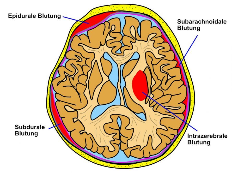
Brain hemorrhages are named based on the meninges adjacent to them © songkram | AdobeStock
What are the causes and symptoms of these types of brain hemorrhages?
All four types of brain hemorrhages mentioned can result from trauma. Intracranial hemorrhages involving the meninges (epidural hematoma and subdural hematoma) are particularly common following head trauma. These include
- traffic accidents, sports injuries, and workplace accidents,
- violent crimes involving blows or strikes to the head.
Intracerebral hemorrhage
High blood pressure is the most common cause of an intracerebral hemorrhage. Elevated blood pressure over an extended period damages the blood vessels. This can cause them to rupture, leading to a brain hemorrhage.
Other risk factors that increase the likelihood of a brain hemorrhage include:
- advanced age,
- tobacco smoking,
- excessive alcohol consumption,
- hard drug use,
- high blood lipid levels,
- lack of exercise,
- brain tumors, and
- vascular malformations.
Intracerebral hemorrhage presents with symptoms similar to those of a stroke. This is caused by a disruption in blood flow. The symptoms include
- sudden, severe headaches,
- nausea and vomiting,
- speech difficulties,
- visual disturbances, or
- paralysis on one side of the body.
Loss of consciousness is also possible.
Subarachnoid hemorrhage
A head injury often results in a subarachnoid hemorrhage of varying severity. In some cases, however, a ruptured aneurysm is responsible for this type of bleeding.
An aneurysm is a sac-like dilation of a blood vessel (arterial or venous). It develops due to a localized weakness in the vessel wall at the site of an arterial bifurcation, often associated with
- high blood pressure,
- long-term tobacco use, and
- genetic predisposition.
A subarachnoid hemorrhage is characterized by the sudden onset of a particularly severe headache. It is accompanied by neck pain or a stiff neck.
The symptoms usually begin during intense physical exertion. Other symptoms include
- nausea,
- vomiting, or
- impaired consciousness.
Subdural hemorrhage
Subdural hematomas can occur in two forms:
- acute, as a recent hemorrhage, or
- chronic, as a long-standing hemorrhage.
Chronic subdural hematomas primarily affect older adults. The cause is a decrease in brain volume and the resulting increased tension on the so-called bridging veins. Chronic subdural hematomas develop slowly over several weeks.
In most cases, a minor head injury is the trigger; in some cases, there was no significant trauma at all. Blood-thinning medications increase the risk of a chronic subdural hematoma.
Chronic subdural hematomas can persist for a long time without causing any clinical symptoms. Possible symptoms include
- headaches,
- a feeling of pressure in the head,
- dizziness, or
- newly developed cognitive changes.
Focal neurological deficits or impaired consciousness are also possible. An acute subdural hematoma is usually caused by a severe accident in which blood vessels in the subdural space are injured.
The clinical symptoms of an acute subdural hematoma include
- headaches,
- dizziness, or
- mental confusion.
Specific symptoms include
- speech disorders,
- seizures, or
- hemiplegia.
These are observed particularly when pressure is exerted on the brain. These deficits are very similar to the symptoms of epidural hematomas.
Epidural hemorrhage
The most common cause of epidural hemorrhage is a skull fracture accompanied by injury to an artery in the dura mater. Very often, an accident is the trigger. The hematoma spreads only within the cranial sutures.
Epidural hemorrhage is usually accompanied by loss of consciousness immediately following a traumatic injury.
Although rare, a characteristic finding is when the patient regains consciousness after a head injury and experiences relatively few symptoms for a few minutes. This is referred to as the “clear interval.” Shortly thereafter, the patient typically develops
- headaches,
- nausea,
- vomiting, and
- neurological deficits, followed by impaired consciousness.
How is a brain hemorrhage treated?
The effects of a brain hemorrhage can be fatal. That is why prompt action is essential. The later the patient receives treatment, the more likely the brain hemorrhage is to be fatal. Therefore, an emergency medical service should be called at the slightest suspicion!
A CT (computed tomography) scan of the head reveals the location of the hemorrhage. For further treatment, a hospital with a neurosurgery department should be consulted.
With brain hemorrhages, there is always a risk of brain swelling. The resulting increase in intracranial pressure affects the entire body. This leads to a lack of blood supply to the brain. Various measures can be taken to counteract this:
- mechanical ventilation,
- lowering blood pressure with medication, or
- diuretic infusions
are the first steps in treatment. These measures, or external cerebrospinal fluid drainage, can regulate the volume of cerebrospinal fluid and thus also intracranial pressure. Larger hemorrhages cause extreme intracranial pressure and are therefore usually surgically removed.
During the operation, the surgeon opens the skull at the affected site. There, the surgeon removes the hematoma and stops the source of the bleeding. Despite prompt treatment, patients may still die in some cases. This means that all intracranial hemorrhages are serious conditions. They require emergency treatment.
What are the possible consequences of a brain hemorrhage?
Depending on the location of the brain hemorrhage, various neurological impairments can result. Hemiplegic symptoms—including reduced sensation and weakness on one side of the body—are particularly common. Some patients experience only mild unsteadiness when walking. Others, however, struggle with extensive paralysis.
Incomplete hemiplegia (hemiparesis) affects not only the arms and legs but also the face.
Other possible consequences include
- impaired coordination of movement (ataxia),
- difficulty with complex motor sequences (apraxia), and
- reduced awareness on one side of the body
may occur.
Follow-up Care and Rehabilitation After a Brain Hemorrhage
Prompt treatment in the hospital is usually followed by neurological rehabilitation. This is intended to improve the prognosis following emergency treatment.
The various affected areas, such as
- speech,
- vision,
- concentration, or
- movement
require close collaboration among therapists from different medical disciplines:
- Occupational Therapy
- Physical Therapy
- Physical Therapy
- Speech therapy
- Neuropsychology
- Case Management (Social Services)
The prognosis for recovery depends, among other things, on the size and extent of the hemorrhage. The patient’s level of consciousness upon admission to the hospital is also a significant factor.
Some patients recover completely after a cerebral hemorrhage. However, the majority of patients can only partially manage their daily lives on their own, or are only able to do so after several months or years.
FAQ
What is a cerebral hemorrhage?
A cerebral hemorrhage is bleeding within the brain, the meninges, or between the skull bones and the dura mater. Doctors distinguish between different types of cerebral hemorrhages, such as intracerebral hemorrhage, subarachnoid hemorrhage, subdural hemorrhage, or subdural hematoma. The bleeding can damage brain tissue and is considered life-threatening.
What are the symptoms of a brain hemorrhage?
Typical symptoms of a brain hemorrhage include sudden severe headaches, neurological deficits, nausea and vomiting, or impaired consciousness. Many patients with a brain hemorrhage also develop paralysis, speech difficulties, or dizziness. The signs of a brain hemorrhage vary significantly depending on the location of the hemorrhage and the area of the brain affected.
What are the causes and risk factors for a brain hemorrhage?
The most common cause of a brain hemorrhage is high blood pressure. Other causes and risk factors include aneurysms, blood-thinning medications, traumatic brain injury, or damaged blood vessels in the brain. Traumatic bleeding or vascular changes can also trigger an intracranial hemorrhage.
How is a brain hemorrhage diagnosed?
CT, MRI, magnetic resonance imaging, or computed tomography are used to diagnose a brain hemorrhage. In addition, angiography and neurological examinations help determine the cause and location of the hemorrhage. Rapid diagnosis is particularly important when a stroke or subarachnoid hemorrhage (SAH) is suspected.
How is a brain hemorrhage treated?
Treatment for a brain hemorrhage depends on the size and location of the hemorrhage. Some intracerebral hemorrhages require surgical or neurosurgical intervention to stop the bleeding and reduce pressure inside the skull. Acute care is followed by rehabilitation and specialized therapy to limit long-term damage and improve the prognosis.
Share this article
Sources
- Deutsche Gesellschaft für Unfallchirurgie
- flexikon.doccheck.com/de/Hirnblutung