Exostoses are benign bony growths that occur primarily near joints and on long bones. The condition can present as a single exostosis or in the form of multiple cartilaginous exostoses. In cases of hereditary multiple exostoses, several osteochondromas often develop simultaneously. Many exostoses remain asymptomatic; however, depending on their location, they can cause symptoms and restrict movement.
The bony growths usually develop in the area of the growth plates and can affect bone growth. In rare cases, there is a risk of malignant transformation. Imaging techniques such as MRI or CT are used for diagnosis. Surgical treatment may be appropriate if the exostoses cause pain or impair surrounding structures.
What are the different types of exostosis?
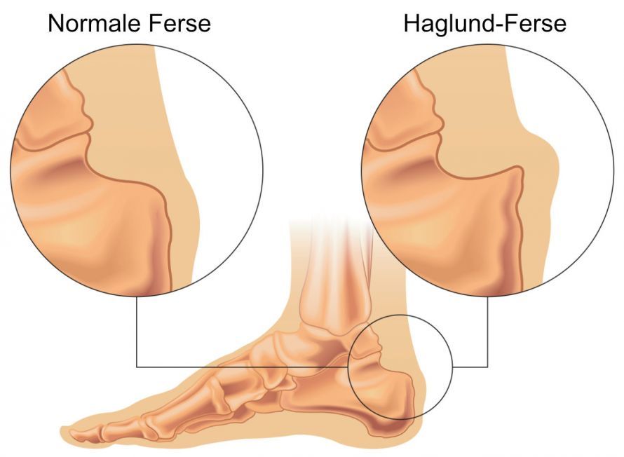
The metatarsal bones and the back of the hand are particularly commonly affected. A fairly common form of exostosis is known as Haglund’s exostosis. In this condition, a portion of the heel bone protrudes significantly. As a result, a bony protrusion develops in the area where the Achilles tendon attaches.
Cartilaginous exostosis, also known as an osteochondroma, occurs primarily on the long bones. This type of bony protrusion forms, for example, on the thighbone or upper arm bone. The knee and ankle joints can also be affected.
If there are several bony protrusions in these areas, the condition is referred to as multiple cartilaginous exostoses.
Other forms of exostoses include:
- subungual exostoses under the fingernails or toenails
- exostoses on the pelvis (spiny pelvis)
- exostoses of the ear canal
- exostoses on the lower jaw (mandibular exostoses)
Hallux valgus (pseudoexostosis) must be distinguished from true exostosis. It is not an actual bony protrusion, but merely a joint misalignment. However, due to its shape, hallux valgus on the foot can easily be mistaken for a true exostosis.
What are the symptoms?
The symptoms vary depending on the location and severity of the exostosis.
For example, Haglund’s exostosis eventually leads to Haglund’s syndrome. This is characterized by
- inflammation of the bursa at the Achilles tendon insertion or
- inflammation of the Achilles tendon
. The main symptoms of Haglund syndrome are stabbing pains in the heel bone, which are primarily triggered by pressure or weight-bearing. Because of the pain, patients limp.
An exostosis in the ear canal can cause symptoms such as
- hearing loss,
- tinnitus, or
- inflammation of the ear canal (otitis externa) with pain and itching in the ear canal area
.
Exostoses on the tubular bones can lead to
- leg length discrepancies,
- asymmetries in the shoulder and pelvic girdles, as well as
- hip dislocations
. Similarly, the bone structures
- compress nerves,
- blood vessels, or
- tendons
in the immediate vicinity. This can result in circulatory disorders and/or sensory disturbances.
How does an exostosis develop?
In general, several causes or risk factors can contribute to the formation of an exostosis. Chronic pressure and irritation on the bone are often the cause. For example, shoes with inflexible edges can irritate the bone tissue, thereby contributing to the development of Haglund’s deformity.
Another risk factor for an exostosis on the foot is being overweight. In overweight individuals, the heel bone must bear a particularly heavy load.
Other conditions also pose a risk, such as chronic joint wear (osteoarthritis). Silverskjöld exostosis, a bony protrusion on the top of the foot, is usually caused by osteoarthritis.
The cause of an osteochondroma is not yet fully understood. Genetic factors are thought to play a decisive role.
How does a doctor diagnose an exostosis?
If exostosis is suspected, the doctor usually performs an X-ray examination. The bone changes are usually clearly visible on the X-ray. Computed tomography (CT) and magnetic resonance imaging (MRI) are also suitable for further diagnosis.

X-ray image of an exostosis on a femur © J. Lengerke - Dr. Jochen Lengerke’s Practice, CC BY-SA 3.0 de, Link
How is an exostosis treated?
Just like the symptoms, treatment depends primarily on the location and severity of the bony protrusion.
Treatment of Haglund’s exostosis
Treatment can be conservative and orthopedic or surgical. The goal of treatment is to relieve pain in the foot and improve the structure’s ability to withstand stress during daily activities and sports.
Open-toed shoes or heel-raising insoles reduce pressure on the exostosis and thus relieve pain. Anti-inflammatory and pain-relieving medications containing active ingredients such as ibuprofen or diclofenac are used as adjunctive therapy.
Physical therapies are also suitable for treating Haglund’s exostosis. These include, for example,
- shock wave therapy and
- electrotherapy.
If conservative methods are ineffective, surgery to remove the exostosis may be advisable.
Treatment of (multiple) cartilaginous exostoses
Treatment includes
- individualized pain management,
- physical therapy, as well as
- surgical procedures to remove the exostosis.
However, some people suffer from numerous, i.e., multiple, exostoses. In such cases, multiple surgeries are often necessary. Unfortunately, a satisfactory outcome cannot always be achieved in these situations.
Treatment of External Auditory Canal Exostosis
Surgery for external auditory canal exostosis is often associated with complications. For this reason, surgery is only performed when symptoms occur regularly. An indication for surgery in cases of external auditory canal exostosis would be, for example, recurrent ear canal infections.
Conclusion on Exostosis
The presentation and treatment options for exostoses are extremely diverse. The general rule is: The earlier symptomatic bony protrusions are treated, the lower the risk of (permanent) complications.
FAQ
What are exostoses?
Exostoses are benign bony growths, also known as osteochondromas. They usually form near joints and often develop at the growth plates of the bones. A single exostosis often causes no symptoms, while multiple cartilaginous exostoses can affect several bones. The condition is one of the most common benign bone tumors.
What are hereditary multiple exostoses?
Hereditary multiple exostoses is a genetically caused exostosis disorder characterized by the occurrence of numerous osteochondromas. It is often caused by mutations in the EXT1 or EXT2 genes. In cases of multiple cartilaginous exostoses, 15–18 exostoses or more may develop. The condition often affects individuals throughout their lives and can influence bone growth.
What symptoms can exostoses cause?
Many exostoses remain asymptomatic. However, depending on their location, exostoses can lead to restricted movement, deformities, axial deviations, or leg length discrepancies. If muscles or tendons are irritated by the bony growths, pain and functional limitations may occur. Bone spurs can impair mobility, especially when they are located near joints.
When is surgical treatment recommended?
Surgical treatment is particularly advisable when exostoses cause pain or compromise blood vessels, nerves, and surrounding structures. Surgical removal of the exostoses is performed and can be combined with correction of misalignments. In some cases, corrective osteotomies are necessary. Follow-up physical therapy is important after the surgical procedure.
Can exostoses become malignant?
Exostoses are generally benign; however, there is a small risk of malignant transformation, particularly in cases of hereditary multiple exostoses. In such cases, an exostosis can develop into a malignant tumor. Warning signs include renewed growth after the end of growth or increasing symptoms. Therefore, regular checkups and lifelong follow-up care are recommended, especially in cases of multiple exostoses.
Share this article