Pleural diseases affect the costal and parietal pleura, which together form the pleura and are essential for the free movement of the lungs within the chest cavity. Conditions such as pneumothorax, pleural effusion, pleurisy, or pleural mesothelioma can significantly impair the function of the lungs and lead to serious symptoms such as shortness of breath, coughing, or chest pain.
These conditions are often caused by inflammation, tumors, metastases, pneumonia, or reactive fluid accumulation in the pleural space. Since some cases—particularly those involving malignant tumors—can be severe, prompt diagnosis in thoracic surgery or pulmonology is crucial. University hospitals also often play a central role when complex procedures, drainage, or surgical interventions become necessary.
A pleural effusion, pleurisy, or pneumothorax directly affects breathing, as the lungs can no longer expand freely. Depending on the cause, treatment options vary widely and range from puncture to more extensive surgical procedures. A precise diagnosis is essential, as benign, malignant, or inflammatory conditions require entirely different treatment approaches.
What Are Pleural Diseases? – Basics of the Pleura and Typical Changes
Pleural diseases affect the pleura, the membrane that lines the lungs and the chest cavity. A thin film of fluid lies between the two layers, allowing for low-friction breathing. If this area is disrupted, the lungs have difficulty expanding, which often causes shortness of breath. In addition, chest pain occurs, especially when taking a deep breath. Nonspecific symptoms such as coughing may also develop.
Typical conditions include pleural effusion, pneumothorax, and inflammatory processes such as pleurisy. A pleural effusion occurs when fluid accumulates in the pleural space and exerts pressure on the lungs. In a pneumothorax, air enters the chest cavity and causes the lung lobe to collapse. Pleurisy usually triggers severe pain that worsens with breathing. All of these conditions significantly impair lung function.
Tumorous changes, such as pleural mesothelioma or metastases, can also affect the pleura. Such processes sometimes spread extensively throughout the pleura on one side of the chest. While benign changes are less common, malignant tumors often lead to progressive shortness of breath. In addition, the chest may become hardened or tender. These developments require prompt medical evaluation.
Causes and Triggers of Pleural Diseases—Inflammation, Pneumothorax, and Tumors
One of the most common causes is an inflammatory change in the pleura, often triggered by pneumonia or an infectious disease. This causes the pleura to become inflamed, leading to severe pain that worsens with breathing. Sometimes a fever or cough may also develop. The inflammatory changes can be acute or chronic. In some cases, the inflammation spreads to the diaphragm.
Pneumothorax is another major cause. In this condition, air enters the space between the pleura and the lung, causing the lung to partially or completely collapse. A tension pneumothorax occurs when air cannot escape and a valve mechanism forms—this is a medical emergency. Causes include injuries, spontaneous ruptures, or diseases of the lung tissue. This severely restricts breathing.
Tumors such as pleural mesothelioma or metastases from other organs can also cause pleural diseases. These tumors often spread over a large area and impair the mobility of the chest. Malignant tumors can form metastases, particularly in cases of breast cancer or lung cancer. This often leads to fluid accumulation in the pleural space. Shortness of breath and chest pain then progressively worsen.
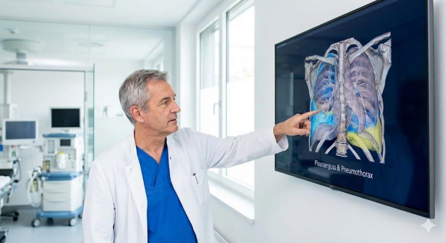
Precise Diagnosis in Thoracic Surgery: A detailed analysis of changes in the pleura is crucial for selecting the appropriate treatment.
Pleural diseases with symptoms such as shortness of breath and inflammation of the pleura
Typical symptoms—shortness of breath, chest pain, and impaired lung function
Shortness of breath is the most common symptom of pleural diseases, as the lungs can no longer expand freely. Shortness of breath occurs abruptly, particularly in cases of pleural effusion and pneumothorax. A feeling of tightness in the chest is also typical. Many patients also experience shallow breathing. The symptoms may worsen with physical exertion.
Chest pain is another key symptom, particularly in cases of pleurisy. This pain usually worsens when breathing in, coughing, or moving. In the case of a pneumothorax, the pain may be sudden and stabbing. Tumor-related changes often lead to dull, persistent discomfort. Pain in the diaphragm or chest wall may also occur.
Impaired lung function often manifests as rapid fatigue and reduced physical performance. Breathing becomes shallower and less effective. With large effusions, a weakened breath sound may sometimes be heard. Coughing may accompany these symptoms, especially in inflammatory conditions. In the long term, an untreated functional disorder can significantly impair quality of life.
Diagnosis and Treatment in Thoracic Surgery
Diagnostics in Thoracic Surgery and Pulmonology—From X-rays to Ultrasound
Diagnosis typically begins with an X-ray, which reveals pneumothorax, effusions, or tumor-like changes. A pneumothorax, for example, is indicated by a lack of lung opacity. Fluid, on the other hand, accumulates near the bottom of the chest cavity. Displacement of structures can also be detected. The X-ray is a quick and important first step.
Ultrasound allows for a more precise visualization of fluids in the pleural space. Doctors can use it to decide whether a pleural effusion should be drained. Ultrasound is also well-suited for monitoring progress. Pulmonologists use the procedure to assess the extent of the effusion. It is painless and can be performed quickly.
If the findings are unclear, CT or MRI scans are used. These reveal detailed abnormalities such as tumors, inflammatory processes, or adhesions in the pleura. Tension pneumothorax or complex effusions can also be reliably diagnosed. In some cases, a diagnostic puncture is performed to analyze the fluid. Collaboration between pulmonology and thoracic surgery is crucial in this process.
Benign changes in the pleura, recurrent pleural effusions, or unclear masses often require specialized evaluation at experienced centers such as the University Hospital of Würzburg or the University Hospital of Freiburg. There, not only is the underlying condition comprehensively diagnosed, but an interdisciplinary assessment is also conducted to determine whether conservative therapy, a puncture, or surgical intervention is necessary. The expertise in thoracic surgery and pulmonology ensures that both harmless and serious causes are reliably distinguished. This allows for early intervention to prevent further deterioration of breathing and lung function.
Treatment Options—Conservative Therapy, Drainage, Puncture, and Surgical Procedures
Conservative treatments are used for mild, benign, or inflammatory conditions. These include pain medications, anti-inflammatory drugs, and antibiotics for infectious processes. Breathing exercises can also help the lungs regain their full capacity. For smaller effusions, observation is often sufficient. Treatment is always based on the underlying condition.
A puncture is recommended when a large amount of fluid has accumulated in the pleural space. During the procedure, the fluid is removed to facilitate breathing. The procedure is performed under ultrasound guidance. In cases of recurrent effusions, a chest tube may be necessary. This continuously drains fluid from the chest cavity.
Surgical interventions are used when conservative methods are insufficient. In cases of pneumothorax, a drain is often inserted to remove air from the chest cavity. For tumors or pleural mesothelioma, thoracic surgery performs more extensive procedures. The goal may be to remove affected areas or to stabilize the lung. Minimally invasive procedures are also used.
Pleural Mesothelioma – A Malignant Disease of the Pleura
Pleural mesothelioma is a rare but aggressive form of cancer affecting the pleura. It is often caused by asbestos exposure and develops over many years. The tumor spreads extensively across the pleura. Typical symptoms include shortness of breath, chest pain, and fatigue. Treatment requires close collaboration between thoracic surgery, oncology, and pulmonology.
In early stages, surgeons attempt to surgically remove the affected sections of the pleura. In advanced cases, treatment often involves a combination of surgery, chemotherapy, and radiation therapy. Modern approaches such as immunotherapy are also used. The prognosis depends heavily on the tumor stage and extent of the disease. Regular follow-up care is absolutely essential.
A CT scan is essential for diagnosis to assess the extent of the disease. If the condition is suspected, a tissue sample is often taken. Ultrasound and X-rays complement the imaging. It is also important to take a detailed occupational history, particularly with regard to asbestos exposure. Treatment is determined on an individual basis.
An Overview of Pleural Diseases: Comparing the Pleura, Thoracic Membrane, and Pleural Lining
The pleura consists of the inner lung membrane and the outer chest membrane. Together, they form the pleura, which lines the entire chest cavity and enables the lungs to move with minimal friction. When these structures undergo pathological changes, the condition is referred to as pleural disease. This includes inflammatory processes, fluid accumulations such as a pleural effusion, or air-related problems such as a pneumothorax. Each form differs in terms of cause, course, and treatment options.
The pleura is sensitive to inflammation, changes in pressure, and mechanical stress. In tension pneumothorax, the pleura detaches from the chest wall, leading to acute shortness of breath. In a pleural effusion, fluid accumulates in the pleural space—often due to inflammation or a tumor. Tumorous changes, metastases, or pleural mesotheliomas can spread extensively throughout the pleura on one side of the chest and significantly impair chest function. The precise localization of the lesions is crucial for any treatment.
A comparison of these conditions highlights the wide range of therapeutic options, which range from conservative therapy to drainage, punctures, and surgical interventions. Pulmonology and thoracic surgery work closely together in these cases, especially when large areas of the pleura or adjacent organs are affected. Some changes are benign, while others are severe or malignant. Precise diagnosis—often using X-rays, ultrasound, or CT scans—is essential for detecting complications early and maintaining stable breathing.
Depending on the cause, pleural diseases can present in very different ways and may also affect neighboring structures. In cases of inflammatory pleural effusion or tumor-related masses, the disease can spread extensively throughout the pleura and affect surrounding organs such as the pancreas or the pericardium. The condition also becomes dangerous when accompanying complications such as a pulmonary embolism occur, which further exacerbate shortness of breath and pain. Therefore, in cases of persistent symptoms, unexplained pain in the rib or chest area, or increasing shortness of breath, prompt medical evaluation is always advisable to prevent serious complications in a timely manner.
FAQ
How dangerous are pleural diseases, and when do they require emergency treatment?
Pleural diseases can start out harmless but may take a severe course in some cases—especially if air becomes trapped in the intercostal space, creating negative pressure that compresses the trachea or blood vessels. A tension pneumothorax must be treated immediately as an emergency so that the lung can re-expand. With prompt drainage, shortness of breath usually subsides quickly. The Department of Thoracic Surgery specializes in such acute situations.
Can a pleural disease spread to other structures in the chest cavity?
Yes, certain conditions can spread along the side of the chest and, in severe cases, affect the pericardium or adjacent rib structures. Tumors or severe inflammation can be the underlying cause. Such processes must be detected early to prevent complications. Imaging and thoracic surgical diagnostics are crucial in this regard. Trained thoracic surgeons are able to initiate the appropriate measures.
What symptoms indicate a dangerous accumulation of air or fluid in the pleura?
Typical symptoms include increasing shortness of breath, stabbing chest pain, and the sensation that the lung cannot fully expand. An accumulation such as a pleural effusion or air in the pleural space compresses the lung, which exacerbates the symptoms. If shortness of breath increases rapidly, immediate medical evaluation is necessary. Treatment depends on the cause and aims to relieve pressure on the chest cavity.
Can inflammation of the pleura also affect other organs, such as the pericardium?
In rare cases, yes. Severe or infectious inflammation can spread and cause pericarditis. This usually occurs only in isolated cases when the inflammatory process is not treated in a timely manner. Symptoms such as increased pain or worsening shortness of breath should be taken seriously. Early treatment usually prevents the condition from spreading.
When should you definitely seek medical help for pleural diseases?
If shortness of breath worsens rapidly, pain intensifies, or there is suspicion of air or fluid accumulation, a doctor should be consulted immediately. In particular, if there are signs of a tension pneumothorax, emergency treatment is required to prevent further compression of the lung. Recurring symptoms also require evaluation to identify the underlying condition. Prompt action effectively prevents complications.
Share this article