Although skull base tumors are predominantly benign, they can destroy bony structures and invade the cranial nerves and blood vessels. Due to the complexity of skull base tumors, their treatment should be performed at experienced centers. With appropriate surgical planning and perioperative monitoring, these complex procedures can be performed with an acceptable level of risk.
Here you will find further information as well as a selection of specialists and centers for skull base tumors.
What is the skull base?
The base of the skull is the lower part of the cranial vault. It thus forms the boundary between the brain and the spaces containing cerebrospinal fluid. The 12 cranial nerves and blood vessels pass through the base of the skull into the facial skull or the neck.
The entire skull base can be divided into three major regions:
- anterior skull base (frontobase): Located above the nose and paranasal sinuses as well as the orbit (eye socket) and covers the anterior cranial fossa downward
- lateral skull base (laterobasis): Located above the ear and bordering the middle skull base
- portions that border the posterior cranial fossa.
Due to its proximity to the face and neck, the skull base also forms a boundary in the spectrum of medical treatment. This is where
. For this reason, interdisciplinary collaboration is often necessary to ensure optimal care for affected patients.
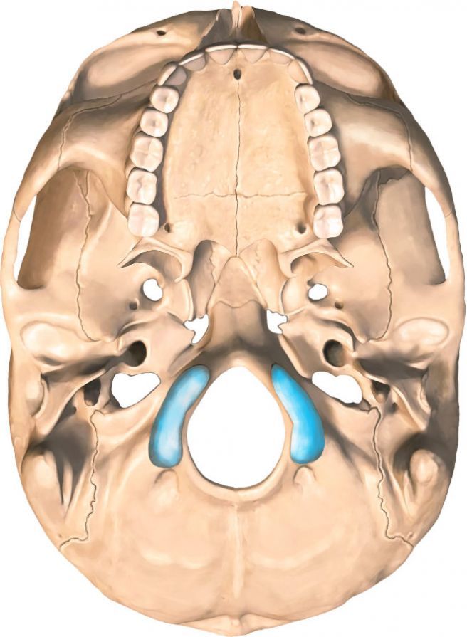
Skull base tumors and other skull base disorders
Congenital defects can manifest at the skull base. Other skull base disorders occur following accidents that result in fractures of the skull base. These can lead to the leakage of cerebrospinal fluid (cerebrospinal fluid fistula).
In addition, benign and malignant tumors as well as inflammatory conditions can develop at the skull base. Common skull base disorders include, among others,
- meningiomas,
- neurinomas,
- carcinomas,
- pituitary adenomas,
- esthesioneuroblastomas,
- chordomas,
- chondrosarcomas, and/or
- cholesteatomas.
The vast majority of these tumors are benign space-occupying lesions, such as meningiomas or neurinomas. Nevertheless, they can destroy bony structures and invade the cranial nerves and blood vessels. Skull base tumors are often not detected until they have reached a considerable size.
Symptoms of Skull Base Tumors
Patients’ symptoms depend on the tumor’s specific growth pattern and location. They are therefore also related to the tumor’s proximity to the cranial nerves passing through the skull base and to the brainstem.
Typical symptoms include (with indication of the affected nerves):
- Disturbances of smell and taste (olfactory nerve, N. olfactorius),
- Visual disturbances (optic nerve, N. opticus),
- Double vision (oculomotor nerve, trochlear nerve, and abducens nerve),
- Facial pain or abnormal sensations in the face (trigeminal nerve, N. trigeminus),
- Facial muscle paralysis (facial nerve, N. facialis),
- Hearing loss,
- numbness,
- ringing in the ears (tinnitus) (cochlear nerve, vestibular nerve),
- Dizziness,
- Unsteady gait,
- difficulty swallowing, and
- hoarseness.
Other symptoms such as
- unsteadiness,
- paralysis, and
- sensory disturbances
in certain parts of the body can be caused by pressure on the brainstem.
However, narrowing or blockage of the cerebrospinal fluid pathways (ventricles), which leads to impaired cerebrospinal fluid circulation (hydrocephalus), can also cause
- gait disturbances,
- memory problems,
- bladder dysfunction (incontinence), and
- signs of increased intracranial pressure such as headaches, nausea, and vomiting, as well as impaired consciousness
.
Large tumors that press on
- the frontal lobe or
- the temporal lobe of the brain
can also trigger personality changes and seizures.
Similarly, hormonal disorders are observed when pressure is applied to the pituitary gland (hypophysis).
Preoperative Evaluations
Before surgery to treat a skull base tumor, a series of examinations must first be conducted. This is referred to as preoperative diagnostics.
Doctors need reliable information about
- the size of the mass,
- the exact location of the lesion, and
- its extent within the skull base.
Various radiological procedures are used for preoperative diagnostics. Computed tomography (CT) can be used to assess bone involvement and destruction. Magnetic resonance imaging (MRI) allows for a better assessment of soft tissue structures as well as possible intracranial involvement and infiltrations.
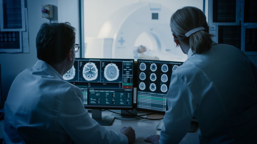
In addition, for some tumors, vascular imaging is performed using
- MR angiography or
- digital subtraction angiography
to prepare for surgery.
To minimize intraoperative blood loss, in cases of high tumor blood flow,
- selective embolization (occlusion) of the blood vessels supplying the tumor may be performed beforehand, or
- in rare cases, permanent vascular occlusion
be performed.
Furthermore, as part of the preoperative evaluations, testing of cranial nerve function—such as hearing, vision, and balance tests—is recommended. These can be supplemented by electrophysiological examinations (e.g., brainstem evoked potentials, AEP).
Treatment of Skull Base Tumors
Surgery on the skull base primarily involves the surgical removal of tumors of the anterior, middle, and posterior skull base. The surgical field extends from the orbit at the front, through the paranasal sinuses, to the foramen magnum.
Without treatment, permanent functional deficits may occur, potentially leading to damage to vital centers in the brainstem. In the case of slow-growing tumors, there is often no immediate time pressure. This allows for targeted and detailed surgical planning.
When planning treatment for skull base tumors, interdisciplinary collaboration among
- neurosurgery,
- ENT,
- oral and maxillofacial surgery, and
- occasionally ophthalmology
is advisable.
It is not always possible to completely remove skull base tumors. For this reason, radiation therapy also plays a significant role in the interdisciplinary treatment decision and follow-up therapy.
Surgery is advisable when other treatments, such as drug therapy or radiation therapy,
- are less effective,
- cause excessive side effects, or
- are not feasible.
Another indication is to confirm the diagnosis when the nature and origin of a mass are unclear.
Procedure for Skull Base Tumor Surgery
The goal of any skull base tumor surgery is to
- to eliminate the cause of existing symptoms and
- to prevent or delay the onset of new symptoms.
The choice of surgical approach and surgical method depends on the nature and location of the mass. The goal of planning is to keep the surgical approach as minimally invasive as possible.
The procedures are performed microscopically and/or endoscopically. When indicated, the surgery is computer-assisted, meaning it is performed with the aid of a navigation system. Additionally, cranial nerve function can be monitored during the operation.
Thanks to interdisciplinary collaboration and modern methods, even major surgeries at the skull base can be performed without causing permanent damage or with only minimal impairment to the patient.
Possible Complications and Risks of Skull Base Tumor Surgery
Some surgical risks can be explained by the anatomical proximity to cranial nerves, blood vessels, and/or the brainstem. If a tumor compresses or encases a cranial nerve, it can cause primary damage to that nerve.
Surgery may lead to a deterioration or complete loss of function in the affected nerve or neighboring cranial nerves. Consequently, cranial nerve deficits—ranging from
- visual disturbances ranging up to blindness,
- impaired sense of smell and taste,
- facial and eye muscle paralysis with double vision, and
- other symptoms already listed
are among the common potential complications of skull base surgery.
Since skull base tumors are often highly vascularized, bleeding and postoperative hemorrhage may occur. Furthermore, blood vessels may be invaded by tumors, which can lead not only to bleeding but also to vascular occlusions with infarcts during dissection.
In addition, in cases where the process extends beyond the dura mater, it can be difficult to reconstruct it. This can result in the leakage of cerebrospinal fluid—a so-called cerebrospinal fluid fistula—which requires further surgical treatment. There are also general risks such as infections, impaired wound healing, thrombosis, and embolisms.
Follow-up Care After Skull Base Tumor Surgery
Due to the often extensive tumor growth (e.g., into the cavernous sinus), complete surgical resection is not possible in some patients. Depending on the diagnosis and the tumor’s growth pattern, follow-up treatment is performed after surgical tumor debulking.
For skull base tumors, radiation therapy is the primary treatment. Depending on the tumor type, this may be delivered as intensity-modulated radiation therapy (IMRT) or heavy-ion therapy. In cases of carcinoma, chemotherapy and/or radiation therapy are often administered in conjunction with treatment for the underlying disease.
Share this article