Aortic valve reconstruction is a cardiac surgical procedure in which a leaky or narrowed aortic valve is repaired rather than replaced with a prosthesis. The goal is to permanently stabilize the function of the patient’s own aortic valve and maintain natural blood flow between the left ventricle and the aorta. Compared to complete aortic valve replacement, reconstruction often yields better long-term outcomes and reduces the need for lifelong anticoagulation. Depending on the findings, different techniques—such as those described by David or Yacoub—are used.
What is the aortic valve?
The aortic valve is a valve that closes off the aorta at the point where it connects to the left ventricle of the heart. It is attached to the aortic wall at the beginning of the aorta and consists of three leaflets that, when unfolded, fill the diameter of the aorta and close it off. Oxygen-rich blood leaves the heart here and is pumped through the aorta into the systemic circulation.
The heart’s pumping action forces blood against the valve, causing it to open. When the pressure subsides, the valve closes, preventing blood from flowing back into the heart.
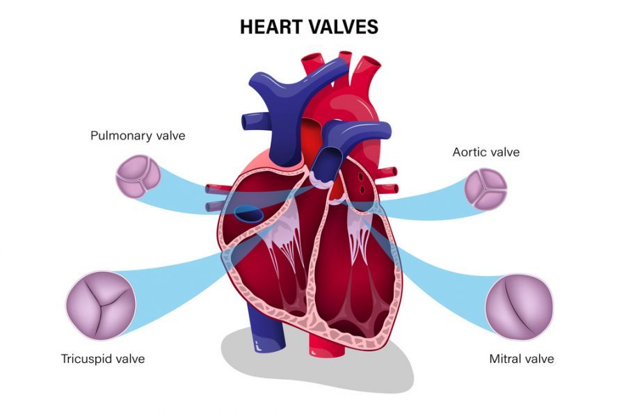
The four heart valves @ Dee-sign /AdobeStock
Aortic valve diseases
Every year in Germany, more than 12,000 people require surgery on a severely diseased aortic valve. About 20% of these surgeries are necessary because the valve has severe insufficiency, i.e., it leaks.
The heart relies on the coordinated action of the heart muscle and heart valves to function properly. The most important heart valves are those of the left side of the heart: the mitral valve (inlet valve) and the aortic valve (outlet valve).
The most common disorders affect the aortic valve. If it does not close completely, a leak occurs, and this is referred to as aortic valve insufficiency. In this case, blood flows back from the aorta into the heart.
The opposite occurs when the aortic valve does not open wide enough. It is narrowed, thereby preventing sufficient blood from entering the systemic circulation. This narrowing is called aortic valve stenosis.
It is also possible for both conditions to occur together.
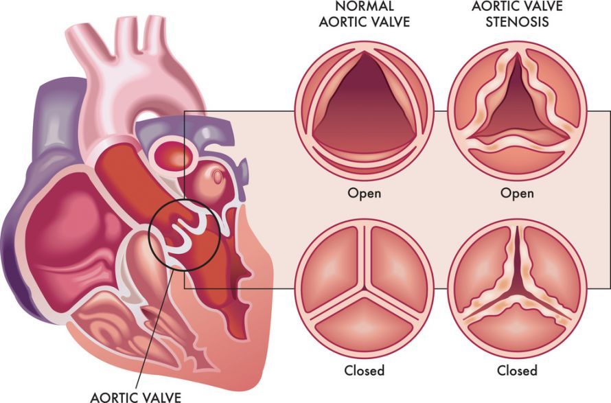
Aortic valve stenosis is the most common heart valve defect @ rob3000 /AdobeStock
In 50% of cases, this is accompanied by a dilation of the ascending aorta (aorta ascendens), which causes the leaflets of the aortic valve to no longer make sufficient contact with one another and thus to no longer close tightly. Very often, a deformation of one or more leaflets—which disrupts the valve’s geometry—is seen either as a contributing factor or as the sole cause of the insufficiency.
Causes and Symptoms of Aortic Valve Insufficiency
Various causes can lead to aortic valve insufficiency (leakage). In people under the age of 50, the most common cause is a congenital malformation of the aortic valve, in which the valve consists of only 2 leaflets instead of 3. This can lead to deformation of the valve, which then becomes leaky over the course of years or decades.
In people over the age of 50, deformities of the leaflets due to connective tissue degeneration are the primary cause; this is often accompanied by or caused by an enlargement of the aorta (the main artery). Shrinkage of the valve—for example, as a result of rheumatic fever—is rare today.
The primary symptom of aortic valve insufficiency is shortness of breath, which initially occurs during exertion and later also at rest. Since people tend to adapt to gradual changes, the shortness of breath—or the resulting decline in physical performance—is often first noticed by others in the patient’s immediate environment.
Increasing fatigue as a result of physical work is occasionally experienced. Chest pain attributed to the heart is less common. Some people experience no symptoms despite significant strain on the heart, and in these cases, only echocardiography (heart ultrasound) clearly reveals the extent and significance of the valve damage.
In most affected individuals, aortic valve insufficiency is tolerated over a long period of time. The left ventricle (heart chamber) becomes more muscular and enlarges to compensate for the backflow of blood.
After years, changes then develop that indicate the heart’s reserves are running low. This often means that the patient or someone close to them notices that physical performance is slowly declining or that shortness of breath occurs more frequently during the same level of exertion.
In other cases, even in the absence of self-reported symptoms, echocardiography reveals that the left ventricle has reduced contractile strength or has become markedly enlarged. All of these changes indicate that the valve defect is no longer harmless; rather, there is now a risk of heart failure, and life expectancy is reduced.
At this point, surgery on the diseased aortic valve becomes necessary. The surgery should be performed by an experienced cardiac surgeon.
Goals of Aortic Valve Reconstruction
The goal of the surgery is to restore the aortic valve to its normal form and function, either completely or at least to a large extent. This normalizes heart function; patients almost always notice an improvement in their physical performance and a reduction or elimination of symptoms.
Compared to aortic valve replacement, these goals can be achieved with a lower likelihood of long-term side effects.
The David or Yacoub Procedures
Two main cardiac surgery procedures are used in aortic valve reconstruction: the David procedure and the Yacoub procedure. Both aim to preserve the function of the patient’s own aortic valve and ensure its long-term stability. The difference lies in the way the aortic root and sinus are reconstructed. Both methods are considered established alternatives to aortic valve replacement in cases of aortic valve insufficiency or aortic stenosis.
| Characteristics | David procedure | Yacoub procedure |
|---|---|---|
| Objective | Preservation and stabilization of the patient’s own aortic valve within a conduit | Restoration of the natural aortic root and sinus structure |
| Procedure | The aortic valve is secured within a vascular prosthesis to stabilize the valve annulus | All portions of the aortic wall, including the sinuses, are replaced; formed sinuses are implanted into the vascular prosthesis |
| Advantage | Excellent stability and low reoperation rate | Natural mobility of the aortic root is preserved |
| Disadvantage | Technically demanding; slightly stiffer valve apparatus | Slightly higher risk of recurrent valve leakage |
| Anticoagulation | No lifelong anticoagulation required | No lifelong anticoagulation required |
| Typical application | For a morphologically intact aortic valve and dilated aortic root | For an intact valve with an aortic wall structure worth preserving |
According to recent long-term studies, such as those published in the Ann Thorac Surg, both procedures yield excellent results and allow for the long-term preservation of the patient’s own aortic valve without the need for lifelong anticoagulation. The choice of the appropriate procedure depends on the patient’s individual anatomy, the structure of the aortic wall, and the experience of the cardiac center.
Information Prior to Aortic Valve Reconstruction
The preoperative consultation covers all significant advantages and disadvantages, including potential risks specific to the individual patient. Even if reconstruction is highly likely to be feasible, unexpected issues may arise during surgery that require a new decision.
It is therefore important to discuss one of the possible replacement procedures as a backup plan in addition to the planned aortic valve reconstruction.
Procedure for Aortic Valve Reconstruction
The Surgery in Detail: The goal of the surgery is to restore the shape and function of the patient’s own aortic valve. This must be done with precision to bring the deformed aortic valve as close as possible to its normal shape and, consequently, its normal function.
For the surgery, the chest is opened through the sternum, and after anticoagulation, the patient is connected to a heart-lung machine. The heart is stopped by injecting a cold solution.
For valve reconstruction, the aorta and aortic valve must first be stretched so that they resemble normal conditions as closely as possible.
There are now standard measurements for various dimensions of the aortic valve. These are measured to determine an individualized plan for restoring normal function. In many cases, this plan involves correcting the overstretching of a pocket in the aortic valve by means of “sutures.” Appropriate surgical procedures exist for other problems as well.
After the body has warmed up again and blood flow to the heart has resumed, the function of the heart and the reconstructed aortic valve is carefully assessed using transesophageal echocardiography (TEE).
This is followed by weaning from the heart-lung machine. Blood clotting is normalized, wound drains are inserted, and the chest is closed.
Preoperative Evaluations for Aortic Valve Reconstruction
Echocardiography can objectively confirm the presence of aortic valve insufficiency. The most accurate information can be obtained via transesophageal echocardiography (TEE). If there is dilation of the aorta, this can be precisely documented using computed tomography (CT) or magnetic resonance imaging (MRI).
If there is an increased risk of coronary artery narrowing due to age, family history, or specific risk factors, a cardiac catheterization may be necessary.
Alternatives to Aortic Valve Repair
A leaky aortic valve can currently only be treated with surgery. Large studies have shown that medication offers no benefits in cases of severe regurgitation.
In principle, there are various alternative surgical procedures, all of which have specific advantages and disadvantages, and their use in individual patients must be carefully weighed.
Aortic valve replacement is the standard treatment for advanced aortic regurgitation. Both mechanical and biological prostheses are safe. Mechanical prostheses can lead to clot formation on the valve, requiring lifelong use of anticoagulants. Nevertheless, there remains a small but real risk of clotting and bleeding complications.
Biological prostheses have only a low tendency to form clots, so anticoagulants are required for only 3 months. However, they wear out over time, and this wear progresses more rapidly the younger the patient is.
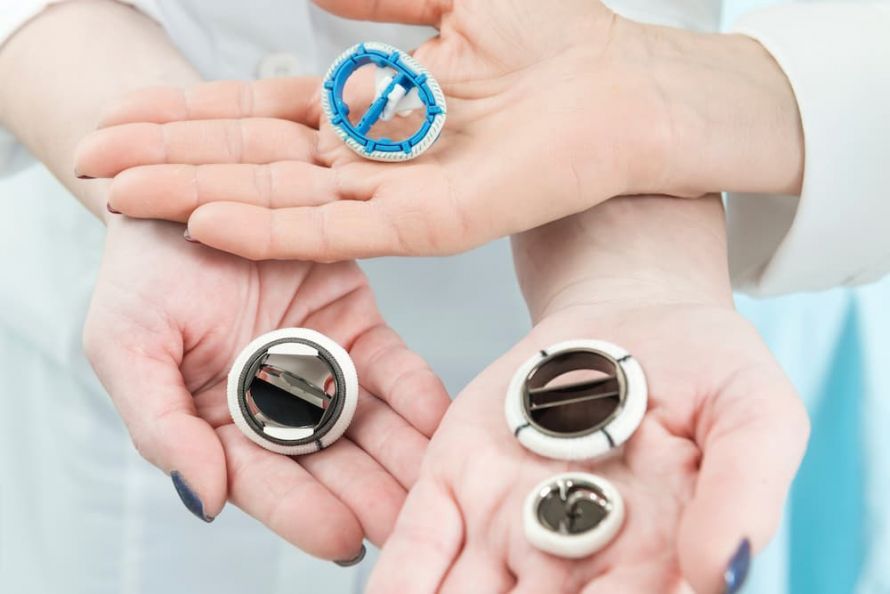
Mechanical heart valves @ pirke /AdobeStock
In younger patients, surgeons sometimes take advantage of the fact that the pulmonary valve is very similar in shape to the aortic valve. The pulmonary valve is then removed from its original location and implanted into the aorta (Ross procedure).
A biological valve is implanted in place of the original pulmonary valve, as it is subjected to less stress than in the aorta. The long-term outcome of this procedure is good, and anticoagulant therapy is not necessary. However, the procedure involves two heart valves, even though initially only one is diseased.
All procedures carry specific risks, including the possible need for repeat surgery.
Over the past 5 years, new procedures have been developed that allow a heart valve to be implanted using catheter technology to replace the aortic valve. These procedures are currently only suitable for selected patients.
Complications and Risks Associated with Aortic Valve Reconstruction
Overall, the risk associated with aortic valve reconstruction is low. As with any heart surgery, unwanted complications—such as bleeding, cardiac arrhythmias, or wound healing problems—may occur. The most significant complication is the recurrence of aortic valve regurgitation, which, depending on its severity, may require another surgery.
The individual risk associated with the surgery depends largely on age, the presence of other medical conditions, and pre-existing damage to the left ventricle. The surgeon is best able to explain this during a personal consultation.
Postoperative Care Following Aortic Valve Reconstruction
The surgery is followed by a stay in the intensive care unit, where circulation is carefully monitored, especially during the first few hours after surgery. Once circulation is stable and oxygen uptake by the lungs is normal, the endotracheal tube can be removed. Patients are often able to be transferred to a general ward within 1 to 2 days. This is followed by a gradual recovery.
The patient can speed up their recovery by getting out of bed more often and walking deliberately—that is, exercising the body in moderation—as early as the first few days after surgery. Climbing stairs is often possible as early as 5 or 6 days later.
Among the various medical procedures performed is echocardiography, which is used to monitor the function of the aortic valve even several days later in order to objectively assess the success of the treatment.
The 6- to 10-day hospital stay is followed by rehabilitation. Full recovery can be expected after about 8 weeks.
Medication After Aortic Valve Reconstruction
Medications taken prior to surgery are usually continued both before and after the procedure. After surgery, the treating surgeon will determine whether the medications need to be adjusted to account for the postoperative condition. Following reconstruction, taking aspirin for 2 months is often recommended.
Findings on aortic valve reconstruction
Traditional aortic valve replacement is, in principle, a safe procedure; however, it is associated with the long-term drawbacks mentioned above. Reconstruction is not always feasible for a leaky aortic valve. If it is possible and the shape of the aortic valve is correctly restored, fewer problems are expected in the long term.
The risk of blood clot formation is negligible, and taking blood-thinning medications is usually not necessary. The susceptibility to bacterial infections of the valve (endocarditis) is significantly lower than after mechanical or biological replacement.
In principle, however, aortic valve reconstruction is a procedure for which experience is limited to the last 10 to 15 years, so no data can yet be provided regarding a 20- or 30-year follow-up period.
Conclusion on Aortic Valve Reconstruction
Aortic valve insufficiency is one of several pathological changes affecting the aortic valve and is often characterized by shortness of breath during exertion or a decline in physical performance.
Accurate diagnosis and careful evaluation are essential for planning the best possible treatment.
Traditionally, the valve is replaced with a mechanical or biological prosthesis. Aortic valve reconstruction is an effective treatment for this condition. Compared to valve replacement, it carries the lowest risk of valve-related complications.
FAQs on Aortic Valve Reconstruction
What is the goal of aortic valve reconstruction?
The goal of aortic valve reconstruction is to restore normal function to the patient’s own aortic valve. During the procedure, the valve is surgically stabilized to prevent blood from flowing back into the left ventricle. Reconstruction allows the morphologically intact portions of the valve and annulus to be preserved, leading to better long-term outcomes than complete valve replacement.
How is the David or Yacoub procedure performed?
In both procedures—the David procedure and the Yacoub procedure—the aortic root is opened to reconstruct all parts of the aortic wall, including the sinuses. In the David procedure, the patient’s own aortic valve is secured within a vascular or tubular prosthesis (conduit), thereby stabilizing it. The Yacoub procedure, on the other hand, preserves the natural sinuses by implanting formed sinuses into the vascular prosthesis. Both procedures require the highest precision in cardiac surgery.
When is aortic valve reconstruction possible?
Reconstruction is particularly indicated when the valve is morphologically intact and the valve’s inability to close properly or an enlargement of the valve annulus can be surgically corrected. If there is concomitant dilation of the aortic root or changes in the coronary artery origins, the surgeon can address these issues during the procedure.
What risks and complications are possible?
As with any heart surgery, bleeding, arrhythmias, or wound healing complications may occur. Recurrent regurgitation may require a follow-up operation if the tissue structure is unfavorable. The individual risk depends on the condition of the aortic valve, the patient’s age, and any comorbidities. However, if the valve is implanted within a stabilizing tubular prosthesis, the long-term outcomes are usually very good.
Is anticoagulation necessary after the surgery?
Following a successful reconstruction of the patient’s own aortic valve, lifelong anticoagulation is generally not necessary. Anticoagulation medication may be used for a short period until the reconstructed aortic valve is fully stabilized. This distinguishes reconstruction significantly from heart valve replacement, in which anticoagulation remains necessary permanently.
What are the long-term outcomes after aortic valve reconstruction?
Studies and case reports to date from leading cardiac centers—including those published in Ann Thorac Surg—show very good long-term outcomes. Patients benefit from the preservation of their own aortic valve, physiological valve function, and a low risk of infection. Compared to prosthetic valves, stable results are observed over many years.

About the medical author
Prof. Hans-Joachim Schaefers
Medical Author · Director
Univ. Prof. Dr. med. Hans-Joachim Schäfers – Author: Discover expert medical articles at Leading Medicine Guide.