A pituitary adenoma is a tumor of the pituitary gland—also known as the hypophysis—that is usually benign. Pituitary adenomas originate in the adenohypophysis and are among the most common types of endocrine tumors in the region of the sella turcica. Pituitary adenomas are benign tumors, but their location can cause clinically significant symptoms. Depending on hormone production, they are classified as hormonally active or hormonally inactive adenomas.
A hormone-active pituitary adenoma can increase the secretion of hormones such as prolactin, ACTH, TSH, or growth hormone. Other pituitary adenomas remain hormone-inactive and are only noticed due to their size or pressure on surrounding tissue. When a pituitary tumor grows and compresses the optic chiasm or the optic nerves, headaches and visual disturbances often occur. Today, the condition is usually diagnosed using imaging techniques such as MRI (magnetic resonance imaging).
Definition: What are pituitary adenomas?
The pituitary gland is a gland about the size of a cherry and consists of
- an anterior lobe (adenohypophysis) and
- a posterior lobe (neurohypophysis).
The posterior lobe stores hormones from the hypothalamus, a nearby part of the brain. It releases these hormones as needed.
The anterior lobe of the pituitary gland, on the other hand, produces hormones that are important for the body, such as
- growth hormone,
- follicle-stimulating hormone (FSH),
- prolactin,
- thyrotropin (TSH), and
- adrenocorticotropic hormone (ACTH).
Pituitary adenomas are benign forms of pituitary tumors, i.e., tumors of the pituitary gland. About 10–15% of all intracranial tumors (tumors inside the skull) are pituitary tumors.
In terms of their benign or malignant nature, pituitary tumors can be classified into
- typically benign adenomas (approx. 85% of cases)—benign,
- more aggressive atypical adenomas (approx. 15%)—benign but problematic, and
- pituitary carcinomas (approx. 0.1%)—malignant
.
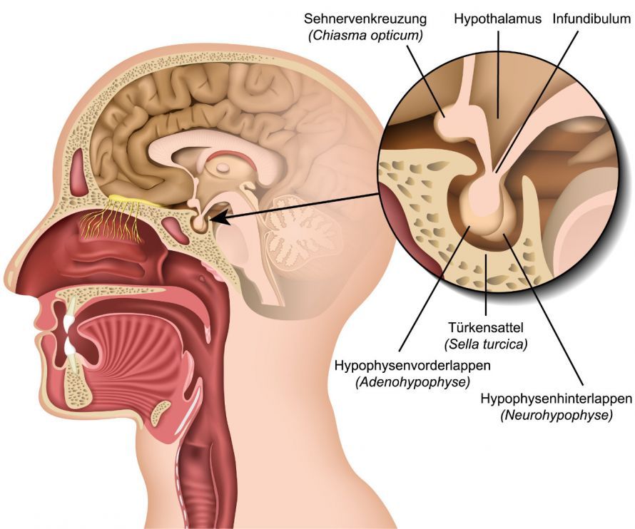
Pituitary adenomas, abbreviated as H-adenomas, grow in the sella turcica. This is a bony cavity near the point where the two optic nerves cross.
Doctors classify pituitary adenomas into different types based on two characteristics:
- Their size: Microadenomas are smaller than 1 cm; macroadenomas are larger than 1 cm.
- Their hormonal activity: hormonally active (produces hormones) or hormonally inactive (does not produce hormones).
Among the hormonally active adenomas are, among others,
- prolactinomas (which produce the hormone prolactin, accounting for approximately 50% of all adenomas),
- growth hormone-producing adenomas (about 22%),
- ACTH-producing adenomas (about 5%), and
- TSH- and FSH-producing adenomas (less than 1%)
. Depending on the hormone produced, these are then classified, for example, as
- somatotropinomas,
- corticotropinoma, or
- thyrotropinomas
. About 23% of adenomas are hormonally inactive.
A pituitary carcinoma usually develops from a prolactinoma. The more aggressive atypical adenomas grow into the surrounding brain tissue. Once this happens, they can no longer be completely removed.
How does a pituitary adenoma develop?
Changes in the genetic material of a pituitary cell can lead to uncontrolled cell division. This results in the development of a mass, the pituitary tumor. The exact mechanisms are still unclear.
In some patients, the pituitary adenoma occurs in association with multiple endocrine neoplasia (MEN-1 syndrome). This is an inherited endocrine disorder.
Symptoms and Diagnosis of Pituitary Tumors
The signs of a pituitary tumor depend on hormonal activity and, if present, which hormones are involved. Depending on whether there is an excess or a deficiency of hormones, the symptoms can vary greatly.
For example, prolactinomas in women can cause
- menstrual irregularities,
- infertility, and
- in non-pregnant women, milk discharge from the breasts
. In men, it can
- lead to the development of female-like breasts (gynecomastia),
- libido disorders, and
- erectile dysfunction
.
Growth hormone-producing adenomas lead to gigantism (excessive growth) even before puberty. In adults, this results in changes
- in physical appearance,
- metabolism, and
- in internal organs (a condition known as acromegaly).
Patients may experience a wide range of symptoms. The most common are
- headaches,
- menstrual irregularities,
- impotence,
- visual disturbances,
- excessive sweating,
- loss of libido,
- Sleep disorders,
- carpal tunnel syndrome, and
- joint disorders.
In the case of ACTH-producing adenomas, the excess ACTH leads to Cushing’s disease. Typical symptoms include, for example,
- moon face and buffalo hump,
- trunk-predominant obesity,
- gonadal dysfunction,
- osteoporosis,
- muscle weakness,
- edema,
- acne,
- diabetes, and
- high blood pressure.
As they grow, non-hormone-secreting pituitary tumors displace surrounding hormonal or nervous tissue. This can lead to hypopituitarism as well as visual and visual field impairments.
Depending on which hormone is being produced in insufficient quantities, this can result in
- in growth (e.g., short stature, lipid metabolism disorders, reduced physical performance),
- of the gonads (e.g., menstrual irregularities, infertility, erectile dysfunction, small, soft testicles, abnormal underarm and pubic hair growth),
- thyroid function (e.g., weight gain, fatigue, lethargy, personality changes), or
- the adrenal cortex (e.g., pale skin, weakness, fatigue, weight loss, nausea, hypotension)
.
How is a pituitary adenoma diagnosed?
Since pituitary adenomas grow very slowly, the first signs of a pituitary tumor often do not appear until many years later.
The diagnosis is based on
- a discussion with the patient about their symptoms and medical history (anamnesis),
- measurement of hormone levels in urine, saliva, and blood by an endocrinologist,
- imaging techniques (particularly magnetic resonance imaging, MRI), and
- an ophthalmological examination.
The pathologically altered tissue is clearly visible on an MRI. The radiologist can see exactly how large the pituitary adenoma is, where it is located, and whether it contains calcifications.
Saliva, blood, and urine tests reveal which hormone is present in excess or in insufficient quantities.
A corticotropinoma can be detected using the dexamethasone test. Dexamethasone is a corticosteroid medication often used to reduce brain swelling.
An ophthalmological examination is also recommended, particularly visual field testing or computer-assisted perimetry. This helps determine whether the adenoma is pressing on or invading structures of the visual pathway. This would manifest as a restricted visual field.
How is a pituitary tumor treated?
Treatment of a pituitary adenoma depends on
- the symptoms,
- the effects on hormonal metabolism, and
- the tumor’s hormonal activity.
Some pituitary adenomas do not cause any symptoms. In such cases, it is usually sufficient to monitor the tumor’s growth at regular intervals using imaging techniques.
Treatment is only initiated if the tumor causes symptoms in the patient.
Some types of pituitary adenomas generally do not require surgery: prolactinomas, for example, are typically treated with medication (dopamine agonists). In certain cases, however, surgical removal is also considered for prolactinomas. Conditions for this include, for example, when
- drug therapy is ineffective,
- the patient cannot tolerate the medication,
- vision loss continues to progress despite treatment, or
- the patient wishes to have children.
The treatment of choice for growth hormone- and ACTH-producing adenomas is pituitary surgery. The prognosis is very good.
Since microadenomas are very small, they can be removed through the nose. After pituitary adenoma surgery, no visible scar remains on the head. In 9 out of 10 cases, these small tumors can be removed without leaving any residual tissue.
For macroadenomas, an opening in the skull is required.
Pituitary tumors are removed by a neurosurgeon. Invasively growing adenomas cannot be completely removed surgically. In such cases, the patient often has to take hormones for the rest of their life.
Radiation therapy is used very rarely for these tumors, such as in cases of very large H-adenomas. In such cases, doctors attempt to shrink the tumor with radiation therapy prior to pituitary adenoma surgery.
If hormonal imbalances are too severe, the patient is treated with medication prior to surgery.
The tumor-induced hypopituitarism must be compensated for before and after surgery by administering hormones. This is referred to as hormone replacement therapy. Depending on the specific hormone deficiency, this may involve taking
- hydrocortisone,
- L-thyroxine,
- sex hormones (such as androgens, estrogens),
- growth hormones, or
- desmopressin
.
FAQ
What symptoms does a pituitary adenoma cause?
The symptoms of a pituitary adenoma depend on its size and hormonal activity. A hormonally active pituitary adenoma can, for example, trigger typical symptoms such as acromegaly, Cushing’s disease, or hyperthyroidism due to prolactin, ACTH, or GH. As the tumor grows, headaches and visual disturbances may occur because the optic chiasm is affected.
Is a pituitary adenoma benign?
Pituitary adenomas are benign tumors and are not classified as malignant brain tumors. Nevertheless, a pituitary tumor can cause significant complications due to its location in the sella turcica. In rare cases, a macroadenoma may grow toward the internal carotid artery or adjacent brain structures.
How is a pituitary adenoma diagnosed?
If a pituitary adenoma is suspected, comprehensive diagnostic testing and treatment planning are conducted in the endocrinology department. In addition to hormone tests for TSH, ACTH, prolactin, or ADH, MRI and, if necessary, CT scans are used. Magnetic resonance imaging (MRI) reveals microadenomas and macroadenomas, as well as any possible enlargement of the sella turcica.
How is a pituitary tumor treated?
The treatment of pituitary adenomas depends on the type of adenoma. Prolactinomas and other prolactin-secreting adenomas are usually treated with dopamine agonists. For hormone-inactive adenomas or cases involving compression of the optic nerves, surgical treatment—often transsphenoidal via the skull base—is necessary; in rare cases, a craniotomy is performed. Radiation therapy may also be required as an adjunct.
What is the prognosis for a pituitary adenoma?
The prognosis for a pituitary adenoma is good in most cases. A pituitary adenoma is a benign tumor that is easily treatable if diagnosed early. Key factors include the tumor’s size, hormonal activity, and whether the pituitary adenoma is growing or was diagnosed early. However, long-term endocrinological monitoring remains necessary.
Share this article

About the medical author
Dr. Claus Puhlmann
Medical Author · Medical journalist
View full expert profile →Sources
- Petersenn, S. (2018) Hypophysenadenome – nicht immer benigner Natur. Der Onkologe 24: 133. https://link.springer.com/article/10.1007/s00761-017-0319-2?error=cookies_not_supported&code=6e2e99e7-3864-4806-a641-b26cf4e36119
- Stalla G., Dimopoulou C. (2014) Hypophysentumore. In: Lehnert H. et al. (Hrsg) SpringerReference Innere Medizin. Springer, Heidelberg