Skip to content
Leading Medicine Guide logo

Treatment · Spinal Surgery

Neuronavigation: Information & Neuronavigation Specialists

Here you will find selected medical experts and specialists in clinics and medical practices for the diagnosis, treatment, surgery and rehabilitation in the medical field Neuronavigation. All listed physicians are specialists in their field and have been carefully selected for you according to strict guidelines.

Author of this articleLeading Medicine Guide editorial team

Neuronavigation is a computer-assisted, software-controlled procedure used in neurosurgery. It is primarily used during surgeries on the brain or spine. The goal of neuronavigation is to ensure better surgical planning and improved orientation during the procedure.

Here you will find further information as well as a selection of specialists in neuronavigation.

Definition: What is neuronavigation?

Neuronavigation is a computer-assisted procedure in neurosurgery. This means that computers assist in the procedure to support the surgeon and minimize errors and complications.

In a broader sense, computer-assisted procedures are part of the instrumental techniques used in intraoperative diagnostics. This refers to diagnostic methods employed during a surgical procedure. These techniques have become an indispensable part of everyday neurosurgical practice.

Neuronavigation allows the surgeon to see “more” than just the surgical site directly in front of them. On a monitor, they can view three-dimensional images of tissue, nerves, and other structures. The computer also displays the current position of the microsurgical instruments being used. This allows the surgeon to orient themselves precisely.

Synonyms for the term “neuronavigation” include

  • frame-less stereotaxy,
  • interactive image-guided navigation, and
  • computer-assisted surgery in general.

The first description of intraoperative navigation dates back to 1986 and was provided by the American neurosurgeon D. Roberts. Almost simultaneously, similar systems were developed in Europe (Mösges, Reinhardt) and in Japan (Watanabe).

In recent years, the technology has continued to evolve. Neuronavigation is no longer used exclusively in brain tumor surgery. It is increasingly being applied to other conditions such as

The range of applications and clinical indications for neuronavigation are the same for all systems. The potential sources of error and the limitations of the method are also independent of the specific components selected.

The clinical purpose of neuronavigation is

  • preparation for surgery (image data acquisition, preparation, and planning),
  • primary intraoperative correlation (registration), and
  • the actual application during the procedure (implementation of the access plan, intraoperative orientation).

Advantages of Neuronavigation

The spectrum of intraoperative diagnostics ranges from neuronavigation to intraoperative imaging and intraoperative neurophysiological monitoring. All of these methods improve surgical procedures. Their goal is to

  • make the procedure more efficient by providing the surgeon with additional information, e.g., through improved tumor resection.
  • reduce the risk of complications during surgery. As a result, fewer neurological disorders occur.

Neuronavigation achieves this by providing enhanced spatial orientation capabilities during the surgical procedure. Neurophysiological monitoring provides more functional data from the areas of the brain being operated on.

Technical Foundations of Neuronavigation

Neuronavigation is based on stereotaxy. This is a procedure for the spatially precise, targeted guidance of radiation therapy or surgical procedures. Compared to stereotaxy, neuronavigation lacks

  • the so-called frame of the stereotactic device,
  • the stereotactic ring, and
  • the associated target bracket system.

Both methods are based on the principle of precisely localizing the structures to be treated on a patient’s CT or MRI images within the surgical field. To do this, it is necessary to align the two coordinate systems—that of the patient and that of the image data—with one another. This means the system must know which point on the image corresponds to which point in reality. This process is called registration (see below).
Neuronavigation1
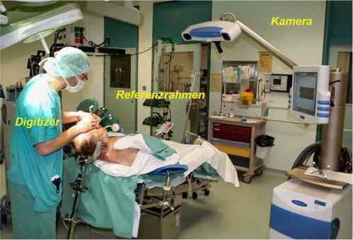
Figure 1: Performing preoperative registration to correlate the real neuroanatomy with the virtual image data (MRI): The patient’s head is secured in the head mount. The reference frame is detected by the camera. A digitizer is used to perform the preoperative and intraoperative alignment of the two coordinate systems.

Neuronavigation operates without a frame. A three-dimensional digitizer serves as the link between the patient’s anatomical reference structures and the image data. This digitizer is used to define individual points within the workspace. For example, the position of an instrument tip requires x, y, and z coordinates. In this way, the surgical field and the three-dimensional image of the patient are digitized.

In intraoperative neuronavigation, the patient’s image data is available as three-dimensional data sets. Individual points—for example, inside the skull—are precisely defined along the x, y, and z axes. Increasingly powerful computer technology can process and display the data from the digitizer and the enormous volumes of image data more quickly.

Components of the navigation system in neuronavigation

The individual system components of navigation units used in the neuronavigation procedure are largely similar:

  • navigation computer and workstation containing the patient data,
  • monitors for intraoperative visualization,
  • the fixed reference frame at the surgical site, and
  • the digitizer, whose spatial location is tracked and which correlates image data with the patient.

During neuronavigation, the system continuously evaluates the spatial position and coordinates of the digitizer. It transmits the processed data so that the digitizer’s location can be displayed on the image data at all times. This makes it possible to determine the position and display the location of the digitizer continuously throughout the entire operation. The digitizer can also be defined intraoperatively via the microscope’s focal point.

Various systems serve as digitizers in neuronavigation. Optical systems are currently established as a sort of standard in neurosurgery. Infrared or visible light, which is detected by cameras, is used to track the digitizer or the surgical microscope.

Magnetic sensor systems are currently experiencing a resurgence. They use the distortion of a magnetic field emitted by the system to determine position. For a time, they were hardly used at all.

Image Data Acquisition, Preparation, and Planning for Neuronavigation

The choice of method used for neuronavigation ultimately depends on the nature of the process to be visualized.

For example, a bone-related process is best visualized using a CT scan. This is particularly true for spinal neuronavigation. For most indications in tumor surgery, however, MRI plays a more significant role. Functional data, such as the mapping of speech functions, can be more easily implemented here (so-called matching; see also Figure 3).

Regardless of the imaging modality chosen, physicians place markers in the surgical area prior to the procedure. These markers are used for subsequent registration. In addition, a volumetric dataset of the surgical area—such as the skull—is created. A volumetric dataset consists of multiple images taken at different heights within the skull, which can later be assembled into a three-dimensional model.

The datasets are then transferred to the navigation system. There, in preparation for neuronavigation, registration is performed using the markers, and a 3D image is reconstructed.

Afterward, the surgeons can plan the surgical approaches and finally define the tumor boundaries in the dataset.

System Registration and Intraoperative Use of Neuronavigation

At the start of the surgery, the patient is positioned and their head is secured. The reference frame is typically attached directly to the headrest (Figure 1). This ensures that it remains correctly aligned with the patient’s head even as the operating table moves.

Next, the dataset is registered in relation to the patient. Once both coordinate systems have been aligned using the digitizer, neuronavigation can be initiated.

The basis of registration in neuronavigation is the correlation of identical points in both coordinate systems. This is achieved either by

  • the use of the previously applied markers or
  • digitizing the skin surface and correlating it with its reconstruction from the image data.

The accuracy of the registration is critically dependent on the intraoperative deviation, which then allows for precise localization of the target area during neuronavigation.

Once registration is complete, the navigation system is ready for use.

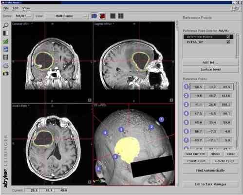
The surgeon can plan the approach and the size and location of the cranial opening. This is important for minimizing surgical trauma (Figure 2).

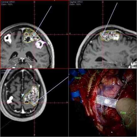
During this phase of the procedure, the available image data is typically displayed through the microscope’s focal point. The microscope then functions as a digitizer (Figure 3). This allows normal structures to be distinguished from pathological ones under microscopic visualization. Once the imaging signals from the tumor margins are detected, the progress of the surgery can be monitored using neuronavigation.
Neuronavigation2
Figure 2: Screenshot of a Stryker-Leibinger navigation system in the three spatial axes—sagittal, coronal, and axial: The brain tumor with a large necrotic component is highlighted in yellow. The crosshairs indicate the shortest access route to the center of the lesion. In the 3D view, the tumor contours are projected onto the surface of the skull.
Neuronavigation3
Figure 3: Intraoperative navigation with a surgical microscope, showing the course of a sarcomatous tumor in the axes of 3D space: Additionally, functional MRI image data is overlaid, allowing for the integration of functionally important areas related to the hand and foot regions. In addition, electrophysiological monitoring—specifically, motor cortex stimulation (using a patch electrode)—is performed to reliably define and preserve functionally important areas.

Limitations and Sources of Error in Neuronavigation

Despite numerous innovations, applications such as neuronavigation cannot replace the operating surgeon’s knowledge of neuroanatomy.

The neurosurgeon alone bears responsibility for the plausibility of the navigation data and for any erroneous information during neuronavigation. Known sources of error include

  • insufficient or incorrectly placed markers,
  • incorrect import of image data (side convention!),
  • failure to fix the reference frame, and
  • incorrect registration (e.g., displacement of markers).

The most significant weakness of neuronavigation is that the surgeon alters the anatomical structures during the procedure. Neuronavigation, however, continues to use the images created beforehand. In other words, the image on the monitor shows an outdated view from before the operation.

This error, commonly referred to as “brain shift,” can be significant. It can only be corrected through the use of intraoperative imaging, such as MRI or ultrasound.

Clinical Significance of Neuronavigation

Despite the widespread use of neuronavigation in neurosurgery, there is no evidence-based data to suggest that it is absolutely necessary.

The research group led by C.R. Wirtz (Wirtz) conducted a comparative study on the effectiveness of tumor resections with and without the use of neuronavigation. In this study, he demonstrated an increase in the extent of resection without being able to demonstrate a significantly better outcome.

Only for deep-seated lesions is there broad consensus that neuronavigation should be used. As before, there are still no legal consequences resulting from the failure to use neuronavigation during neurosurgical procedures. The surgeon’s liability also remains unchanged.

Sources
  • Jakola AS, Unsgård G, Solheim O: Quality of life in patients with intracranial gliomas: the impact of modern image-guided surgery. Neurosurg, 2011, Feb 11. [Epub ahead of print]
  • Mösges R, Schlöndorff G: A new imaging method for intraoperativen therapy control in skull base surgery. Neurosurg Rev 11, 1988, 245-247.
  • Raabe A, Krishnan R, Wolff R et al.: Laser surface scanning for patient registration in intracranial image-guided surgery. Neurosurgery 50, 2002, 797-801.
  • Reinhardt H, Meyer H, Amrein E: A computer-assisted device for the intraoperative Ct-correlated localization of brain tumors. Eur Surg Res 20, 1988, 51-58.
  • Roberts DW, Strobehn JW, Hatch JF et al.: A frameless stereotactic integration of computerized tomographic imaging and the operating microscope. J Neurosurg 65, 1986, 545-549.
  • Watanabe E, Watanabe T, Manaka S et al.: Three-dimensional digitizer (Neuronavigator): new equipment for computed tomography-guided stereotactic surgery. Surg. Neurol 27, 1987, 543-547.
  • Wirtz CR, Albert FK, Schwaderer M et al.: The benefit of neuronavigation for neurosurgery analyzed by its impacts on glioblastoma surgery. Neurol Res 22, 2000, 354-360.

Verified expertise

Recommended specialists

29 specialists

1 / 3

Medical spectrum

Related medical topics