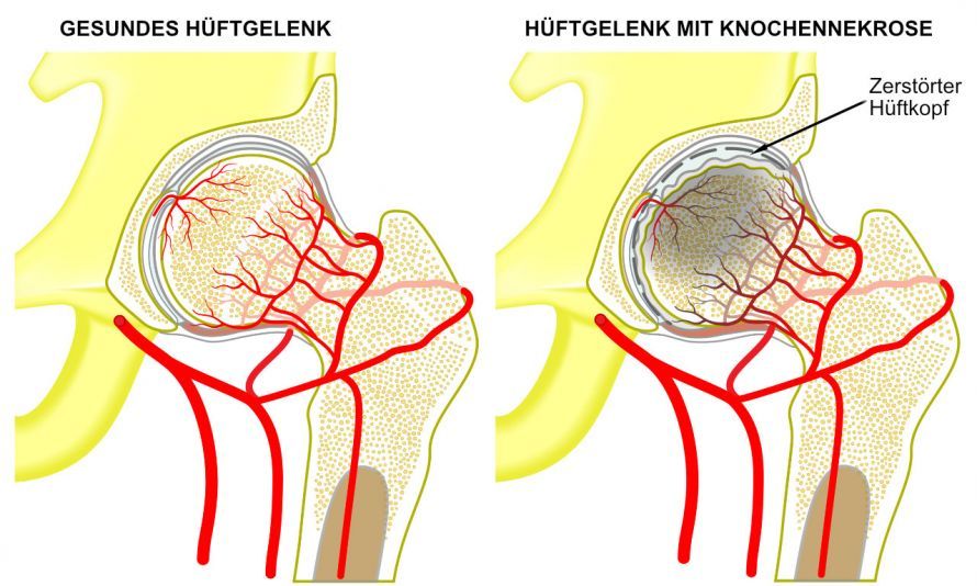
Bone necrosis, also known as osteonecrosis or avascular necrosis, is a condition in which bone tissue dies due to a disrupted blood supply. Joint structures such as the hip, knee, or foot are particularly commonly affected. Aseptic bone necrosis occurs without a bacterial infection and is one of the most common forms of osteonecrosis.
Typical symptoms include pain, limited mobility, and discomfort in the affected joint. In the early stages, an MRI often already reveals changes, while an X-ray may initially appear normal. Conditions such as Ahlbäck’s disease or Köhler’s disease I and II are specific forms of bone necrosis.
If left untreated, osteonecrosis can progress and lead to osteoarthritis or the need for joint replacement. Treatment for osteonecrosis may be conservative or surgical and depends on the stage, location, and course of the disease.
Causes of Osteonecrosis
Depending on the cause, a distinction is made between septic and aseptic osteonecrosis.
Septic osteonecrosis develops as part of osteomyelitis, which is a bacterial infection of the cortical bone and bone marrow.
Aseptic osteonecrosis can have various causes. These include, among others,
- trauma,
- vascular occlusions,
- endocrine and metabolic disorders,
- vascular and coagulation disorders,
- cortisone medication, and
- alcoholism.
Bone necrosis can also have an unknown cause. This is referred to as spontaneous (idiopathic) osteonecrosis. In cases of epiphyseal necrosis during growth, the growth plate and adjacent parts of the metaphysis may also be affected (osteochondral necrosis). Primary necrosis of the articular cartilage does not occur due to the cartilage’s lack of its own blood supply. However, the cartilage can become affected secondarily.
Juvenile Aseptic Osteochondronecrosis
Juvenile aseptic osteochondrosis can occur in
- epiphyses,
- apophyses, and
- short bones
. It is caused by local circulatory disturbances of unknown origin. It heals spontaneously through tissue repair but may leave behind permanent structural and functional abnormalities.
Juvenile aseptic osteochondronecrosis follows a staged course with
- an initial stage,
- condensation stage,
- the fragmentation stage, and
- the repair stage.
Major forms of juvenile aseptic osteochondronecrosis
- Necrosis of the femoral head (Perthes disease): The most common condition in this group, affecting children between the ages of 3 and 7, primarily boys. Symptoms include pain, limping, and progressive deformity. Later, it progresses to deforming osteoarthritis. Treatment: Weight-bearing relief. If necessary, corrective osteotomy.
- Necrosis of the tibial apophysis (Schlatter’s disease): Primarily boys between the ages of 12 and 15. Pain when the quadriceps tendon is stretched and when pressure is applied (when kneeling!), joint not involved. Swelling. Treatment: Rest (avoid sports!), heat therapy. Plaster cast with neutralization of quadriceps tension.
- Necrosis of the navicular bone in the foot (Köhler I disease): Children between the ages of 3 and 8. Pain, occasional swelling. Treatment: Orthotic insoles.
- Necrosis of the metatarsal heads (Köhler’s disease type II, Freiberg’s disease): Most commonly girls around puberty. Pain, possibly swelling in the forefoot. Treatment: Orthotic insoles, forefoot bandage with metatarsal pad, orthopedic shoe modifications. If unsuccessful: Surgery.
- Calcaneal apophysitis (Haglund’s disease): Ossification disorder in the dorsal calcaneal apophysis. Pain when walking and upon tension of the Achilles tendon. Treatment: Heel lift, possibly a walking cast in a toe-up position for 4 weeks.
- Osteochondrosis dissecans: Detachment of a bone/cartilage fragment from a joint surface, most commonly at the knee, during and after puberty. Corpus liberum (joint mouse). Pain, effusion, possibly impingement. Treatment: Rest (from sports), if necessary, a plaster cast or orthosis for 6–12 weeks, arthroscopy. Attempt at refixation or removal of the loose body. In cases of extensive cartilage damage, Pridi drilling; for the hip joint, possibly a realignment osteotomy.
Aseptic osteonecrosis in adults
- Adult femoral head necrosis: Predominantly men between the ages of 25 and 30. Alcohol, corticosteroids, diabetes, hyperlipidemia, liver disease, divers, post-traumatic femoral head necrosis following a femoral neck fracture. Pain, limping, progressive loss of range of motion. In cases with pronounced clinical presentation, typical X-ray findings. Early diagnosis: scintigraphy, magnetic resonance imaging (MRI). Treatment: initially, possibly a weight-bearing orthosis, flexion osteotomy, or femoral head resection with arthroplasty.
- Osteonecrosis of the femoral condyle (Ahlbäck’s disease): Primarily women over 60 years of age, following corticosteroid therapy; pain on the inner side of the knee; effusion. X-ray: Flattening and sclerosis, later dissection. Treatment: Conservative management, as with knee osteoarthritis, offers little hope; valgus osteotomy, arthroplasty.
- Necrosis of the humeral head (Hass disease): Similar to femoral head necrosis. Treatment: Arthrodesis or alloplastic arthroplasty.
- Lunate osteonecrosis (Knieböck’s disease): Necrosis of the lunate bone of the hand. Possibly an occupational disease; pain upon use of the hand. Treatment: Immobilization. Possibly surgical debridement or arthrodesis.
- Necrosis of the sesamoid bones beneath the metatarsophalangeal joint of the big toe: pain with weight-bearing and pressure, and pulling pain during passive dorsiflexion of the big toe. X-ray: nodular destruction. Treatment: pressure-relieving orthotic or excision.
- Posttraumatic aseptic necrosis of a vertebral body (Kümmel-Verneuil disease): Pain and progressive kyphosis months after a vertebral fracture. X-ray: Densification, flattening, and progressive wedge-shaped deformity with preservation of the adjacent intervertebral spaces. Differential diagnosis: Tumor! Treatment: physical therapy, rehabilitation, possibly a brace. Spondylodesis as a last resort.
Conclusion
Bone necrosis, or osteonecrosis, is a serious disease of the bones and joint structures in which impaired blood supply leads to the death of bone cells. Aseptic bone necrosis and avascular necrosis, in particular, frequently affect bones in the knee and hip regions or manifest as bone necrosis in the foot.
Conditions such as Ahlbäck’s disease or femoral head necrosis demonstrate how varied the course of osteonecrosis can be. Typical symptoms of osteonecrosis include pain, limited mobility, and joint discomfort, which may worsen depending on the stage of the disease. In the early stages, changes are often detectable only on an MRI, while X-rays typically do not reveal abnormalities until later. If left untreated, the necrosis can progress and lead to osteoarthritis, cartilage damage, or even joint replacement.
Diagnosis of osteonecrosis involves orthopedic examinations, MRI, X-rays, and a precise assessment of the location and extent of the avascular bone necrosis. Bone infarction, bone marrow edema, and ischemic bone necrosis must also be considered. Risk factors such as alcohol, corticosteroids, oxygen deprivation, or circulatory disorders of the blood vessels play an important role in many cases of osteonecrosis.
Treatment for osteonecrosis may involve conservative measures such as weight-bearing relief and medication, or it may require surgery. In advanced stages, surgical procedures from orthopedics and trauma surgery are used. Early diagnosis is crucial for the treatment of osteonecrosis and other forms of bone necrosis in order to preserve the affected bones for as long as possible and prevent the disease from progressing.
FAQ
What is bone necrosis?
Bone necrosis is a condition in which bone cells die due to a lack of blood supply. Osteonecrosis is also known as avascular bone necrosis or ischemic bone necrosis. Joint areas such as the hip, knee, or bones in the foot are particularly commonly affected.
What are the symptoms of osteonecrosis?
Typical symptoms of osteonecrosis include joint pain, pain with activity, and limited mobility. In advanced stages, osteoarthritis and permanent damage to the affected joint may develop. Many patients initially notice the symptoms only at rest or during physical activity.
How is osteonecrosis diagnosed?
Osteonecrosis is diagnosed through medical history, physical examination, and imaging tests such as MRI, X-ray, or radiography. MRI is particularly well-suited for detecting aseptic osteonecrosis in its early stages. In addition, doctors assess the extent of the osteonecrosis and any possible bone marrow edema changes.
What are the causes and risk factors?
The most common causes include circulatory disorders, injuries to blood vessels, excessive alcohol consumption, or long-term corticosteroid therapy. Other potential causes, such as bone infarction, oxygen deprivation, or circulatory disorders, can also trigger osteonecrosis. Conditions such as Ahlbäck’s disease or femoral head necrosis are considered specific forms of aseptic bone necrosis.
How is osteonecrosis treated?
Treatment for osteonecrosis depends on the stage and location of the disease. In the early stages, conservative treatment, weight-bearing relief, and medication may be considered. In cases of advanced osteonecrosis, surgical intervention or joint replacement may be necessary.
Share this article

About the medical author
Sabine Schneider
Editor-in-Chief
Sabine Schneider – medical author: Explore expert articles and medical expertise in the Leading Medicine Guide.
View full expert profile →