Of all the organs, the human skin is where the most new tissue growths (tumors) occur. In most cases, however, these tumors are benign. Skin cancer refers to malignant tumors. Skin cancer is one of the most common types of cancer in Germany and can occur on any part of the skin, whether hairy or hairless. Here you will find further information as well as a selection of skin cancer specialists and centers.
Types of skin cancer (squamous cell carcinoma, basal cell carcinoma, malignant melanoma, benign tumors)
Mostly benign, rarely malignant
Not every red spot is skin cancer. The human skin has a wide variety of spots of different colors and shapes. Most of them are benign.
Malignant tumors have the ability to:
- to invade neighboring tissue (invasive growth) and
- to spread through the bloodstream to other organs and form secondary tumors (metastases) there
Benign tumors do not possess this ability. In rare cases, a benign skin tumor can develop into malignant skin cancer.
Benign skin tumors or skin lesions include, for example:
- Various forms of epithelial and melanocytic nevi (including birthmarks, freckles, and liver spots)
- A wide variety of skin cysts, and
- Seborrheic keratosis (also known as a seborrheic wart or age spot)
Seborrheic keratosis is the most common skin tumor.
Skin cancer is not skin cancer
When it comes to malignant skin tumors, doctors generally distinguish between non-melanoma skin cancer and melanoma.
The two most common forms of non-melanoma skin cancer are:
- Basal cell carcinomas, also known as basal cell cancer
- Squamous cell carcinomas, also known as spiny cell carcinomas, pinocellular carcinomas, or spinaliomas. These types of skin cancer are relatively rare and usually develop from actinic keratoses (also called solar keratoses). These are among the most common skin lesions.
- Melanoma is also known as malignant melanoma.
In the case of melanoma, doctors believe it may develop from benign nevi (moles). However, there is no definitive evidence to support this yet.
Basal cell carcinoma, on the other hand, develops on normal-looking skin and therefore has no precursor.
Incidence of skin cancer
The incidence of malignant skin tumors has been rising steadily for several decades. As a result, skin cancer is now one of the most common cancers in humans.
Overall, approximately 19,000 people in Germany are diagnosed with melanoma and approximately 200,000 with non-melanoma skin cancer each year.
Melanoma, which accounts for 4% of all new cancer cases and 1% of cancer deaths in Germany, is very dangerous. Therefore, early detection of skin cancer and targeted treatment are crucial.
Symptoms
- the face (nose, lips, ears, forehead, etc.)
- the scalp
- the neck and
- the forearms and the backs of the hands
However, they can also occur in areas that have only been exposed to the sun occasionally.
With malignant melanoma, changes often appear on areas of skin that were repeatedly affected by severe sunburns during childhood and adolescence.
In principle, however, it can develop on any area of the skin, including:
- the scalp
- the mucous membranes of the eye
- the mouth
- the genitals (penis, testicles, vulva) as well as on
- the feet (including the soles) and
- nails (toenails and fingernails)
Basal cell carcinoma, which is often inconspicuous
Basal cell carcinoma (basalioma) usually begins as a skin-colored to grayish-white, hardened area measuring just a few millimeters in size. It grows very slowly but inexorably.
As a result, basal cell carcinoma can spread extensively and destroy all surrounding tissue. Depending on the type, it can spread superficially or into deeper layers.
Some types grow in an ulcerative or flat manner, while others are nodular, scarring, resemble eczema, and/or cause itching. However, metastasis rarely occurs.
Scaly actinic keratosis and nodular squamous cell carcinoma
The dermatologist identifies actinic keratoses by examining and palpating or swabbing the skin. They are only a few millimeters in size, have sharply defined borders, and feature a rough, scaly, and slightly reddened surface. Over time, brownish-yellow areas of keratinization form, which can be easily scraped off.
In most cases, actinic keratoses do not cause any symptoms.
Sometimes the following may occur:
- Itching
- Sensitivity to touch, or
- Inflammation
Squamous cell carcinomas often develop at the base of these keratotic lesions as nodules—rapidly growing lesions. They rupture in the center and can also form metastases.
Melanoma doesn’t have to be black
Melanoma is also characterized by its diverse manifestations:
The skin lesions are often:
- brownish to reddish-blue
- blackish or grayish-white, and
- often asymmetrical
In addition, it may ooze and form scabs. As it progresses, it may become nodular or spread across the surface. If it grows deep into the skin, the melanoma can form metastases in other organs relatively quickly.
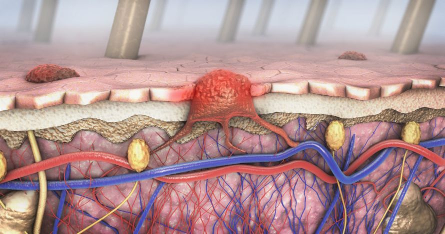
Take skin changes seriously!
If you notice any changes in your skin, you should definitely talk to your doctor about them.
Signs and symptoms of skin cancer:
- The skin spot bleeds or itches
- The texture and surface of the skin spot changes
- Scabs form
- The spot is getting larger
- The mole changes color
These changes and symptoms do not necessarily mean that a skin spot is skin cancer. However, you should take them seriously and have them examined by an experienced dermatologist.
Causes and Risk Factors
UV radiation as the main risk factor
Sunlight—more specifically, UV radiation—is considered the main risk factor and cause of skin cancer or its precancerous stages.
UV radiation causes damage to genetic material (DNA). This can lead to uncontrolled cell growth and, ultimately, to skin cancer.
Studies indicate that regular users of tanning beds have an increased risk of skin cancer. The risk of skin cancer is particularly high among people who first used a tanning bed before the age of 20.
Since the skin of children and adolescents is particularly vulnerable, Germany has a ban on minors using tanning salons.
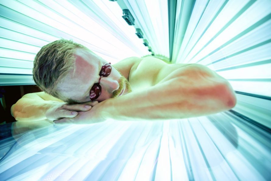
Regular use of tanning beds increases the risk of skin cancer © didesign | AdobeStock
Since skin cancer typically develops over many years, age is also a risk factor.
The likelihood of developing skin cancer increases with age. The risk is particularly high if certain areas of the body have been repeatedly exposed to intense UV radiation. If you have had frequent sunburns in the past, you have an increased risk of skin cancer.
X-rays and certain chemical substances can also cause skin changes that may lead to skin cancer.
These substances include:
- Arsenic
- Tar, and
- cytostatic drugs
Immunosuppressed patients and those with certain genetic predispositions are also at increased risk for skin cancer. There is evidence, particularly in the case of malignant melanoma, that it can also be hereditary.
Risk Groups
The following groups of people are particularly at risk:
- Children (especially infants) and adolescents
- People who burn easily
- People with fair skin, light or red hair, or many lentigines (sunspots, age spots)
- People who have many, conspicuous, or congenital moles
- People with compromised immune systems, such as those who have undergone an organ transplant
- People who have had skin cancer themselves or whose first-degree relatives have had skin cancer
- People who work outdoors or spend their free time in the sun
Examination and Diagnosis
Given the wide variety of possible forms that skin cancer can take, it is strongly recommended that you consult an experienced specialist for evaluation. Unfortunately, what may appear to be harmless eczema or moles often turns out to be skin cancer.
Skin Cancer Screening
More frequent screenings are recommended:
- if you belong to a high-risk group—for example, if you have a large number of moles—and
- if you have had skin cancer before
At the start of the examination, you should point out to the dermatologist any skin changes that you or your family members have noticed. Also let them know if an area of skin is itchy, feels rough or scaly, or has bled.
The dermatologist typically uses a special magnifying glass, called a dermatoscope, for the subsequent examination of the skin.
With the help of this illuminated magnifying glass, the dermatologist can examine suspicious areas more closely. Often, it is already possible at this stage to determine whether the change is merely a harmless skin lesion or not.
In some cases, it makes sense to take a photo of a skin lesion. This makes it easier to detect changes in a skin spot during the next follow-up examination.
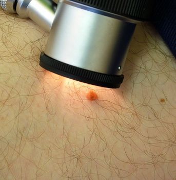
Examination of a suspicious skin lesion with a dermatoscope © Keith Frith | AdobeStock
A specialized technique called confocal laser scanning microscopy can distinguish melanomas from other skin changes. However, statutory health insurance plans do not cover the cost of this examination.
Is it melanoma or not? The ABCDE rule
Using the ABCDE rule, a dermatologist can make an initial assessment of whether a lesion is a melanoma.
The letters stand for the following terms:
- A (Asymmetry): The skin lesion is irregular in shape (not round); its texture is heterogeneous.
- B (Border): The skin lesion has irregular borders, some sharp and some blurred.
- C (Color): The color of the skin lesion is brown, black, blue, red, white, or gray; the pigmentation is uneven.
- D (Diameter): The skin lesion is larger than five millimeters.
- E (Elevation or Evolution): The skin lesion protrudes above the skin’s surface or is growing in size.
You can also apply the ABCDE rule to your moles yourself to detect any changes. If at least one of these characteristics applies, you should have it examined by a dermatologist as soon as possible.
However, you should see a dermatologist immediately if the mole:
- bleeds or itches
- is getting larger, or
- changes its shape or color
Further Tests
If a histological examination confirms the suspicion of malignant skin cancer, further tests will be conducted.
Imaging tests check whether metastases have already formed.
These include:
- Ultrasound
- X-ray
- CT
- MRI, and
- PET
In addition, a physical examination is performed and, if necessary, tumor markers are measured in the blood.
General Information on Treatment
Surgery is usually not required for actinic keratosis
To prevent actinic keratosis from progressing to squamous cell carcinoma, the doctor must treat it.
Doctors often treat individual cases of actinic keratosis using cryotherapy. During this procedure, the doctor freezes the skin lesion for several seconds using a cold spray or by applying the cryogen directly to the skin. This destroys the tissue.
You may feel some localized pain during the targeted treatment. Afterward, the skin area may swell slightly, and a blister may form.
In some cases, scarring or hyperpigmentation or hypopigmentation of the treated area may occur.
If there are multiple actinic keratoses, the doctor may treat the affected skin area daily with a fluorouracil cream for several weeks.
After a period of inflammation, the actinic keratoses will eventually disappear. The tissue usually does not scar afterward.
In addition, there are other substances that you may need to apply for several months.
Light therapy (photodynamic therapy) can effectively treat actinic keratoses. In this procedure, a cream containing so-called photosensitizers is applied to the skin.
These photosensitizers then accumulate in the actinic keratoses. They destroy the lesions when exposed to either sunlight or a specific artificial light source.
Surgical removal of actinic keratoses is necessary if:
- the actinic keratoses do not resolve using the methods described above
- they are particularly raised, or
- there is suspicion that they have already invaded adjacent tissue
Skin cancer is surgically excised whenever possible
Doctors completely remove white skin cancer surgically. To ensure that the surgeon has removed all of the cancerous tissue, the margins of the incision are sent for histological examination.
If any tumor residue remains, the dermatologist must remove it.
Tumors cannot always be removed in a single operation. Sometimes they are located in areas where surgical removal is not possible.
Some patients are also not candidates for surgery.
In such cases, the following treatments may be used:
- radiation therapy or chemotherapy
- for some tumors, cryotherapy or curettage (“scraping”)
If doctors detect a melanoma in its early stages, the disease can be cured by completely removing the melanoma surgically.
If necessary, doctors cover the wound with a skin graft. If the lymph nodes are already affected, doctors must also remove them surgically.
If metastases are present in other organs or if the melanoma cannot be removed, the following treatments are used:
- Radiation therapy
- Chemotherapy and/or
- Other drug therapies
Course and Prognosis
As a general rule, however, the earlier doctors detect skin cancer, the better the chances of a cure. In such cases, doctors can remove it before it spreads to surrounding tissue or before metastases form.
According to estimates, about five to ten percent of actinic keratoses progress to squamous cell carcinoma.
The chances of recovery from basal cell carcinoma are about 95 to 99 percent; the 5-year survival rate for melanoma is currently about 85 percent.
Depending on the type of skin cancer and the stage of the tumor, regular follow-up examinations are recommended. During these examinations, doctors carefully inspect the site where the tumor was located and the surrounding skin.
They palpate the lymph nodes, and imaging techniques may also be used if necessary. The doctor also measures tumor markers.
This allows them to detect and treat any tumor that may have regrown or appeared in a different location at an early stage.
Prevention
Avoid sunburn and excessive sun exposure!
UV radiation is the main risk factor for skin cancer and its precancerous stages. Therefore, it is important to protect yourself from intense sunlight and prevent sunburn.
Consistently wear clothing that covers your skin (don’t forget your head!) and apply sunscreen with an adequate SPF to unprotected areas. You should also protect areas of skin with actinic keratosis with sunscreen.
You must apply sunscreen generously, evenly, and repeatedly (for example, after swimming). Keep in mind, however, that sunscreen is not intended to allow you to stay in the sun longer—not even with repeated applications!

Wearing sunscreen while in the sun is an effective way to prevent skin cancer © AustrianImages.com | AdobeStock
Your skin can acclimate to the sun more effectively if you spend short periods of time in the sun on a regular basis. Your skin should not become red during this time.
This is especially important in the spring and at the start of your vacation in regions with higher UV radiation (mountainous areas or the south).
In strong sunlight, you should wear appropriate sunglasses. Never look directly into the sun—not even if you’re wearing sunglasses.
Since people who frequently use tanning beds have an increased risk of skin cancer, you should avoid regular visits to tanning salons. Children and adolescents are prohibited from using tanning salons anyway.
Prevention starts in childhood
Also make sure your children do not get sunburned. Infants should ideally not be exposed to the sun at all.
When the sun is strong, dress your children in clothing that covers their skin, including their arms and legs.
A wide-brimmed hat protects your child’s face and neck, and appropriate sunglasses protect their eyes
FAQ
How does skin cancer develop?
The development of skin cancer can be promoted by long-term damage to skin cells, particularly from intense UV radiation. If the genetic material is damaged, this can lead to uncontrolled cell growth.
Who is at increased risk for skin cancer?
People with fair skin, many moles, or frequent, intense sun exposure are particularly at risk. Genetic factors or a weakened immune system can also increase the risk of skin cancer.
How can skin cancer be detected?
Skin cancer can be detected when new skin changes appear or existing moles change. Signs to watch for include an increase in size, changes in color, irregular borders, or wounds that do not heal.
How is skin cancer diagnosed?
To diagnose skin cancer, a dermatologist thoroughly examines the skin, often using a dermatoscope. If skin cancer is suspected, a tissue sample is taken and examined histologically.
Share this article

About the medical author
Dr. Claus Puhlmann
Medical Author · Medical journalist
Dr. Claus Puhlmann – medical author: expert articles, professional insights and medical knowledge in the Leading Medicine Guide.
View full expert profile →Sources
- Arbeitsgemeinschaft der Wissenschaftlichen Medizinischen Fachgesellschaften e. V., Deutsche Krebsgesellschaft, Stiftung Deutsche Krebshilfe (2019) Melanom. Eine Leitlinie zur Untersuchung, Behandlung und Nachsorge des schwarzen Hautkrebses. Patientenleitlinie. Leitlinienprogramm Onkologie, Berlin
- Arbeitsgemeinschaft der Wissenschaftlichen Medizinischen Fachgesellschaften, Deutsche Krebsgesellschaft, Deutsche Krebshilfe (2020) S3-Leitlinie Aktinische Keratose und Plattenepithelkarzinom der Haut. Kurzversion 1.1. AWMF-Registernummer: 032/022OL. Leitlinienprogramm Onkologie, Berlin
- Arbeitsgemeinschaft der Wissenschaftlichen Medizinischen Fachgesellschaften, Deutsche Krebsgesellschaft, Deutsche Krebshilfe (2020) S3-Leitlinie Prävention von Hautkrebs. Konsultationsfassung. Leitlinienprogramm Onkologie, Berlin
- Arbeitsgemeinschaft der Wissenschaftlichen Medizinischen Fachgesellschaften, Deutsche Krebsgesellschaft, Deutsche Krebshilfe (2020) S3-Leitlinie Diagnostik, Therapie und Nachsorge des Melanoms. AWMF-Register-Nummer: 032/024OL. Leitlinienprogramm Onkologie, Berlin
- Sterry W, Stockfleth E (2018) Maligne epitheliale Tumoren. In: Plewig G, RuzickaT, Kaufmann R, Hertl M (Hrsg.) Braun-Falco's Dermatologie, Venerologie und Allergologie. Springer Berlin
- Fritsch P, Schwarz T (2018) Dermatologie Venerologie. Kap. 17 Neoplasien (Tumoren) und tumorähnliche Läsionen der Haut. Springer, Berlin
- Zouboulis CC, Röhrs H (2005) Kryochirurgische Behandlung aktinischer Keratosen mit evidenzbasierter Übersicht. Hautarzt 56:353–358