The malignant changes in melanoma originate in the pigment-forming cells of the skin, the melanocytes. These skin cells react particularly sensitively to UV radiation. Therefore, the German Cancer Centre considers excessive sun exposure as one of the main causes of skin cancer.
Heavy UV exposure and recurrent sunburns in childhood and adolescence massively increase the risk of melanoma. Particularly at risk of sunburn and also skin cancer are people with
- light skin and light eye colour and
- red or blond hair.
Other risk factors are:
- previous cancers,
- many or conspicuous moles,
- melanocytic tumours of the skin formed before birth,
- freckles and
- XP disease (xeroderma pigmentosum).
In most patients, the tumour develops from a pre-existing birthmark. A melanoma is therefore usually darkly pigmented.
Only rarely are so-called amelanotic melanomas found that do not show any pigmentation. These occur mainly on the hands or feet.
At the time of diagnosis, the majority of skin cancer patients have symptoms. Most tumours are diagnosed during screening examinations. Only sometimes do patients notice a slight itching or a little bleeding beforehand.
Basically, malignant melanoma can be divided into different types. The distinction are based on
- the exact type of tumour,
- the thickness of the tumour and
- the exact localisation.
The following types of melanoma are distinguished:
- Superficial spreading melanoma (SSM) develops rather flatly.
- Nodular melanoma (NM) grows nodular and bleeds frequently.
- Lentigo-maligna melanoma (LMM) tends to grow slowly and occurs mainly on the face in older people.
- Acro-lentiginous melanoma (ALM) develops mainly under the nails and on the palms of the feet and hands.
Mucosal melanomas, choroidal melanomas in the eye and melanomas of the meninges are also possible, but rather rare.
It is not always clear at first glance whether it is just a normal birthmark or a melanoma. However, a first rough distinction is possible with the help of the so-called ABCDE rule:
- A for asymmetry: A melanoma is unevenly shaped.
- B for Limitation: The border is rather blurred and irregular.
- C for colour Colour mixtures of brown, blue, red, white and black appear.
- D for diameter: The birthmark has a diameter of more than 5 millimetres.
- E for sublimity: A melanoma usually protrudes above the normal skin level.
If one or more criteria of the ABCDE rule are fulfilled, you should urgently consult a dermatologist.
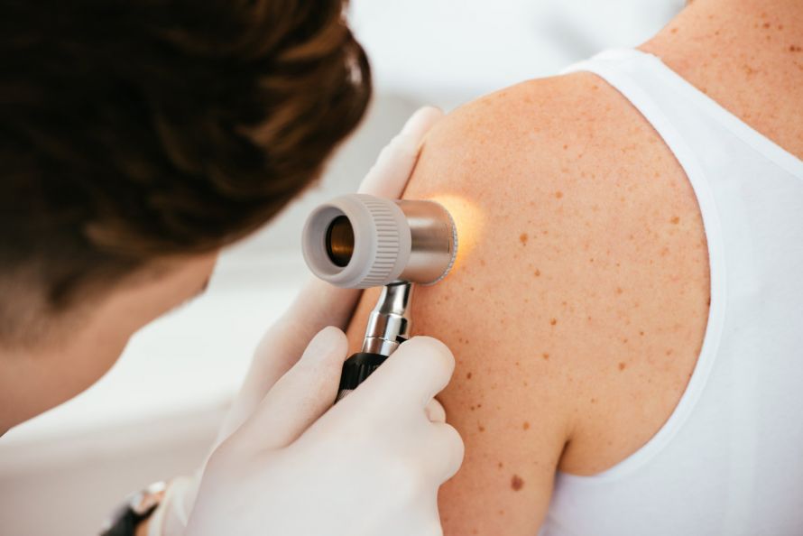
It is also advisable to have a cancer screening every two years of your skin performed by your family doctor or dermatologist. The doctor then examines the suspicious moles with a dermoscope. This allows him to assess the exact pigment structure of the marks.

Dermatologists check the patient's existing moles during skin cancer screening © LIGHTFIELD STUDIOS | AdobeStock
If the suspicion of cancer is confirmed, the affected area is surgically removed under local anaesthesia and examined under the microscope.
If the diagnosis of "melanoma" is then confirmed, the tumour thickness according to Breslow must be determined. This is the most important prognostic factor of skin cancer.
If the tumour is thicker than one millimetre, a spread diagnosis must be made. It has the aim of preventing metastases in other organs and lymph nodes at an early stage. In addition, there are
which can be performed.
The treatment depends mainly on the diagnosed stage of the cancer.
The most important form of treatment is surgical removal of the malignant melanoma. The tumour is always removed as a whole to prevent metastasis. When removing them, a sufficiently large safety distance of one to two centimetres should also be ensured.
From a tumour thickness of one to 0.75 millimetres, the surgeon also removes the so-called sentinel lymph nodes. These are the lymph nodes that first come into contact with the lymph fluid from the tumour area. If the sentinel lymph node is also affected by the cancer, the surrounding lymph nodes are also removed.
If the tumour is thicker and affects the lymph nodes, the patient is recommended adjuvant treatment. The patient then receives interferon alpha over a period of 18 months. This is a chemical substance that is supposed to prevent a possible tumour recurrence (recurrence of the cancer).
If the tumour has already metastasised, a complete cure is rather unlikely. The treatment then aims to prolong life. As well as chemotherapy with cytostatic drugs (cell growth inhibitors),
- further surgical interventions are used to reduce the tumour mass as well as
- radiotherapies.
Another therapy method for the treatment of malignant melanoma is the stimulation of the immune system with antibodies. If this is successful, the immune system can fight the degenerated cells itself. Various immunotherapeutic procedures are also currently undergoing clinical trials.
Malignant melanoma is the most aggressive and malignant tumour of the skin. Compared to other skin cancers, malignant melanoma metastasises to other organs at a fairly early stage. The chances of recovery therefore depend on various factors.
The extent to which the disease has already progressed at the start of treatment is particularly decisive for the prognosis. The tumour thickness according to Breslow and the penetration depth according to the so-called Clark Level play an important role. If the melanoma is still small and tends to grow more on the surface, the prognosis is quite good.
The chances of a cure worsen if the tumour has already spread beyond the dermis (middle layer of skin). If there are metastases in the
a cure is virtually impossible.
The ten-year survival rate of malignant melanoma is around 80 percent. This means that 80 percent of all patients are still alive ten years after diagnosis.
For lymph node metastases and metastases in other organs, on the other hand, the ten-year survival rate is only ten to 20 percent.


