When every step is painful and hip mobility is severely limited in daily life, a diagnosis of hip osteoarthritis is often suspected. When conservative treatments such as physical therapy and pain medication no longer help, hip replacement surgery (total hip arthroplasty) is often the only way to regain quality of life. An artificial hip joint completely replaces the damaged joint. Modern orthopedics now offers minimally invasive procedures that spare the muscles and surrounding tissues. Whether a cemented or cementless hip prosthesis is chosen depends on the patient’s bone quality and age. Learn everything here about the hip surgery procedure, the durability of the implants, and rehabilitation so that you can get back on your feet quickly.
Uses of a hip replacement—most commonly for osteoarthritis
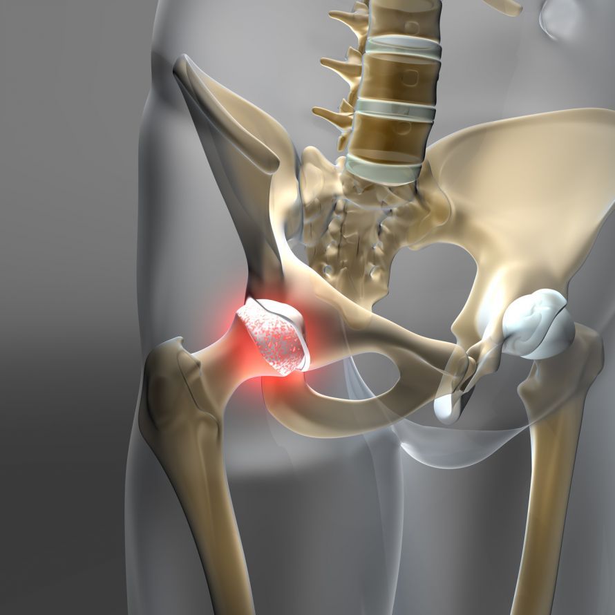
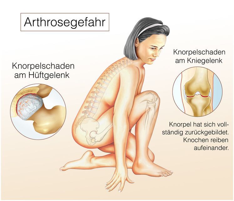
In most cases, a total hip replacement is performed when the natural hip joint is damaged by osteoarthritis of the hip. Osteoarthritis refers to age-related or strain-induced wear and tear of the cartilage layer in the joint. The cartilage wears away, causing the bones to rub directly against one another. This degenerative joint disease of the hip often develops in patients aged 55 and older.
Coxarthrosis, as the condition is also known, is therefore generally a sign of aging. About 10 percent of women and 17 percent of men receive this diagnosis, and in one-third of those affected, both hip joints are affected. The damage already caused by hip osteoarthritis is irreversible; in other words, it cannot yet be reversed through conservative treatment. In most cases, surgery is necessary in the end stage.
Conservative treatments can therefore often only provide temporary relief. It is therefore important to discuss the option of surgery and the use of an artificial joint with your doctor in a timely manner.
It has been shown that 77% of patients diagnosed with coxarthrosis already had a pre-existing hip joint condition. In such cases, physicians refer to it as secondary osteoarthritis or osteoarthritis of secondary origin. Typical forms of secondary osteoarthritis include:
- Subluxation coxarthrosis/dysplasia-related coxarthrosis
- Coxarthrosis following Perthes disease, juvenile epiphyseolysis of the femoral head, or idiopathic avascular necrosis of the femoral head
- Protrusion-related coxarthrosis
- Post-coxitis coxarthrosis
- Posttraumatic coxarthrosis
In these cases, hip-related symptoms can develop early in life. It is not uncommon for young patients to require hip treatment as a result. Hip joint surgeries are also performed significantly earlier in these cases than in primary degenerative osteoarthritis.
Artificial Hip Joint and Total Hip Replacement (THR): Surgery and Implantation of a Hip Prosthesis for Coxarthrosis and Hip Osteoarthritis
The Three Different Types of Pain Associated with Hip Osteoarthritis
Hip osteoarthritis, similar to osteoarthritis of other joints, presents three distinct types of pain:
- Stress pain—the most common type of pain in hip osteoarthritis. The pain occurs during movement and weight-bearing and persists throughout
- Start-up pain—pain at the beginning of movement. For example, the pain occurs after getting out of bed and then subsides with continued movement.
- Rest pain—discomfort without joint movement. The pain is always present, even without movement.
The pain is usually referred to the groin area, the region of the greater trochanter on the side of the hip joint, or the buttocks. However, the pain often radiates into the thigh or even down to the knee joint.
Due to the pain originating from the affected hip, the patient often adopts a compensatory posture. This unnatural posture is intended to reduce the sensation of pain. However, this can weaken certain muscles (muscle imbalance), leading to so-called contractures and further restricting mobility.
During a physical examination by a doctor, standardized movement tests (known as “signs”) are performed that trigger pain in specific positions. In hip osteoarthritis, this occurs particularly when rotating the hip inward or abducting the hip joint. If muscular weakness develops due to a compensatory posture adopted to avoid pain, the Duchenne and Trendelenburg signs may also be positive. This often manifests as a characteristic limp when walking.
In advanced coxarthrosis, significant muscle atrophy (muscle wasting) as well as a flexion-external rotation-adduction contracture may be present. Typically, tasks such as tying shoelaces or cutting toenails become extremely difficult. This malalignment, caused by shortened and weakened muscles and tendons, also results in functional leg shortening.
Diagnosis of Hip Osteoarthritis
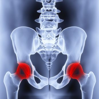
X-rays of the affected hip joint form the basis of diagnosis and are routinely performed. The hip is X-rayed from the front (anterior-posterior) and from the side (lateral). As a rule, a so-called pelvic overview X-ray is taken to assess symmetry and leg length.
In X-ray images, in cases of advanced disease, most of the radiological criteria for osteoarthritis—namely
- narrowing of the joint space,
- sclerosis,
- subchondral cysts, and
- osteophytes
. In rare cases, when anatomical conditions are complex, special X-ray views of the femoral neck (Rippstein view) or the acetabular roof (Faux profile according to Lequesne) may be used; in such cases, a computed tomography (CT) scan (possibly with 3D reconstruction) can provide further insights or be used to create special templates or custom-made implants. Further diagnostic testing using MRI is usually not necessary if there are clear signs on the X-ray, but it can provide additional information in individual cases.
Conservative Treatment of Hip Osteoarthritis
Conservative treatment should be used in the early stages of osteoarthritis.
Sports involving movements without impact, such as
- cycling,
- swimming, or
- gymnastics.
If the patient is overweight, weight loss is strongly recommended.
Additional relief can be achieved through physical therapy exercises to prevent contractures, as well as manual therapy. However, because the onset of symptoms often has a mechanical component in addition to an inflammatory one, it is not uncommon for symptoms to worsen during physical therapy.
If this does not provide adequate pain relief, an injection of a local anesthetic—or cortisone—into the joint capsule may be performed. However, this may only lead to temporary relief of symptoms. In the early stages, treatment with hyaluronic acid may also be attempted.
Long-term pain management with nonsteroidal anti-inflammatory drugs (NSAIDs) is very common, but can be harmful to the gastrointestinal tract or the kidneys due to side effects. More gastrointestinal-friendly selective COX-2 inhibitors are also used, but they are not suitable for every patient due to potential cardiopulmonary side effects.
The prognosis and course of the disease in coxarthrosis vary greatly from patient to patient and are difficult to predict. The majority of patients with coxarthrosis require surgical intervention in the end stage.
Joint-Preserving Surgery for Hip Osteoarthritis
One of the most common congenital hip joint malformations that can lead to secondary hip osteoarthritis is hip dysplasia. This condition is characterized by reduced coverage of the femoral head combined with a rotation of the femoral neck. If these conditions are detected early, joint-preserving surgery may be helpful.
Joint-preserving surgeries are divided into realignment procedures (osteotomies) in the femur and pelvis.
Intertrochanteric femoral osteotomies correct misalignments in the proximal femur, thereby reducing transarticular pressure, realigning the force transmission, and optimizing torque.
Realignment osteotomies can
- be varus-correcting,
- valgus-correcting,
- extending,
- derotating,
- flexing, or
- in combination
. Osteotomies in the pelvic region aim to correct a lack of coverage of the femoral head and the associated localized stress and overload on the articular cartilage. Notable examples here include the Salter and Chiari procedures, as well as the triple osteotomy.
It should be noted that these surgical techniques provide lasting benefit only to patients who still have intact articular cartilage. Thus, third- and fourth-degree osteoarthritis with significant cartilage damage precludes joint-preserving surgery.
Joint Replacement: Total Hip Arthroplasty (THA)
In total hip arthroplasty (THA)—that is, complete replacement of the hip joint with an endoprosthesis—the surgeon selects the surgical method, the implants, and their fixation mechanisms.
In making this decision, the surgeon should
- the severity of osteoarthritis,
- the anatomy of the hip,
- comorbidities,
- age, and
- the patient’s lifestyle and environment
of the patient. The following sections provide an overview of open and minimally invasive surgical techniques as well as various prosthesis designs.
Approaches to the hip joint: conventional or minimally invasive
Typically, access to the hip joint can be achieved through several approaches, which, depending on the incision, require the patient to be positioned in the supine or lateral position.
With the lateral approach, the patient can be operated on in either the supine or lateral position. Here, the incision is made over the area of the greater trochanter (trochanter major) and requires transection of the iliotibial tract as well as part of the gluteal muscles (gluteus medius and minimus).
With the posterior (dorsal) approach, the incision is made dorsally to the trochanteric mass, which requires the patient to be positioned on their side. Here, too, muscles (piriformis muscle) must be detached and later reattached.
In modern hip arthroplasty, these approaches and procedures are generally avoided today.
Minimally Invasive Implantation Procedures
In contrast, newer minimally invasive implantation techniques preserve soft tissue structures such as the iliotibial tract and the important gluteal muscles.
They are typically performed from the front (AMIS) or from the front-lateral (ALMIS) through so-called muscle gaps. They are characterized by the complete avoidance of partial, subtotal, or total transection or detachment of the muscles, thereby playing a crucial role in helping patients regain mobility more quickly after surgery and preventing muscular insufficiency.
Watch the video below to see how a total hip replacement is performed:
Prosthesis Types and Materials for Total Hip Replacement
The individual implants differ in a variety of parameters, such as:
- Material (titanium and its alloys, cobalt-based alloys, steel)
- Cemented or cementless
- Coating (pure titanium, hydroxyapatite)
- Surface texture (micro- or macrostructure)
- Shaft-neck angle (CCD angle)
- Shape (short stem, straight stem, anatomical stem)
- Distance from the center of rotation to the stem axis
Artificial Hip and Prosthesis: Cemented Fixation, Follow-up Care, and Complications in Patients Who Have Undergone Hip Replacement Surgery
Cementless vs. Cemented Total Hip Replacement
In terms of acetabular cups, a distinction is generally made between cemented and cementless types, with the cemented variants typically being polyethylene monoblock designs, whereas cementless cups feature a metallic cup shell and are implanted using press-fit or screw fixation.
A variety of material combinations can be used for the bearing pair (acetabular insert/prosthesis head), each with its own advantages and disadvantages. In modern hip arthroplasty, highly cross-linked polyethylene (PE)/ceramic and ceramic/ceramic bearing pairs are primarily used as standard.
Cementless Arthroplasty for Total Hip Replacement (THR)
With this implantation technique, a press fit or form fit is achieved by appropriately preparing the cortical-cancellous prosthetic socket using a drill (prosthetic cup) or so-called impactors (prosthetic stem), followed by the insertion of the prosthesis. This “locking” of the total hip replacement (THR) achieves primary stability, which allows for secondary ingrowth of cancellous bone structures. Immediate weight-bearing on the prosthesis is possible.
With this type of anchoring, a healthy bone matrix is a prerequisite for good long-term results. However, osteoporosis or bone metabolism disorders of any origin are contraindications, meaning that the endoprosthesis cannot be implanted cementlessly in such cases.
In the case of the hip stem, the design of the prosthesis determines the force transmission and thus the load on the bone surrounding the prosthesis and its remodeling processes.
Short-stem prostheses transmit forces as early as the femoral neck region, resulting in a significant increase in bone density in this area, whereas with straight-stem prostheses, the force transmission occurs well below the femoral neck, leading in the long term to bone loss in the region of the femoral neck and the trochanters due to inactivity. This can lead to problems during revision surgery. Furthermore, the short and round design of short-shaft prostheses allows for optimal use of minimally invasive approaches to the hip joint.
Cemented Endoprosthetics for a Hip TEP
In this procedure, a synthetic material (bone cement) serves as the interface between the bone matrix and the total hip replacement (THR). Once implanted, this THR can immediately bear full weight.
This implantation technique is used for patients in whom a cementless prosthesis is not indicated due to reduced bone quality or density, such as in cases of osteoporosis.
Hybrid Endoprosthetics for Hip TEP
Here, a hybrid approach combining cementless and cemented endoprosthetics is used, which combines the respective advantages of both implantation techniques.
The acetabular cup is often implanted cementlessly, as bone metabolism disorders involving a decrease in bone density do not manifest in the acetabular region until a later stage. The stem, on the other hand, is cemented, and the bone cement ensures an immediate weight-bearing connection, even to bone that has already deteriorated in quality. Full weight-bearing after surgery is also well possible in this case.
Which doctors perform hip replacement surgery?
In hip arthroplasty, specialists in orthopedics and trauma surgery typically focus exclusively on the hip joint and its surgical treatments, such as joint replacement and total hip arthroplasty (THA).
FAQ: The 8 Most Important Questions About Total Hip Replacement
How long does an artificial hip joint last?
The longevity of a modern hip prosthesis is very good today and, statistically speaking, ranges from 15 to 20 years—often even longer. Factors such as body weight, activity level, and the quality of bone anchoring influence its lifespan. In younger patients, a revision surgery may be necessary after this time, as the implant can become loose.
When is the right time for hip replacement surgery?
The right time for total hip replacement (THR) varies from person to person and depends on the severity of symptoms. If hip pain occurs even at rest, walking distance is significantly reduced, and quality of life is severely impaired, surgery is often unavoidable. The orthopedic surgeon uses an X-ray to assess how far the wear and tear of the joint cartilage (osteoarthritis) has progressed.
What is the difference between cemented and cementless implants?
With a cementless hip prosthesis, the implant is inserted into the bone using a press-fit technique and has a rough surface that promotes bone ingrowth. This option is often chosen for younger patients with good bone quality. A cemented prosthesis is fixed in the medullary canal using bone cement, which allows for immediate full weight-bearing and is often used for older patients with osteoporosis.
How long does rehabilitation take after total hip replacement (THR)?
Rehabilitation usually begins the very next day after surgery in the hospital. The hospital stay lasts about 5 to 7 days. This is followed by a rehabilitation period that typically lasts three weeks and can be conducted on an outpatient or inpatient basis. The goal is to strengthen the muscles and restore a normal gait. It usually takes 3 to 6 months to regain full weight-bearing capacity and the ability to participate in sports.
What sports are allowed with an artificial hip?
After recovery, low-impact sports such as swimming, cycling, walking, or hiking are ideal. Sports involving high impact or abrupt changes in direction—such as soccer, tennis, or jogging—should be avoided, as they can increase wear on the polyethylene liner and raise the risk of loosening.
What complications can occur?
As with any surgery, there are risks. Specific complications of total hip replacement (THR) include dislocation of the joint, infections, leg length discrepancies, or loosening of the prosthesis over time. However, these risks can be minimized through minimally invasive procedures and experienced surgeons. It is important to have any signs of inflammation evaluated by a doctor immediately.
How is minimally invasive hip surgery performed?
In minimally invasive surgery, the surgeon uses natural gaps between muscles to access the hip joint instead of cutting through muscles. This preserves tissue, reduces pain, and speeds up wound healing after surgery. Patients often regain mobility more quickly and experience fewer movement restrictions in the first few weeks.
When can I sleep on my side again?
In the first few weeks after surgery, patients should generally sleep on their backs, possibly with a pillow between their legs to prevent dislocation. Sleeping on the side that wasn’t operated on is often possible again after about 6 weeks, once the joint capsule has healed sufficiently and is stable. Lying on the operated leg may still be uncomfortable at first.

About the medical author
Prof. Dr. med. Karl Philipp Kutzner
Medical Editor · Mainz
Prof. Dr. med. Karl Philipp Kutzner – medical author: expert articles, professional insights and medical knowledge in the Leading Medicine Guide.