A wide range of scientifically proven treatment options is now available for damage to the cartilage in the knee joint. These cartilage treatments aim to regenerate the cartilage and thus halt its deterioration. Treatment plans are tailored to each patient based on the extent of the cartilage damage, its cause, and the patient’s specific needs. The goal of cartilage therapy is to regenerate damaged knee cartilage and thereby restore cartilage function in the long term.
Below you will find further information on the treatment options as well as a selection of specialists in knee cartilage therapy.
Why should cartilage damage be treated as early as possible?
Articular cartilage performs important functional roles in the joints. It acts as a cushion between the joint bones, preventing them from rubbing against each other. Cartilage injuries can be caused, for example, by an accident or a sports injury.
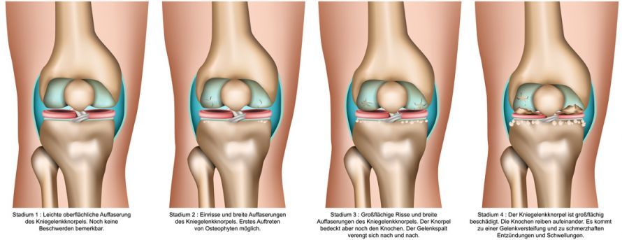
Without adequate treatment, osteoarthritis (joint wear) can develop in the affected joint. As a result, the cartilage in the joint gradually breaks down, which can lead to severe pain. Severely worn cartilage cannot be restored: the damage is irreversible. In severe cases, only a joint replacement can help.
Just like the hip, the knee joint is very commonly affected by osteoarthritis.
The following video shows how bones rub directly against each other in cases of severe osteoarthritis:
With timely cartilage regeneration procedures performed in accordance with clinical guidelines, early-stage osteoarthritis can be prevented or delayed. In recent years, a wide range of successful therapeutic approaches for treating localized traumatic cartilage defects have become established.
That is why early and appropriate treatment is so important. If treatment is delayed, cartilage regeneration is no longer possible.
Basic Requirements for Knee Cartilage Therapy
The selection of the appropriate treatment method for each individual patient depends, among other factors,
- the type and size of the cartilage damage (including defect depth, whether caused by an accident or wear and tear, defect location, etc.),
- the patient’s expectations, as well as
- any additional joint injuries that may be present, such as ligament instability, meniscal tears, and deviations in the mechanical axis of the leg (misalignments such as bowlegs and knock-knees)
.
Any accompanying injuries must be corrected as part of a multimodal regenerative joint therapy. Such joint injuries increase the risk of knee osteoarthritis. Therefore, their treatment is necessary to ensure a sustainable, positive outcome for cartilage regeneration.
The German Society for Orthopedics and Trauma Surgery (DGOU) collects information and publishes it in the German Cartilage Registry. According to this data, well over 50 percent of treated cases of knee cartilage damage are associated with concomitant damage or misalignments.
Conservative Treatments for Knee Cartilage Damage
Conservative therapies refer to all approaches that do not involve surgery. For cartilage damage in the knee, these primarily include
- physical therapy,
- orthopedic aids (e.g., insoles and joint braces),
- physical therapy (cold and heat therapy, lymphatic drainage, ultrasound)
. These help relieve pressure on the affected joint area and promote the resolution of associated inflammatory changes.
Non-steroidal anti-inflammatory drugs (NSAIDs), such as diclofenac, are used for medication-based treatment. They reduce joint pain and inflammation. These medications are a particularly good therapeutic adjunct, especially in the acute phase following injury or surgery.
In addition, a wide variety of cartilage supplements and dietary supplements are available. In particular,
- vitamin D,
- chondroitin, and
- glucosamine, as well as
- other vitamins and substances
play a role here. These also appear to have a positive effect on tissue regeneration even after surgical cartilage treatment. They can temporarily relieve symptoms and are therefore suitable supplements. However, they cannot cause the damaged cartilage in the knee to regenerate.
Furthermore,
- joint injections with PRP (platelet-rich plasma), which is produced from the patient’s own blood through centrifugation, and
- hyaluronic acid, even in combination with PRP,
have a positive effect on cartilage regeneration following surgical cartilage therapy.
Experimental / Obsolete Surgical Cartilage Therapies for the Knee
The DGOU Working Group on Clinical Tissue Regeneration is currently the largest scientific committee in Europe dedicated to cartilage regeneration. It includes cartilage specialists from German-speaking European countries.
It regularly issues updated recommendations on cartilage therapy based on the latest available literature. These recommendations are published internationally and are therefore widely recognized. Many cartilage centers worldwide follow these recommendations.
Articular Cartilage Refixation
Injuries such as patellar dislocation often result in the shearing off of cartilage-bone fragments. In acute cases, if these fragments are well-preserved, they can be fitted into the defect. The surgeon secures them with self-dissolving pins or screws. Successful healing depends on a solid connection between the cartilage-bone fragment and the bony defect base.
Cartilage reattachment, as well as many other cartilage treatment therapies, can be performed arthroscopically. The video demonstrates how minimally invasive arthroscopy is performed:
BMS (Bone Marrow Stimulation), microfracturing, nanofracturing, microdrilling
According to the 2021 recommendations of the DGOU Working Group on Clinical Tissue Regeneration, these procedures are suitable for defects up to 2 cm².
These techniques were developed as early as the 1980s. They are performed during arthroscopy. During the procedure, the surgeon removes the diseased cartilage tissue, exposing the healthy cartilage down to the bone lamella.
The surgeon then perforates the bone layer beneath the defective cartilage using drills or chisels, similar to scarifying a lawn. This is called microfracturing, nanofracturing, or Pridie drilling (in which only thin drill wires of approximately 1.2 mm are used). Nanofracturing stands out among these techniques because it allows for a defined and standardized depth and thickness of the holes.
The fracturing causes bleeding. The goal is to introduce regenerative cells (stem cells) from the bone marrow into the defect area.
However, the number of stem cells obtained from the drill holes in the defect area is often very low. In addition, there is a fairly high risk of complications: Damage to the delicate bone lamella in the cartilage defect caused by the techniques described above leads to bone overgrowth within the defect in 30 to 60 percent of cases. This bone overgrowth can then cause problems again after a short time.
Furthermore, the treatment results are not long-lasting. After only two to three years, the cartilage deteriorates again.
For this reason, specialists are performing this therapy less and less frequently.
Matrix-supported bone marrow stimulation (mBMS), AMIC®, BMAC, microfracturing, nanofracturing
According to the 2021 recommendations of the DGOU Working Group on Clinical Tissue Regeneration, these procedures are suitable for defect sizes ranging from 1.0 to 4.5 cm².
The use of additional matrices has improved BMS techniques. These are membranes made of collagen, hyaluronic acid networks, and other materials. They are designed to provide a three-dimensional structure for cells that have the potential to form cartilage. This refers to chondrogenic cells, such as stem cells. As a result, these cells can adhere more effectively and thus better fill the defect.
The use of these membranes leads to better and longer-lasting cartilage regeneration results than BMS alone.
This technique can often be performed arthroscopically, depending on the size and location of the lesion to be treated. To date, very good mid-term study results and initial long-term results for this technique are available.
Whether this procedure will prove effective in the long term (> 12–15 years) cannot yet be conclusively assessed based on the studies available to date. The publication of ten-year results for this technique is expected in 2023.
Osteochondral Transplantation (OCT)
According to the 2021 recommendations of the DGOU Working Group on Clinical Tissue Regeneration, these procedures are suitable for defect sizes ranging from 1.0 to 1.5 cm².
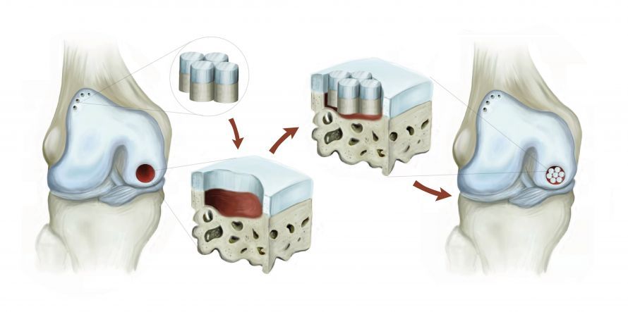
OCT is also known as cartilage-bone transplantation. In this procedure, the surgeon harvests a cartilage-bone cylinder from a low-stress area of the joint. This cylinder is then transplanted into the defect site, which has been drilled to the appropriate size.
The cylinder must contain a sufficiently large bony component to allow for solid integration into the surrounding bone. The cartilaginous portion of the cylinder immediately fills the defect site with high-quality hyaline cartilage. The gaps remaining between the cylinders are covered with inferior scar tissue.
The results are good for small cartilage (bone) defects. However, the destruction of healthy cartilage surfaces is problematic. This can lead to symptoms at the cylinder harvest site that are very similar to those at the original defect site. To minimize these harvest-related symptoms, OCT is recommended only for cartilage defects up to approximately 1.5 square centimeters.
The procedure offers advantages when there are small defects in both the cartilage AND the bone. This is the case, for example, with osteochondrosis dissecans (joint mouse). This procedure allows both tissues to be treated quickly and effectively.
However, it is a challenging procedure: Ideally, the thickness of the harvested cartilage cylinder should match that of the defect area. Otherwise, unfavorable compressive and shear forces will result.
Alternative Methods: Minced Cartilage, Cartilage Chips, Autocart®
According to the 2021 recommendations of the DGOU Working Group on Clinical Tissue Regeneration, these procedures are suitable for defect sizes ranging from 1 to 3.5 cm².
Cartilage cells are normally statically embedded within the cartilage tissue. Under certain circumstances, however, they are able to migrate a few millimeters out of the embedded cartilage and then divide again. In this way, they form new cartilage tissue.
In the method described, the surgeon harvests small pieces of cartilage from the defect site or from low-stress areas of the knee. These pieces are ground into small fragments and then injected into the defect to fill it. Cartilage cells can then migrate from these small cartilage fragments and form new cartilage tissue through division.
By adding
- platelets rich in the body’s own fibrin (PRF) and plasma (PRP),
- membranes/matrices, or
- fibrin glue
are intended to better stimulate the cells to form new tissue.
Some techniques also incorporate microfracturing, microdrilling, or nanofracturing beneath the defect. Several animal studies have achieved better cartilage regeneration using this technique than with microfracturing (BMS) alone.
At present, however, it is unclear which combination of which specific steps in the method might successfully and sustainably generate cartilage tissue. There is therefore no generally accepted standardized procedure that has been proven to guarantee good cartilage regeneration in the medium term. Initial studies currently suggest that a large proportion of the cartilage cells may be destroyed during the pulverization of the cartilage.
Although this procedure has become increasingly widespread in recent years, it is not currently among the scientifically validated and recommended procedures of the Clinical Tissue Regeneration Working Group of the DGOU. Instead, it is considered a “method with potential”! This means that the procedure may have great potential under certain circumstances.
However, this is contingent on studies demonstrating clear results after a follow-up period of at least five years. They must show that a specific form of the minced cartilage procedure is superior to other methods.
This method is currently recommended only for patients participating in a controlled scientific study. From a scientific standpoint, all other described cartilage regeneration procedures—and even BMS/microfracturing—currently yield more reliable results than minced cartilage.
Minced Cartilage, Autocart®, and cartilage chips do NOT constitute a single-stage cartilage cell transplant. According to the current state of scientific knowledge (as of 2023), patients who undergo this procedure still face a high risk. If you nevertheless decide to undergo this therapy, you should be aware of this!
Gold Standard for Knee Cartilage Therapy: Cartilage Cell Transplantation
Autologous chondrocyte transplantation (ACT) refers to the transplantation of the patient’s own cartilage cells. It has been further developed into matrix-induced autologous chondrocyte transplantation (mACT).
Due to its excellent long-term results, only mACT is used in Europe today.
According to the 2021 recommendations of the DGOU Working Group on Clinical Tissue Regeneration, these procedures are suitable for defects measuring 2 cm² or larger.
Since the early 1990s, the transplantation of a patient’s own cartilage cells (autologous chondrocyte transplantation) has been used successfully. It is primarily used for cartilage reconstruction in cases of traumatic defects or osteochondrosis dissecans (OD; osteochondrosis). It is a scientifically proven procedure.
It is now the most scientifically studied procedure for knee cartilage regeneration worldwide. The procedure offers excellent long-term results lasting over 15 years!
In ACT, the cultured cartilage cells are injected under a periosteal flap that is sutured watertight over the cartilage defect. This procedure has been further developed into mACT: Here, the cells are introduced into or onto matrices.
In some cases, the technique can now even be performed arthroscopically via joint endoscopy.
Even for defects larger than 2 square centimeters, this procedure is superior to the previously described cartilage regeneration procedures in the long term.
Requirements for ACT
Patients with localized cartilage damage who have not yet developed osteoarthritis are suitable candidates for ACT. In cases of widespread wear of the articular cartilage—that is, osteoarthritis—ACT is not possible.
The basic requirements for treating cartilage damage described above apply in particular to mACT.
mACT treatment is particularly suitable for patients who have completed growth (puberty) and are up to 55 years of age.
Performing the Cartilage Transplant
This knee cartilage therapy is performed in several steps.
First, the surgeon uses arthroscopy to remove a small amount of cartilage from a non-weight-bearing part of the patient’s knee joint.
In the clean room of a certified laboratory, the cartilage cells are isolated from the cartilage tissue and cultured. Once the required number of cells has been reached, the cartilage cells are
- are seeded into a special three-dimensional collagen sponge structure,
- mixed into a three-dimensional gel, or
- shaped into three-dimensional spheroids that largely correspond to the original biological cellular environment in the cartilage.
There, the cells begin producing new cartilage matrix.
In the laboratory, checks are performed on the
- vitality,
- sterility, and
- the ability of the cartilage cells to form hyaline cartilage.
Only when these values are optimal is the graft delivered to the hospital or the treating physician.
Three to six weeks after the arthroscopic harvest, the second procedure takes place. It can be performed partially arthroscopically. The surgeon transplants the cultured, multiplied cartilage cells into the patient in the form specified by the manufacturer.
First, the surgeon removes the damaged cartilage from the defect area. To do this, he either makes a skin incision approximately 5 cm long or uses an arthroscope. He then fits the graft into the defect. The graft is
- as a cell-seeded membrane,
- as a cell gel, or
- in the form of small, self-adhesive cartilage cell spheres (spheroids)
. While spheroids and the gel are self-adhesive, the matrix is secured with fibrin glue or sutures.
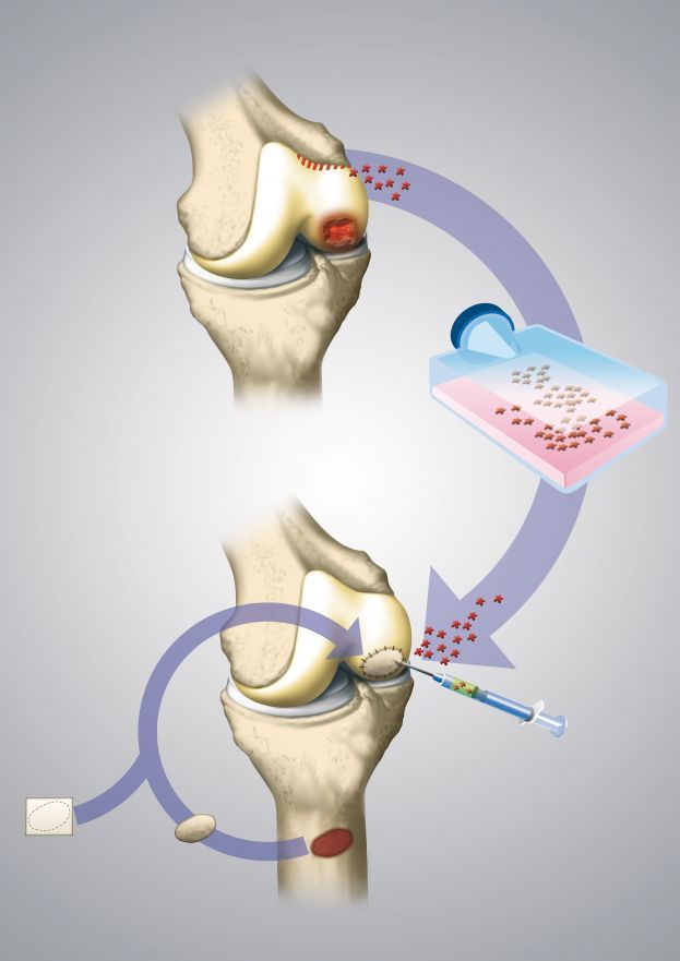
After the second surgery, the mACT, a hospital stay of only 2–3 days is usually required. In favorable cases, the procedure can also be performed as day surgery or on an outpatient basis.
As with all cartilage regeneration procedures, postoperative care is crucial to the success of the treatment.
Costs of mACT
One drawback of the procedure is that it requires two surgical procedures. It is also strictly regulated. As a result, costs can be high, reaching up to €13,000 for cell cultivation alone (as of 2023).
However, international studies show that the therapy can still be worthwhile: When administered in a timely manner, it results in lower long-term costs for a healthcare system than the use of less effective methods. A study published in 2021 shows that mACT can prevent approximately 21 percent of knee replacement surgeries.
For this reason, health insurance providers in Germany cover the procedure on an outpatient basis in certain cases for defect sizes of 2 cm² or larger.
Unfortunately, this does not yet apply in full in Austria and Switzerland. However, a very large number of artificial joints are implanted in these countries, sometimes in young patients. Therefore, according to professional associations, there is an urgent need for action in Austria and Switzerland.
Which treatment is right for me?
As a patient, which cartilage regeneration therapy can you expect to yield the best results for your specific cartilage damage? The recommendations of the DGOU Tissue Regeneration Working Group on cartilage therapy for different defect sizes are based on the current scientific knowledge as of 2021. They provide a good guide.
Cartilage defects up to 3 cm²
For defect sizes between 1 cm² and 3 cm², there are several treatment options. However, if the goal is to achieve the longest-lasting efficacy of the regenerated cartilage, there is little alternative to mACT. The following charts are intended to provide guidance:
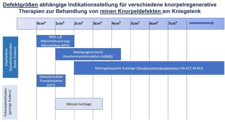
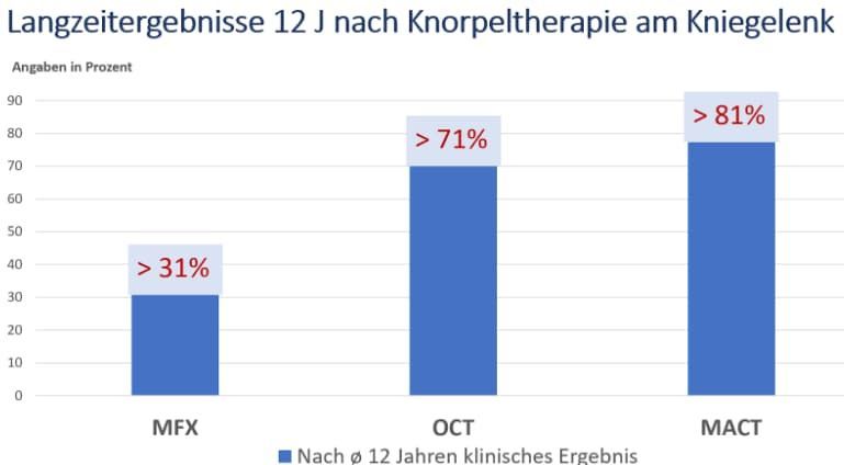
They show how frequently good to very good results can be expected even more than 12 years after knee cartilage therapy. For mBMS, publications are expected in 2023 reporting good to very good study results in over 70% of cases > 10 years.
For the other procedures described, no long-term results exceeding 12 years are currently available.
Cartilage damage with concomitant bone defects
When bone damage is present in addition to cartilage damage, this is referred to as an osteochondral lesion.
The goal of appropriate knee cartilage therapy must be to rebuild this foundation as well. The foundation is the underlying bone. For this purpose, the patient’s own bone from the iliac crest or lower leg is typically used in the form of bone fragments or bone cylinders.
The goal is to restore the boundary layer between cartilage and bone (subchondral lamina) as precisely as possible. Once this has been achieved, any of the described cartilage regeneration procedures—with the exception of BMS—can be applied.
The advantage of OCT for such defects is that it allows for the elegant treatment of cartilage and bone defects up to 1.5 cm².
The use of so-called osteochondral allografts is indicated as a potential method. They are widely used in the United States. These are pieces taken from the knee joints of deceased young patients who have registered as organ donors. They are then measured, milled to the appropriate size, and transplanted into the defect site.
In Europe, this procedure is still rarely used due to the current shortage of available donors.
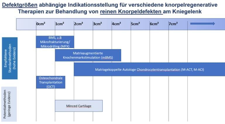
The following diagram summarizes the recommendations of the DGOU Tissue Regeneration Working Group (as of 2021) for different defect sizes. It can be seen that neither BMS nor minced cartilage were included in the recommendations for cartilage-bone defects.
Follow-up Care After Cartilage Treatments on the Knee
The Society for Cartilage Regeneration and Joint Preservation (QKG) provides recommendations for post-treatment care.
Patients should place only partial weight-bearing on the affected leg for approximately ten weeks following any cartilage therapy on the knee. However, the exact duration depends on the size and location of the defect.
Restriction of mobility is sometimes necessary, especially following cartilage regeneration procedures
- behind the kneecap or
- in the patellar glide.
Particularly during the first six weeks after surgery, the use of “continuous passive motion” (CPM) for guided passive or partially active movement of the operated joint on a motorized splint has proven effective. Daily motorized splint therapy lasting several hours is recommended for the first six weeks. This stimulates the cartilage cells to form stable, shock-absorbing cartilage tissue.
During this follow-up treatment, the regenerative cells fill the damaged area with regenerated tissue over the course of the next few months. The quality of the regenerated tissue depends crucially on
- the procedure and the cells used,
- the size of the defect,
- the patient’s age,
- any accompanying damage or misalignments,
- previous surgeries, and
- other factors
. It can take up to two years for the affected cartilage tissue to mature.
There are now also physical therapists who are certified “cartilage specialists” for post-treatment care. They offer sophisticated, phase-based post-treatment programs.
Recently, in addition to cartilage therapy,
- dietary supplements,
- vitamins, and injections such as PRP (platelet-rich plasma) and
- hyaluronic acid
recommended. These can improve the quality of the regenerated cartilage and thus the treatment outcome.
Necessary Treatment of Associated Injuries and Conditions
Successful cartilage therapy can only be expected if concomitant injuries (cruciate ligament tears, meniscus damage) are treated at the same time. If a joint has unstable ligaments, the resulting shear forces usually lead to the loss of the regenerated cartilage. Consequently, the regenerative cartilage procedure fails.
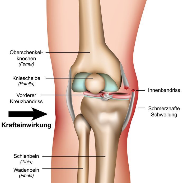
Pathological stress peaks must also be corrected to ensure successful cartilage regeneration. These are caused by misalignment of the leg axis (bowlegs, knock-knees) or the alignment of the kneecaps.
Therefore, in cases of cartilage damage and axial deviation, the appropriate cartilage therapy procedure is combined with a realignment osteotomy. This corrects the axial deviation. In cases of cartilage damage in the patellofemoral joint, a concomitant ligament or bony correction may also be necessary.
Correction of a Leg Axis Deformity
The causes of leg axis misalignment can be varied. In addition to congenital deformities, there are also deformities of the femur or tibia that develop over the course of a person’s life.
One problem arises, for example, when a fracture of the thigh or shin bone does not heal exactly in its original position. The resulting bend in the bone can lead to misalignment of the entire leg axis. This can result in unilateral bowlegs or knock-knees.
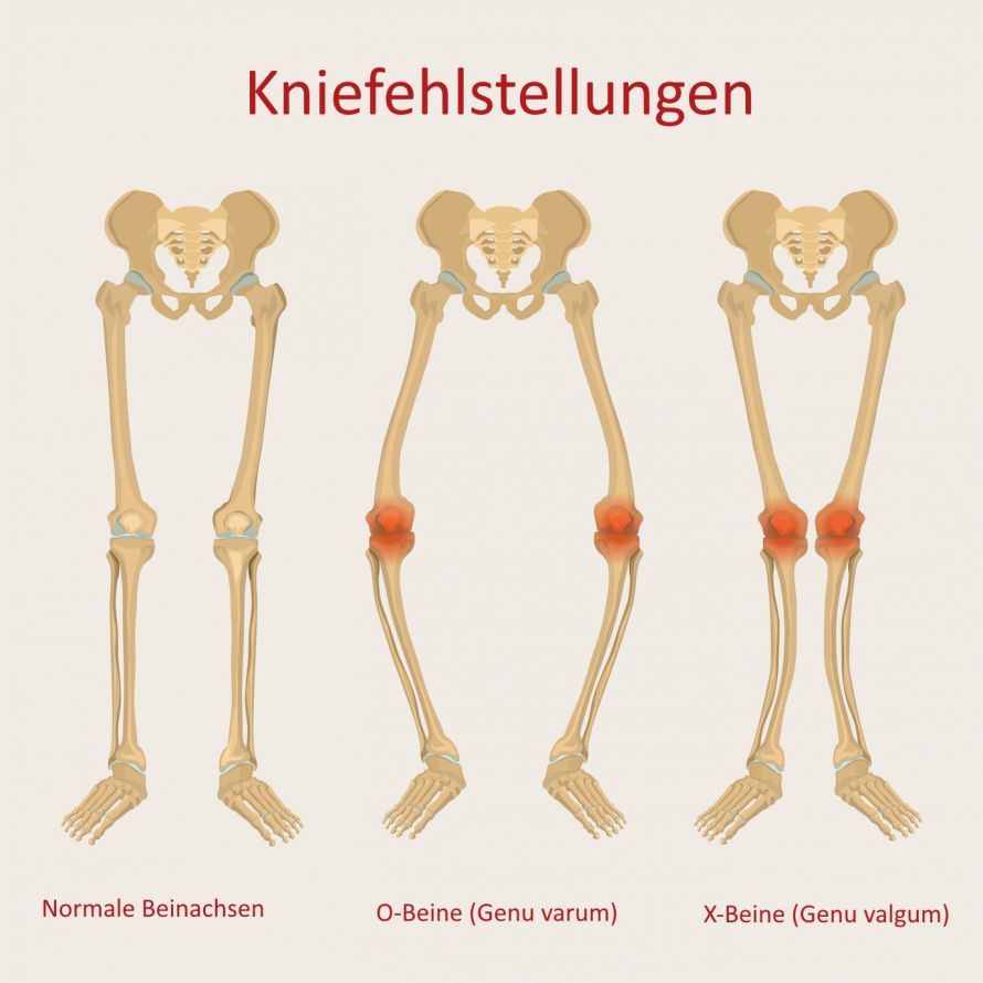
The primary focus is on reducing pain and improving the patient’s quality of life. The goal is to prevent the progression of unilateral joint damage. Ideally, treatment should completely prevent the need for a knee replacement. In most cases, however, the goal is to postpone knee replacement surgery for as long as possible.
In very young patients with minor unilateral cartilage damage in the knee joint, the leg axis is straightened. Cartilage repair procedures can be used to support the damaged side. In cases of severe damage to one side of the joint, the axis is slightly overcorrected. This prevents excessive stress from being placed on the previously healthy side.
For many years, a bone wedge was harvested from the femur or tibia to perform the correction (closing surgical technique, corrective osteotomy). In recent years, new “open” techniques have been developed. The resulting osteotomy gap is held open with appropriate plates. The gap is either left open or filled with autologous, allogeneic, or synthetic bone material.
Navigation devices (e.g., OrthoPilot®, Aesculap) have proven particularly effective for the precise correction of the leg axis. These allow the appropriate correction to be adjusted and monitored intraoperatively.
Correction of Ligament Instabilities
If the ligaments in the knee joint are unstable, this can also lead to increased wear and tear. As a result, cartilage and bone on the unstable side of the knee joint can be damaged.
The cruciate ligaments play a particularly important stabilizing role in this regard. If instabilities are present here, they must be surgically stabilized before or during cartilage therapy. Otherwise, the meniscus and the cartilage layer will be subjected to greater stress due to higher compressive and shear forces.
The meniscus plays a vital role in protecting the knee joint and reducing pressure within it. Therefore, during cartilage regeneration procedures, it should be preserved or sutured whenever possible. Defects affecting more than one-third of the meniscus significantly impair the outcome of cartilage regeneration. Increased cartilage wear can lead to early-onset osteoarthritis. This results in pain, inflammation, and swelling of the knee joint.

About the medical author
Primarius a. D. (DE) Dr. med. Wolfgang Zinser
Sources
- MFX = Mikrofrakturierung (Gudas 2012, Steadman 2003, Goyal 2013, Knutsen 2016, Gobbi 2014, Orth 2019, Ulstein 2014, Pellegrino 2016, Solheim 2020)
- OCT (Ekman 2018, Cognault 2015, Filardo 2015, Solheim 2017, Gudas 2006,Mithofer 2009)
- ACT/mACT ( Aldrian 2014, Niethammer 2020, Kreuz 2019, Ebert 2020, Andriolo 2021, Roffi 2020)