In the past, open hip surgery was often the only way to relieve pain. Today, hip arthroscopy—also known as joint endoscopy—has become the standard minimally invasive procedure. This gentle procedure allows the surgeon to precisely treat hip joint conditions such as femoroacetabular impingement or tears of the labrum without having to make large incisions. Especially for young, athletic patients with hip pain, hip arthroscopy can help delay the onset of hip osteoarthritis and preserve mobility. Learn everything here about the procedure, the indications, and postoperative care following this specialized surgery.
The hip joint is one of the largest joints in the human body. It is a ball-and-socket joint that allows the leg to move in all directions. Hip surgeries are therefore very complex. They must preserve the joint’s function even after the surgery.
Hip arthroscopy is playing an increasingly important role today, particularly for younger and middle-aged patients.
Arthroscopy is a minimally invasive procedure. The surgeon makes only very small incisions in the skin and performs the surgery through them using various very small instruments. This is why it is colloquially referred to as “keyhole surgery.”
The instruments are guided to the surgical site through a tube or cannula and controlled from the outside. A light source, a camera, and special surgical instruments (endoscopy) are used. The camera’s image is transmitted directly to a monitor, allowing the surgeon to view the surgical site magnified.
The video shows how hip arthroscopy works:
Arthroscopies are very commonly used on the knee and shoulder joints. However, hip arthroscopies are also increasingly becoming the treatment of choice.
How is a hip arthroscopy performed?
The procedure is performed under either regional or general anesthesia. During the procedure, the patient lies either on their side or on their back.
It may be necessary to specifically stretch the side to be operated on once more. This opens up the hip joint space for the procedure. A special device is used for this purpose.
After making the incision, the surgeon inserts the necessary instruments into the surgical site.
If necessary, X-rays may also be taken during the procedure.
When is hip arthroscopy necessary?
Arthroscopy is now indicated for many hip conditions.
For example, if the cartilage rim of the hip socket—known as the labrum—is damaged, hip arthroscopy may be performed. The damaged labrum can be sutured, and damaged portions can be removed.
But other conditions, such as
- impingement syndrome (a change in the hip joint space),
- cartilage damage and hip osteoarthritis,
- inflammation of the synovial membrane, or
- loose bodies
are suitable for hip arthroscopy. Thanks to hip arthroscopy, cartilage transplants can now also be performed to a certain extent.
What are the advantages of hip arthroscopy?
Small incisions are sufficient to perform the surgery. Arthroscopy thus minimizes tissue damage and usually does not require cutting through muscles. Since less tissue is damaged, pain is reduced and recovery time is shortened.
Patients who are physically active benefit particularly from hip arthroscopy. Both professional and recreational athletes can return to their previous activity levels more quickly as a result.
After hip arthroscopy, the patient must take it easy for a while. Physical therapy may also be recommended. However, the recovery time is shorter than with open surgery.
In addition, hip arthroscopy offers an advantage from a cosmetic standpoint. The surgical scars are very small and become barely noticeable after a short time.
In summary, hip arthroscopy offers the following advantages over open surgery:
- shorter surgery time,
- less strain on the body,
- faster recovery, and
- smaller scars.
What are the risks associated with hip arthroscopy?
Every surgical procedure carries risks of complications, including hip arthroscopy. These generally include wound healing problems or infections.
In addition, surrounding tissue may also be affected during hip arthroscopy. Nerve injuries, which can lead to numbness or sensory disturbances later on, are also possible. However, these are relatively rare side effects.
What does postoperative care after hip arthroscopy involve?
Postoperative care and rehabilitation always depend on
- the specific indication for surgery,
- the extent of the damage, and
- the patient’s overall condition.
Typically, only a short hospital stay is required. If necessary, forearm crutches are prescribed for the recovery period to provide complete or partial weight-bearing relief.
In many cases, physical therapy begins as early as the first postoperative day. Professional physical therapy is particularly important for people who are active in sports. It ensures that patients can return to their sports as quickly as possible.
This therapy typically includes exercises for mobility, as well as strength and muscle-building exercises. Coordination is also trained through special exercises.
Who performs hip arthroscopy?
Despite all its advantages, hip arthroscopy is a difficult procedure due to the nature of the hip joint. You should therefore consult a specialist. Both surgeons and orthopedic specialists can specialize in minimally invasive hip surgery.
In addition, there are clinics that specialize in hip surgery. These hip centers typically perform several hundred surgeries each year. As a result, the doctors have a wealth of experience with a wide variety of hip surgeries.
Example: Hip Arthroscopy with Cartilage Cell Transplantation
Hip arthroscopy with cartilage cell transplantation is used to treat isolated cartilage damage in the hip joint. Using arthroscopic techniques, cartilage cells—which have been previously cultured in a specialized laboratory—are transplanted into the hip joint.
The goal of cartilage cell transplantation is to prevent progressive damage to the hip joint.
Hip Arthroscopy for the Treatment of Cartilage Damage in the Hip Joint
The treatment of localized cartilage damage in the joint presents a particular challenge.
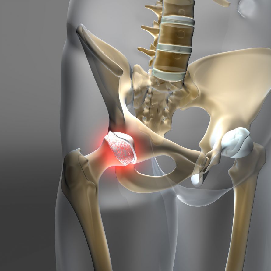
Isolated cartilage damage to the hip joint results from accidents or as a consequence of “impingement syndrome” of the hip. Impingement, caused by bone deformities, leads to a mechanical disruption of the gliding motion of the joint components.
Early signs of such damage include pain when bending or rotating the hip. These symptoms lead to limitations in daily life, e.g.,
- when sitting,
- bending over,
- getting in and out of a car,
- during sports activities, etc.
The doctor diagnoses cartilage damage in the hip joint using X-rays and MRI scans. The exact extent of the cartilage damage is assessed during hip arthroscopy.
Procedure for Treating Cartilage Damage via Hip Arthroscopy
In the case of an isolated, deep cartilage defect without additional osteoarthritis (wear and tear), the surgeon first removes the cause of the cartilage damage. For example, in cases of impingement, the surgeon may shave down bony spurs.
The surgeon then harvests small amounts of cartilage, which are cultured in a specialized laboratory using a 6-week, purely biological process. This process produces so-called spheroids—small “cartilage spheres”—each containing approximately 200,000 viable autologous cartilage cells.
About six weeks later, the surgeon can implant these spheroids via hip arthroscopy. Once in place, they quickly adhere to the cartilage defect. Over the following period, the cartilage cells multiply until the defect is filled with natural cartilage tissue.
Findings on Cartilage Cell Transplantation via Hip Arthroscopy
As a rule, patients regain mobility quickly after a cartilage cell transplant. They simply need to rest the joint and partially offload it for about six weeks.
Younger, active patients in particular benefit significantly from cartilage cell transplantation via hip arthroscopy. In these patients, progressive damage to the hip joint can be prevented.
FAQ: The 8 Most Important Questions About Hip Arthroscopy
What happens during hip arthroscopy?
During hip arthroscopy, the surgeon inserts a camera and fine instruments into the joint cavity through two to three small incisions. First, the joint is gently opened under traction to gain access. Then, any damage can be repaired: A torn labrum is smoothed out or sutured (labrum repair), bone spurs on the femoral head or acetabular rim that are causing problems are shaved down (femoral neck tapering), and inflamed synovial membrane can be removed. Microfracturing to stimulate cartilage regeneration is also possible.
When is hip arthroscopy recommended?
It is primarily indicated for mechanical problems that conservative treatments have been unable to alleviate. These include femoroacetabular impingement (CAM or pincer impingement), in which the bones rub against each other during flexion, as well as tears of the labrum (labrum lesion). Free-floating joint fragments that cause blockages, or early-stage hip osteoarthritis (coxarthrosis) with localized cartilage damage, can also be treated arthroscopically. However, if the wear and tear is far advanced, a hip replacement is often a better option.
How long does recovery take after hip arthroscopy?
The recovery time varies depending on the procedure performed. In the first few weeks after surgery, partial weight-bearing for 2–4 weeks is usually necessary, during which time forearm crutches are used. Physical therapy prevents restricted movement after the procedure and is essential. Light physical activities such as cycling are often possible after 6 weeks, while stop-and-go sports can be resumed only after about 3 to 6 months, once muscle strength has been rebuilt.
What are the risks associated with this procedure?
Although hip arthroscopy is considered safe, there are risks. The traction on the leg can cause temporary numbness in the genital area or on the thigh (nerve stretching). Infections, blood clots, or soft tissue calcifications are rare. An experienced specialist who performs more than 300 hip arthroscopies per year can minimize these risks. In rare cases, arthroscopy may not completely eliminate the symptoms if the cartilage damage was more extensive than anticipated.
What is femoroacetabular impingement?
Femoroacetabular impingement (FAI) is a structural abnormality of the hip joint. In CAM impingement, the transition from the femoral head to the femoral neck is thickened; in pincer impingement, the acetabulum is too deep or tilted incorrectly. During movement, this leads to bony contact (impingement) against the rim of the acetabulum, which damages the labrum and the cartilage. Hip arthroscopy performed as treatment removes these bony protrusions and restores the normal shape.
How long do you have to stay in the hospital?
The hospital stay is usually 2 to 4 days. The surgery itself takes between 60 and 120 minutes, depending on its complexity. Physical therapy begins the day after surgery with passive range-of-motion exercises to prevent adhesions in the joint capsule. The sutures are usually removed after 10 to 12 days.
Can cartilage damage be repaired?
Minor, localized cartilage damage can be treated using hip arthroscopy. One method is microfracturing, in which small holes are drilled into the bone to release stem cells that form replacement cartilage. Cartilage cell transplantation is also possible at specialized centers. However, if osteoarthritis is advanced and the cartilage is worn away over a large area, arthroscopic therapy reaches its limits.
What happens during follow-up care?
Postoperative care is intensive and lengthy. To protect the hip, movements involving more than 90 degrees of flexion and vigorous rotations are often restricted during the first few weeks. Physical therapy helps maintain mobility and strengthen the muscles surrounding the hip. A continuous passive motion (CPM) device can be used at home to passively move the joint. Full weight-bearing on the operated leg is gradually increased, depending on the intraoperative findings and wound healing.
About the medical author
Dr. med. Konrad Körsmeier
Medical writer