Surgical nerve reconstruction may be necessary following nerve tears or severe strains. Such injuries often occur after overstretching (traction injuries), bone fractures, or blunt or sharp trauma. Nerve reconstruction is a microsurgical procedure in which an autologous nerve graft reconnects two nerve ends. In 80 to 90 percent of cases, nerve reconstruction leads to clearly perceptible nerve recovery.
Here you will find further information as well as a selection of specialists and centers for nerve reconstruction.
Definition: What are nerves and nerve fibers?
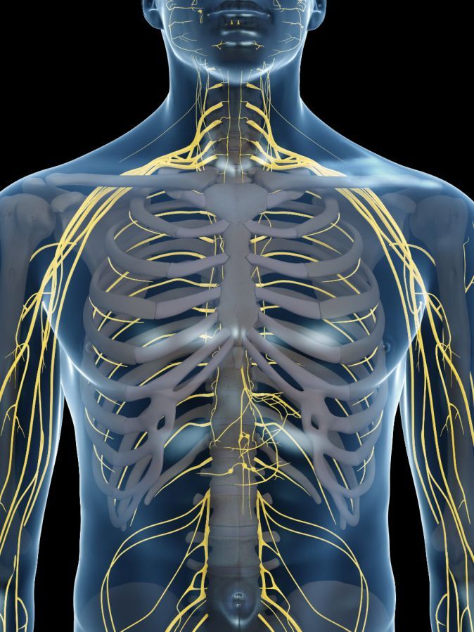
The body has numerous nerves. They are found primarily in the extremities. Nerves range in diameter from that of a pencil lead to that of a pencil; some nerves are even thicker.
The axon (nerve fiber) is simply an extension of the nerve cell, which is located in or near the spinal cord. This extension—the axon—can grow up to two meters long.
Inside a nerve are thousands of nerve fibers that transmit nerve impulses. There are two types of nerve fibers:
- sensory fibers
- motor fibers
Sensory nerve fibers transmit electrical impulses to the spinal cord and brain. There, they are interpreted as pressure, temperature, or pain.
Motor nerve fibers transmit electrical impulses to the periphery, where they cause muscle contraction. The body uses them to control the muscles.
In cases of nerve damage or nerve transection, sensory or motor functions (or both) may be lost.
Severe nerve injuries are caused by
- blunt or sharp force, as well as
- excessive stretching during bone fractures or joint dislocations
. They frequently occur, for example, at the shoulder or knee joints.
How does nerve reconstruction work after nerve injuries?
After such a severe injury, high activity can be observed in the affected nerve cells in the spinal cord. They attempt to regrow their nerve extensions back to where they originally exerted their function.
This regrowth process following a transection forms a connective tissue bridge between the two nerve ends. Nerve fiber regrowth proceeds very slowly, at a rate of about 1 mm per day. The recovery process therefore takes many months.
However, not every nerve fiber eventually reaches its specific target organ. For example, a particular muscle fiber must reconnect to a muscle that performs a specific function. For this reason, the outcome of such nerve reconstruction is inferior to the patient’s previous healthy state.
Consequently, impairments resulting from nerve injuries must be taken into account.
Diagnosis of Nerve Injuries
Identifying nerve injuries based on symptoms
Each peripheral nerve supplies a specific sensory area of the skin and controls specific muscle groups. A doctor can therefore easily deduce which nerve is affected based on the patient’s description of sensory deficits.
The same applies to the loss of motor functions—such as the inability to extend the wrist, fingers and thumb against gravity.
The pattern of symptoms allows for the identification and naming of the damaged nerves. It is often clear where exactly in the limb the damage occurred—for example, in the upper arm, the forearm, or somewhere in between, in the area of the elbow.
It becomes more difficult for the doctor when multiple nerves are damaged and the pattern of deficits is more complicated to identify.
Diagnostic Methods for Nerve Injuries
An electrophysiological examination by a neurologist is always helpful. The neurologist can probe each muscle with a fine needle to determine whether electrical impulses from the brain are reaching the nerves or not.
In the latter case, the muscle itself develops spontaneous electrical activity several weeks after the injury. This indicates a complete nerve injury.
Neurography can also detect nerve injuries. In this procedure, a nerve is stimulated at one point through the skin with a strong electrical current. This nerve impulse is transmitted along the nerve fiber and can be recorded at another point, provided the nerve has not been severed by the injury.
Challenges in Interpreting Test Results
Unfortunately, these tests—which sound very simple—are much more difficult to interpret in everyday medical practice than is described here. After an injury, in the vast majority of cases, no one knows for certain whether a nerve injury involving a tear has actually occurred. It could also simply be a severe strain with temporary loss of function.
This greatly increases the responsibility involved in evaluating neurological and neurophysiological findings. It is certainly possible that a nerve has not been torn and that its connective tissue sheaths have survived. In this case, there is a chance that the nerve fibers—which are only torn internally—will regrow. In such a situation, which is actually favorable, surgery is rather counterproductive.
However, a time-sensitive decision point arises due to the condition of the muscle fibers following a functional nerve loss. Muscle mass diminishes and, after six months, is increasingly converted into connective tissue. This process, known as atrophy, is irreversible.
Nerves can regrow even years later if given the opportunity through surgery. However, the muscles will no longer cooperate by then.
An affected patient must be fully informed about all these complex considerations. This allows them to decide together with their consulting physician whether and when to proceed with nerve surgery.
There are no general rules here, as there are in other areas of medicine, but only discretionary decisions to be made jointly.
Every patient asks whether there is any imaging that can answer all their questions. To this day, the reliability of all these devices remains questionable due to the scar block caused by the accident.
Surgical Techniques for Nerve Reconstruction
The general principle regarding surgical techniques is as follows:
The damaged section of the nerve is not found within the scar. Instead, the nerve is surgically identified in the healthy tissue above and below the scarred area. The damage is then located by tracing the nerve to the scarred region.
The injury results in a soft-tissue-scar block that
- muscles,
- tendons,
- blood vessels, and
- nerves
. If one dissects directly into the scar, one destroys its contents and causes further damage. As a result, the keyhole techniques that are often preferred today are not an option. Instead, based on current knowledge, long incisions are still necessary.
Nerve ends separated by more than two centimeters must no longer be sutured end-to-end. This finding was established in the mid-1950s.
The tensile stress exerted on the reunited nerve ends persists after the surgical procedure. The nerve ends then risk gaping apart internally. Scar tissue develops again between the nerve ends. Consequently, the nerve fibers would be unable to grow past this situation.
Consequently, a few years later in Vienna, H. Millesi developed the concept and technique of nerve transplantation under the surgical microscope.
Procedure for Nerve Transplantation Under the Surgical Microscope
During this procedure, the surgeon makes several small skin incisions. Through these, he removes a nerve from the back of the patient’s lower leg—the sural nerve. This nerve serves exclusively sensory functions on the outer side of the heel. Loss of sensation in that area is the price that must be paid.
The harvested nerve is 30 to 40 centimeters long and has the diameter of a pencil lead. This long segment of nerve now serves as an autologous graft. It can be used in several portions and inserted between the two ends of a main nerve in the arm or leg.
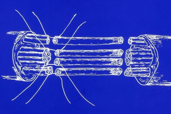
To do this, scar tissue at the nerve ends must first be removed under a surgical microscope. In addition, the microstructure visible in the main nerve must be prepared. This slightly increases the original distance between the two main nerve ends.
Several segments of the harvested nerve are then inserted without tension, taking into account the microstructure of the main nerve.
Threads as thin as a human hair are used for the sutures.
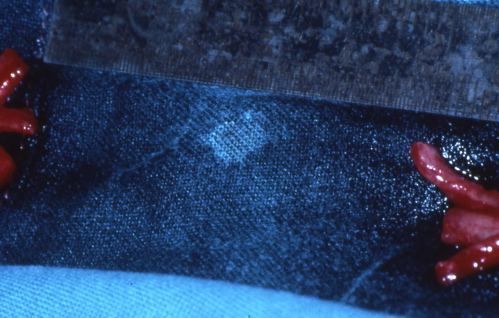
The nerve fibers grow from the reconstructed main nerve into the nerve grafts, through them, and finally back into the remaining peripheral nerve stump.
The use of such a graft leads to better results than simply pulling the ends of severed nerves together.
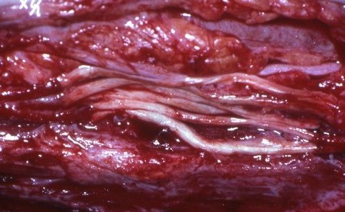
The form of nerve reconstruction described is called “microsurgical autologous nerve transplantation.” “Autologous” means that the transplanted nerve was taken from the patient’s own body. This has the advantage that the body does not reject the graft, as is often the case with allografts.
However, due to the long nerve fiber growth phase, the outcome of such a surgery is not determined until many months later.
Medications for Nerve Reconstruction
To date, the use of medication to promote nerve fiber growth has only been demonstrated in experimental settings.
There are recurring references to vitamins. However, human nerve cells are capable of this on their own, without needing any assistance.
Possible Complications and Risks of Nerve Reconstruction
Surgical exposure of an injured nerve for nerve reconstruction must not result in additional soft tissue injuries, particularly vascular injuries. The risk of vascular injuries increases if vascular reconstruction was already necessary during initial trauma surgery.
In such cases, it may become absolutely necessary to bring erythrocyte concentrates obtained through autologous blood donation prior to nerve reconstruction. Erythrocyte concentrate is a blood product consisting of red blood cells.
After the microsurgical insertion of nerve grafts, the surgical site must not be re-operated on. Otherwise, the grafts would be destroyed.
A prerequisite for planning nerve reconstruction is therefore that no further procedures are necessary in this region. Any bone injuries that occurred simultaneously must heal sufficiently so that they do not require further surgery.
This also applies to implanted osteosynthesis materials—that is, metal components used to treat bone injuries, such as
- screws,
- metal plates,
- wires, or
- pins.
These implants must either be removed during the nerve repair itself or remain in place for life. Their subsequent removal could lead to the destruction of the nerve grafts.
Nerve reconstruction should also be performed as soon as possible. Otherwise, the affected muscles would no longer be functional.
Findings Following Nerve Reconstruction
For the reasons mentioned above, the results of microsurgical nerve reconstruction are always slow to become apparent. Statistical data on healing outcomes can therefore only be obtained at long intervals. If changes are then made to the surgical approach, one must wait many more years for the evaluation of the new results. Progress is therefore very slow.
In the meantime, industrially manufactured artificial nerve grafts are occasionally used. We must wait just as long for the results of these as well.
Artificial nerve grafts lack important cellular components, resulting in problems that remain unresolved to date.
Autologous nerve transplantation leads to noticeable nerve recovery in 80 to 90% of operated cases. A total failure rate of approximately 10% must be expected. This is due to the delicacy and technical vulnerability of the surgical techniques used in nerve reconstruction.
Follow-up Care After Nerve Reconstruction
Physical therapy is used to treat stiffness in joints prone to becoming rigid. It also trains functioning muscles surrounding the affected muscle group so that they can compensate for the lost functions.
Electromyographic monitoring shows whether electrical impulses reappear in the affected muscle group after the nerve fibers have regrown. Physical therapy will then focus specifically on the previously paralyzed muscle group.
During the phase of nerve dysfunction, deformities may develop in the hands or feet. These can be corrected using custom-made splints. The primary goal here is to prevent joint stiffness and overstretching of certain tendons.
Unfortunately, there is no proven method to prevent muscle atrophy. External stimulation of the affected muscle groups using surface electrodes is intended to have a preventive effect here. While scientific proof is lacking, experience suggests it is effective.
It is incorrect to use TENS devices for this purpose, as they are designed for other functions.
Conclusion
Nerve reconstruction following nerve injuries is nowadays a microsurgical, time-consuming, and delicate procedure. Autologous transplantation, involving the harvesting of a cutaneous nerve from the lower leg, yields the best results.
Any return of function following a nerve injury is an invaluable benefit and worth attempting surgery. However, one cannot expect a full restoration of the patient’s previous healthy state.
Determining the optimal timing for surgery is challenging. This decision is left to the discretion of the patient and physician following proper consultation. In cases where nerves are very difficult to access surgically, greater caution will be exercised than with easily accessible nerve pathways.
Before any nerve reconstruction, it must be certain that the surgical site will not need to be reopened for other reasons.
About the medical author
Prof. Dr. med. Götz Penkert
Medical writer