Supinator compartment syndrome is a rare but clinically significant compression syndrome in which the radial nerve is compressed in the supinator compartment at the elbow. The motor branch, the ramus profundus, is primarily affected, which can lead to dysfunction of the extensor muscles in the forearm.
Typical symptoms include exertion-related pain in the forearm, weakness in finger extension, and symptoms that are often mistaken for tennis arm, tennis elbow, or epicondylitis. The nerve compression is often caused by overuse, repetitive rotational movements, inflammatory changes, or constriction by the supinator muscle itself.
Ganglions, anatomical variations, or irritation in the elbow region can also trigger supinator tunnel syndrome. Diagnosis is challenging and, in addition to a clinical examination, often requires imaging techniques such as ultrasound, MRI (magnetic resonance imaging), and measurement of nerve conduction velocity. Depending on the severity, conservative treatment options such as rest, physical therapy, and anti-inflammatory medications, or surgical decompression performed by a hand surgeon, may be considered to relieve pressure on the nerve fibers and prevent lower radial nerve palsy or “drooping hand.”
What is supinator loge syndrome?
Supinator loge syndrome is a nerve compression syndrome. It refers to a nerve impairment that progresses slowly and recurs repeatedly in response to even slight pressure.
The development of supinator loge syndrome requires naturally occurring narrow passages within the extremities (arm or leg). One such narrow passage is located below the elbow joint, beneath the supinator muscle (Musculus supinator). This muscle is responsible for rotating the forearm and hand into a position where we can look down into the open palm from above.
The supinator muscle, functioning in this way, has nothing to do with the affected nerve. It merely indicates the location of the nerve compression in supinator compartment syndrome.
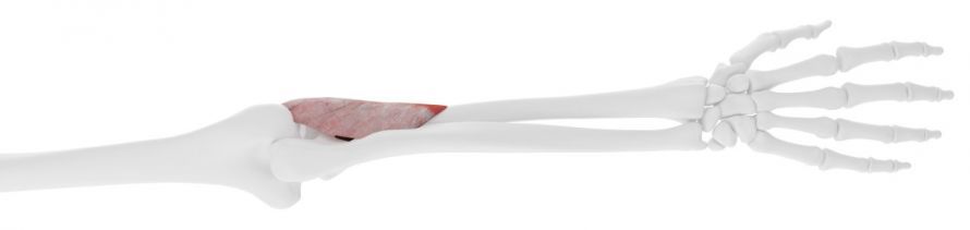
Causes of supinator compartment syndrome
The radial nerve and supinator compartment syndrome
Supinator compartment syndrome affects a branch of the radial nerve (nervus radialis). This nerve controls all the muscles used to extend the wrist, the four fingers, and the thumb.
At the level of the elbow crease, the radial nerve mentioned above divides into two branches:
- a superficial branch (Ramus superficialis) and
- a deep branch (ramus profundus).
The superficial branch primarily contains nerve fibers responsible for sensation
- on the back of the forearm,
- the back of the hand, and
- the backs of the fingers
. This means that this nerve branch transmits touch or pressure on the back of the hand as electrical signals to the brain. The brain processes these signals, allowing us to feel the pressure.
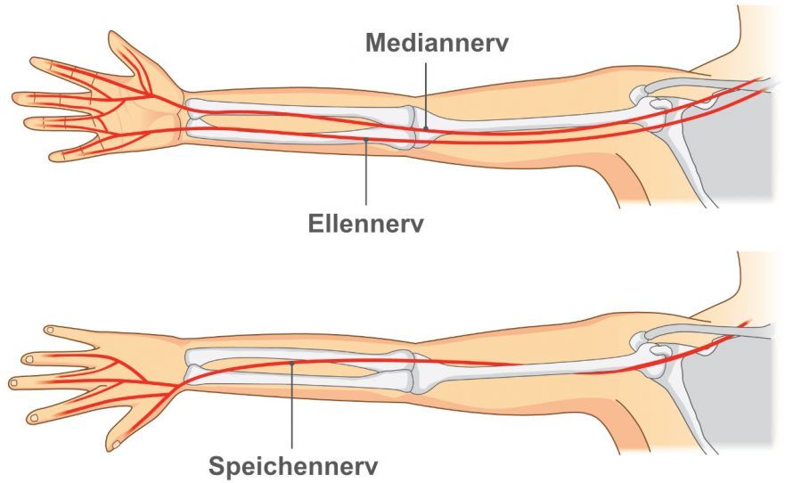
The second branch, located deeper, is responsible for motor function. It transmits electrical signals from the spinal cord to the muscles in the forearm. These muscles are responsible for extending the wrist, fingers, and thumb. It therefore contains no sensory nerve fibers, only motor ones.
A small motor nerve branch represents a regular anatomical exception. It branches off from the superficial, sensory radial nerve branch and leads to the extensor carpi radialis brevis muscle.
This exception is of great significance in classifying the neurological deficit pattern of supinator groove syndrome. It is also important for the surgeon.
Impairments of this nerve branch cause motor deficits (paralysis) in supinator compartment syndrome. Sensory disturbances or pain do not occur in this context.
Structure of a Nerve
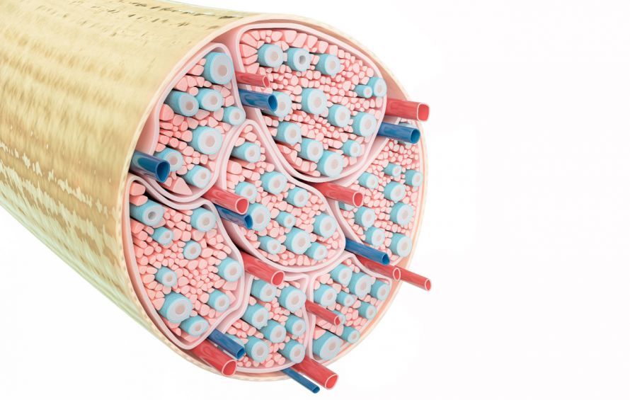
Every peripheral nerve consists of several grouped nerve bundles that are enclosed and held together by a relatively tough sheath, the epineurium. Blood vessels run between the nerve bundles.
The nerve bundles, in turn, consist of thousands of individual nerve fibers. Each of these nerve fibers is surrounded by an insulating layer, similar to the rubber coating on a copper wire. This insulating layer prevents electrical impulses from jumping from one nerve fiber to the next. It also influences the speed at which this nerve impulse is transmitted.
A nerve fiber is a cylindrical cell extension whose parent cell is located in the spinal cord. Within a peripheral nerve, this nerve cell extension can grow up to 2 meters long.
Development of Supinator Compartment Syndrome
The fatty insulating layer surrounding each individual nerve fiber is highly sensitive to pressure. Repeated pressure on a nerve can damage this insulating layer. This reduces the speed at which the nerve impulse travels along the nerve fiber.
This adverse effect then leads to the neurological deficit characteristic of supinator compartment syndrome. Measurements of nerve conduction velocity can confirm the damage.
Symptoms of Supinator Loge Syndrome
Supinator loge syndrome affects the ramus profundus, or deep branch, of the radial nerve. It does not cause pain, but rather motor deficit (paralysis).
In supinator loge syndrome, the four long fingers become increasingly difficult to lift. The same applies to the thumb: its ability to extend away from the back of the hand becomes progressively weaker.
It is noteworthy that, in contrast, the ability to raise the back of the hand remains fairly strong. This can be explained by the aforementioned nerve branch originating from the superficial branch of the radial nerve and supplying the wrist extensor muscles.
The compression of the deep palmar branch (ramus profundus) beneath the supinator muscle in the supinator compartment thus occurs at a point where that small motor branch supplying the thumb-side wrist extensor has already been diverted.
This results in the characteristic presentation of supinator compartment syndrome: weakness of the middle fingers and thumb extensors with preserved wrist extension.
Examination and Diagnosis of Supinator Compartment Syndrome
The characteristic pattern of paralysis seen in supinator compartment syndrome—without accompanying pain—is easily recognized by neurologists. Using electroneurography (measurement of nerve conduction velocity), the neurologist can confirm that the transmission of the electrical impulse responsible for finger extension is slowed.
In addition, testing the muscles of the forearm innervated by the deep branch of the radial nerve reveals changes in evoked potentials. This is assessed using electromyography, which measures electrical muscle activity.
Imaging techniques, such as sonography (ultrasound) or MRI (magnetic resonance imaging), cannot definitively diagnose supinator groove syndrome. However, in individual cases, they can detect or rule out a lipoma (fatty tumor) located beneath the supinator muscle.
For reasons that are currently unknown, a lipoma can develop beneath the supinator muscle. This can contribute to the nerve compression symptoms of supinator loge syndrome.
Procedure for Supinator Loge Syndrome Surgery
Treatment of supinator fossa syndrome can only be performed surgically.
The site of compression is a small, crescent-shaped, tendinous structure from which muscle fibers of the supinator muscle extend. It crosses the course of the deep branch of the radial nerve. When pressure is applied, the edge of this tendon presses into the nerve.
The goal of supinator groove syndrome surgery is to allow the insulating layer surrounding each nerve fiber to fully recover. To do this, the surgeon must make an incision in and sever the tendinous structure that runs across the nerve.
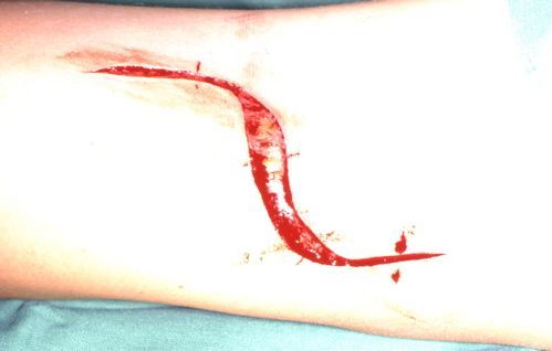
At this point, several small blood vessels also cross the course of the nerve. Furthermore, the nerve branch itself is only as thick as a pencil lead. Therefore, supinator compartment syndrome surgery requires the utmost care to avoid damaging the tiny nerve itself.
Even greater care must be taken to safely identify and preserve the continuity of the aforementioned, even thinner nerve branch—which runs toward the thumb-side wrist extensor—above the beginning of the supinator muscle canal.
If this is not done, this wrist extensor will also fail!
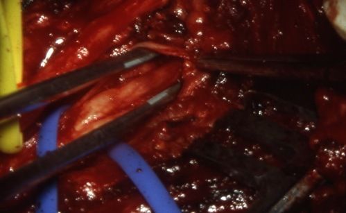
The supinator compartment syndrome surgery is therefore challenging. While it does not require microscopic magnification, the minimally invasive keyhole technique is not an option as a surgical method. The risk of damaging the nerve branch that has just been exposed is too great. It is so thin that any attempt to reconstruct it would likely fail.
The skin incision and choice of approach must therefore be large enough to safely identify and preserve the aforementioned nerve structures.
Medical Treatment of Supinator Compartment Syndrome
There is no effective pharmacological treatment for so-called peripheral nerve entrapment syndromes.
Local corticosteroid injections may, at best, provide temporary relief, but they do not resolve the underlying problem.
Possible Complications and Risks of Supinator Compartment Syndrome Surgery
Postoperative improvement in motor deficits does not occur immediately. The results typically become apparent after weeks or months, depending on the extent of damage prior to supinator notch syndrome surgery.
However, supinator loge syndrome surgery should not lead to a worsening of symptoms due to damage to any anatomical features of the nerve distribution. The patient must, however, be informed of the risk of at least a potential loss of wrist extensor function.
If a lipoma is found beneath the supinator muscle, the planned procedure must also be expanded. However, this is rarely the case.
Postoperative Care Following Supinator Loge Syndrome Surgery
If wrist extensor function is preserved, there is no need to worry that the tendons of the middle fingers and thumb extensors will become overstretched due to constant hanging. Unlike in cases of injury-related radial nerve rupture, a wrist splint is not required in this situation.
Physical therapy or self-directed, mentally guided exercises should focus on the extension movements of the middle fingers and thumb.
Follow-up examinations are also recommended after surgery. For this purpose, electromyography should be performed once or twice.
Conclusion
Supinator loge syndrome is a slowly progressive nerve compression of the deep branch of the radial nerve. The site of compression is located approximately 3 cm below the elbow crease. There is no pain, but there is increasing weakness when extending the fingers and thumb.
In contrast to this paralysis, the ability to extend the wrist remains intact.
Decompression surgery is necessary but requires the utmost care to preserve wrist extension function.
A minimally invasive procedure is currently not recommended due to the specific anatomical situation described.
FAQ on Supinator Fossa Syndrome
What is supinator groove syndrome?
Supinator groove syndrome is a compression syndrome in which the radial nerve—or more specifically, its deep branch—is compressed in the area of the supinator muscle. This nerve compression primarily causes motor deficits in the forearm, without significant sensory disturbances in the palm of the hand.
What are the symptoms of supinator groove syndrome?
Typical symptoms include pain in the forearm and elbow, which may radiate into the arm, as well as weakness in finger extension. In advanced cases, lower radial nerve palsy with a “drooping hand” may occur. Sensory disturbances are usually not the primary concern, as motor fibers are primarily affected.
How is supinator loge syndrome diagnosed?
Diagnosis involves a clinical examination of the elbow, functional tests of the extensor muscles, and measurement of nerve conduction velocity. Imaging techniques such as MRI, ultrasound, or magnetic resonance imaging help to visualize ganglia, a narrowing, or inflammation in the supinator compartment. It is important to differentiate this condition from tennis arm, tennis elbow, or carpal tunnel syndrome.
How is supinator compartment syndrome treated?
Treatment for supinator compartment syndrome usually begins conservatively with rest, physical therapy, anti-inflammatory medications, and avoiding repetitive rotational movements, such as those involved in playing the piano. If symptoms persist or there is a significant impairment of motor function, surgical decompression may be necessary. This procedure involves surgically relieving pressure on the nerve.
When is surgery necessary?
Surgical treatment is recommended if, despite conservative therapy, there is persistent nerve compression, progressive paralysis, or a significant impairment of finger extension. The goal of surgical decompression is to relieve pressure on the radial nerve in order to prevent permanent damage to the nerve fibers and the forearm muscles.
Share this article