Recommended specialists
Brief overview:
- What is hip dysplasia? It is one of the most common congenital skeletal developmental disorders, in which the acetabulum does not sufficiently enclose the femoral head, which can lead to various symptoms.
- Causes: Girls are affected significantly more often. Genetic predisposition, complications during pregnancy or a multiple pregnancy, or neurological diseases usually cause the disease.
- Treatment: The earlier the condition is detected and the less severe it is, the more likely conservative measures such as joint repositioning, retention, and orthoses can be successful. In rare cases, surgery is required.
- Surgery in childhood: An acetabuloplasty between the 18th month and the age of eight repositions a small part of the bone so that the acetabulum can better cover and hold the joint.
- Surgery in adulthood: The highly complex triple pelvic osteotomy fixes the hip with screws after bone repositioning and achieves very good long-term results, significantly reducing discomfort.
- Prevention: The disease cannot be prevented, but early therapy improves the chances of successful treatment. Therefore, medical check-ups during the baby’s first few weeks should be carried out without fail.
Article overview
Background information on hip dysplasia
Hip dysplasia is a malformation of the acetabulum, where the head of the femur is located. Girls are more often affected by this malformation than boys. If hip dysplasia is not recognized, there is a risk of wear and tear (osteoarthritis) in the joint. This can lead to pain and severe difficulty walking.
Hip dysplasia can be reliably detected with the help of a harmless ultrasound examination in the first days of life. Newborns are examined by orthopedists and pediatricians in order to be able to start any necessary brace treatment as early as possible.
This ultrasound examination of the hip joint has proved so successful that it is included in the statutory medical check-up (4-6 weeks after birth).
If hip dysplasia is detected early, there is a very good chance of recovery. With targeted and consistent treatment, healthy hip joints can usually develop. In this way,
- Any operations that may be necessary later,
- A severe hip limp, or
- Premature hip wear
are avoided.
What does hip dysplasia mean?
Hip dysplasia can be congenital or sometimes acquired during life. Often the acetabulum is formed too small and too steep. The femoral head is then not sufficiently covered by the acetabulum, especially laterally and at the front.
The consequence of this maldevelopment can be a dislocation of the hip (hip luxation). In this process, the femoral head moves laterally upward/backward out of the acetabulum, which is too small. Fortunately, however, the proportion of hip dislocations is much lower (only one in about every 15 children with hip dysplasia has a hip dislocation).
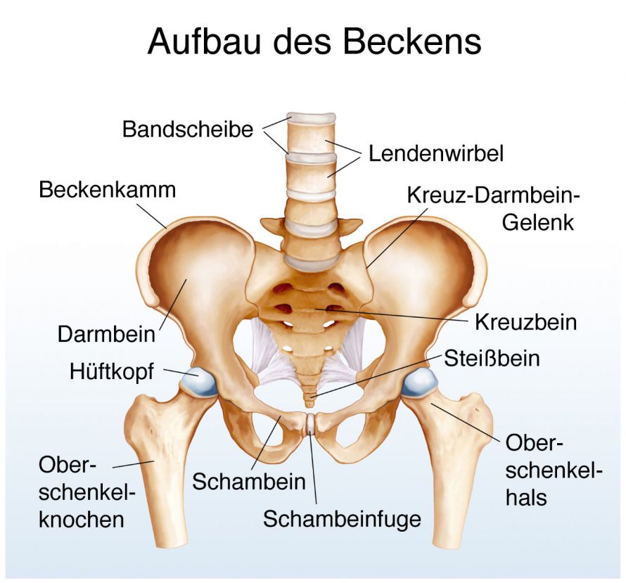
Structure of the human pelvis. The ischium extends below the pubic bone © Henrie | AdobeStock
Causes of hip dysplasia
Hip dysplasia is about 5 to 7 times more common in girls than in boys and is clustered in some families. Often several siblings are affected.
Other possible causes of hip dysplasia include:
- Mechanical factors such as a multiple pregnancy or
- An insufficient amount of amniotic fluid during pregnancy, resulting in relative confinement for the unborn baby.
Also, numerous concomitant neurological disorders, such as
- Spina bifida or
- As a result of early childhood brain damage (cerebral palsy) or
- Rare genetic diseases
can lead to hip dysplasia.
How is hip dysplasia treated conservatively?
The successful conservative (non-surgical) therapy of hip dysplasia is determined by
- The degree of malformation and
- The early start of treatment.
The more severe the dysplasia or the later in a person's life it was diagnosed, the more likely the physician will (have to) resort to surgical methods.
Conservative treatment for severe dysplasia and dislocation is based on three main pillars:
- Reduction (adjustment of the joint into the acetabulum),
- Retention (ensuring that the femoral head remains in the acetabulum) using plaster or splints/braces, and
- Maturation treatment, i.e., consistent orthosis follow-up under ultrasound controls. This allows the joint to develop properly.
In the case of very mild dysplasia and in newborns, it is usually sufficient to give the child a little extra room when swaddling them and leave the baby to their natural urge to move.
In most cases, children then have no more problems in their further development and the hips mature well. This further development of the acetabulum is also monitored by the physician using an ultrasound.
Are there any special preventative measures?
Hip dysplasia cannot be prevented! Even during pregnancy, there are no ways to prevent this.
However, in order to avoid late consequences, hip dysplasia should be diagnosed as early as possible. In Germany, this has been ensured since 1996 by ultrasound exams during the infant’s check-ups at one week and one month at the pediatrician's or orthopedist's office.
Surgical treatment of hip dysplasia in childhood
Despite intensive and long-term conservative treatment, residual hip dysplasia may persist.
The chances of success of conservative treatment deteriorate with increasing age. Severe residual hip dysplasia can no longer be sufficiently improved using conservative methods from the end of the second year of life.
Intensive special physiotherapy using the Vojta method is still very effective in the first year of life. In the second year of life, however, it already loses effectiveness.

Hip dysplasia treatment of a newborn with the help of bands and splints © Marko | AdobeStock
Acetabuloplasty is a proven surgical procedure for treating such residual hip dysplasia. The majority of acetabuloplasties are performed between the ages of 18 months and the age of eight. For this purpose, a small bone wedge is laterally repositioned to better cover the femoral head after the ilium above the acetabulum has been partially separated. The sawed bone wedge is inserted into the resulting gap.
For a few weeks, the children receive aftercare in a plaster cast. Sometimes the hip joints are almost or completely dislocated (luxated). In these cases, surgical, open hip joint resetting is an option after unsuccessful conservative therapy.
This therapy is a combination of femoral shortening and acetabuloplasty. It should be performed in hospitals that have considerable and extensive experience with it.
Surgical treatment of hip dysplasia in adults
Hip dysplasia is the most common cause of early painful hip wear. Likewise, this makes them the main reason for the necessity of an artificial hip joint.
Many hip dysplasia patients between the ages of 20 and 40 have unbearable pain in their hips at times. They are often incapacitated for work in the long term because of this. About 200,000 artificial hip joints are now implanted in Germany every year.
After the conclusion of growth, there is a possibility of correcting severe hip dysplasia with a joint-preserving technique. The procedure is known as a triple pelvic osteotomy (triple osteotomy). It was developed in the Dortmund municipal hospital, Klinikum Dortmund, in the mid-1970s by Prof. Dr. Tönnis and K. Kalchschmidt.
This surgery is one of the largest and most complex pelvic surgeries in orthopedics. It is therefore performed more frequently and in a specialized manner in only a few hospitals in Germany.
Three surgical approaches are required to correct the excessively steep acetabulum using a triple pelvic osteotomy.
- The ischium,
- The ilium, and
- The pubic bone
are severed. These three bones form the acetabulum. Screws are used to fix the bones in the required corrected position.
The operation time is about two ½ to three hours on average.
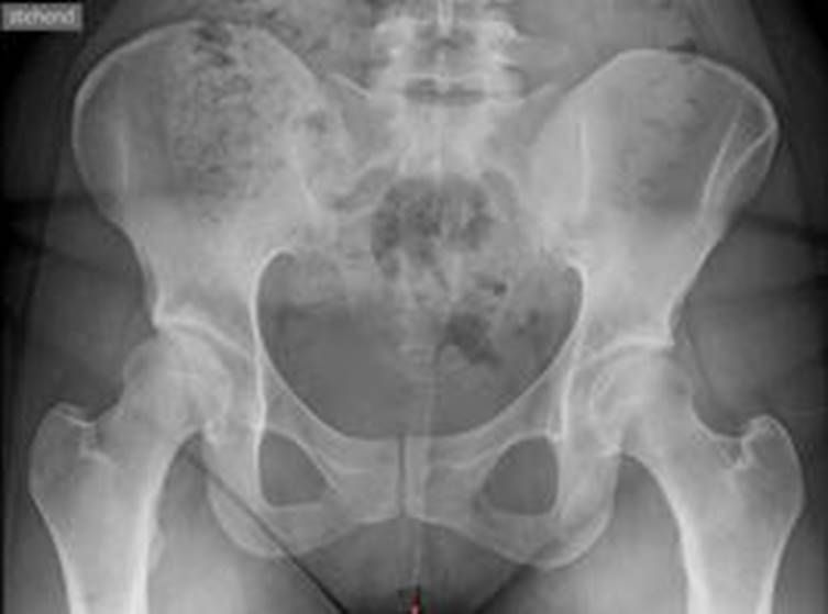
Fig. 1: Severe right hip dysplasia. Orthopedics at the EK-Unna; kindly provided by Dr. med. Pothmann.
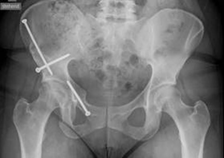
Fig. 2: Hip dysplasia after a triple pelvic osteotomy © Orthopedics at the EK-Unna; kindly provided by Dr. med. Pothmann.
Patients can leave the hospital after about 12 to 14 days. However, the prerequisite for this is that patients can safely take the load off the operated leg by using two forearm crutches.
This operation can completely eliminate the pain from loads and especially groin pain. Most patients can then actively participate in life again without pain or restrictions.
There are over 20 years of long-term results for this operation, which is superior to a PAO (by Axel Küpper et al., Unna/Dortmund). In a POA (periacetabular osteotomy), the acetabulum is chiseled out of the pelvic composite and then rotated.
Nevertheless, PAOs are performed more frequently worldwide because they are technically less complicated. Unfortunately, the long-term results are worse than with a triple osteotomy.


